. 2014 Aug;24(8):2151-9.
doi: 10.1093/cercor/bht065. Epub 2013 Mar 19.

Dissociable functional networks of the human dentate nucleus

Affiliations

Dissociable functional networks of the human dentate nucleus

Jessica A Bernard et al. Cereb Cortex. 2014 Aug.

Abstract

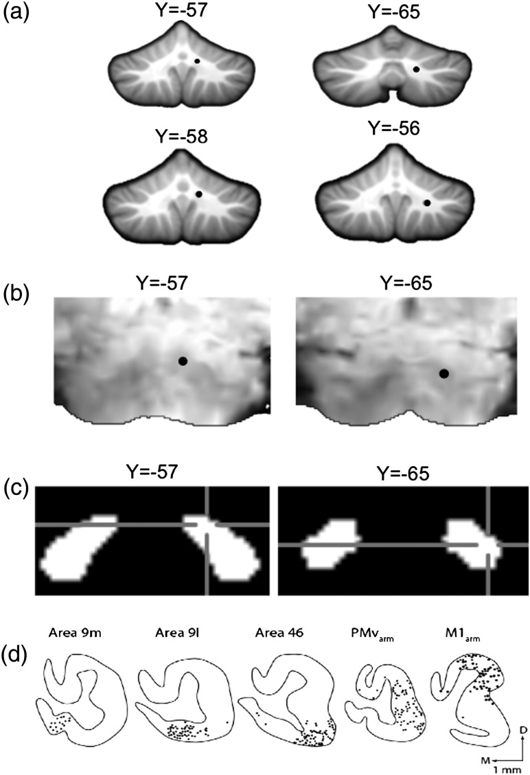
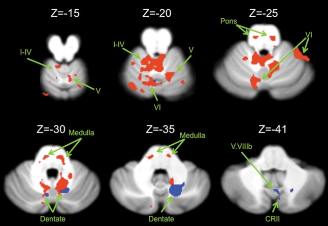
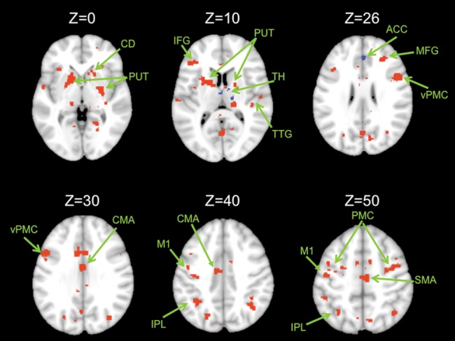
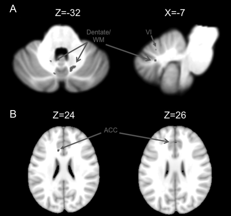
The cerebellar dentate nucleus has been reported to project to motor and prefrontal cortical regions in nonhuman primates from 2 anatomically distinct areas. However, despite a wealth of human neuroimaging data implicating the cerebellum in motor and cognitive behaviors, evidence of dissociable motor and cognitive networks comprising the human dentate is lacking. To investigate the existence of these 2 networks in the human brain, we used resting-state functional connectivity magnetic resonance imaging. The resting-state fMRI signal was extracted from regions of interest in the dorsal and ventral dentate nucleus. We report a "motor" network involving the dorsal dentate, anterior regions of the cerebellum, and the precentral gyrus, and a "cognitive" network involving the ventral dentate, Crus I, and prefrontal cortex. The existence of these 2 distinct networks supports the notion that cerebellar involvement in cognitive tasks is above and beyond that associated with motor response components.

Keywords: MRI; cerebellum; dentate nucleus; resting state connectivity.


Figures

Figure 1.
Figure 1.
Dentate seed locations. (a) Locations of the dorsal (12, −57, −30; top left) and ventral (17, −65, −35; top right) seed regions in the dentate nucleus overlaid on an anatomical image in standardized space. The additional dorsal (10, −58, −31; bottom left) and ventral (20, −56, −37; bottom right) seeds are also presented. The dorsal and ventral seeds were restricted to the dorsal one-third, and bottom half of the dentate nucleus, respectively. (b) The seed locations have also been overlaid on a single subject's functional image, in standardized space, to aid visualization. The dark regions are the dentate nuclei (dorsal, left; ventral, right). (c) The seed locations denoted on the dentate nucleus from the SUIT atlas (Diedrichsen et al. 2011). Blue-cross hairs indicate the seed location. (d) In the monkey dentate, there is a clear topography within the dentate such that the dorsal region projects to the motor cortex, while the ventral region projects to the prefrontal cortex (figure reprinted with permission from Strick et al. 2009).
Figure 2.
Figure 2.
Connectivity maps in the cerebellum. Functional connectivity maps for the dorsal (red) and ventral (blue) dentate in the cerebellum (axial views; both presented at P < 0.00001). The dorsal network includes lobules I–IV, V, and VI in the anterior regions of the cerebellum. The ventral network is predominately made up of more posterior regions including crus II, but also extends into lobule VI. Images are oriented such that the right hemisphere is presented on the right. Roman numerals indicate the cerebellar lobules. CRII: crus II.
Figure 3.
Figure 3.
Connectivity maps in the whole brain. Functional connectivity maps for the dorsal (red) and ventral (blue) dentate seeds in the whole brain (axial views; both presented at P < 0.00001). The dorsal network consists primarily of motor and parietal regions, though there are also prefrontal regions included. The ventral network consists of the anterior cingulate cortex and the caudate. Images are oriented such that the right hemisphere is presented on the right. ACC, anterior cingulate cortex; CMA, cingulate motor area; CD, caudate; IPL, inferior parietal lobule; M1, primary motor cortex; MFG, middle frontal gyrus; PUT, putamen; SFG, superior frontal gyrus; TH, thalamus; TTG, transverse temporal gyrus; PMC, premotor cortex; d, dorsal; v, ventral. Unlabeled areas are extensions from the clusters presented in Table 2.
Figure 4.
Figure 4.
Regions of overlap in the dorsal and ventral networks. The results of our conjunction analysis indicative of overlap between the 2 networks are presented in, (A) the cerebellum, and (B). the whole brain. All images are oriented such that the right hemisphere is presented on the right. Roman numerals indicate the cerebellar lobules. ACC, anterior cingulate cortex.

References

    1. Akkal D, Dum RP, Strick PL. Supplementary motor area and presupplementary motor area: targets of basal ganglia and cerebellar output. J Neurosci. 2007;27:10659–10673. doi: 10.1523/JNEUROSCI.3134-07.2007. - DOI - PMC - PubMed
    1. Alexander GE, Crutcher MD. Functional architecture of basal ganglia circuits: neural substrates of parallel processing. Trends Neurosci. 1990;13:266–271. doi: 10.1016/0166-2236(90)90107-L. - DOI - PubMed
    1. Alexander GE, DeLong MR. Microstimulation of the primate neostriatum: I. Physiological properties of striatal microexcitable zones. J Neurophysiol. 1985a;53:1417–1432. - PubMed
    1. Alexander GE, DeLong MR. Microstimulation of the primate neostriatum: II. Somatotopic organization of striatal microexcitable zones and their relation to neuronal response properties. J Neurophysiol. 1985b;53:1433–1446. - PubMed
    1. Alexander GE, DeLong MR, Strick PL. Parallel organization of functionally segregated circuits linking basal ganglia and cortex. Annu Rev Neurosci. 1986;9:357–381. doi: 10.1146/annurev.ne.09.030186.002041. - DOI - PubMed

Publication types