. 2013 Apr 16;52(15):2649-54.
doi: 10.1021/bi301638h. Epub 2013 Apr 5.

Lipid-induced conformational changes within the cytochrome b6f complex of oxygenic photosynthesis

Affiliations

Lipid-induced conformational changes within the cytochrome b6f complex of oxygenic photosynthesis

S Saif Hasan et al. Biochemistry. .

Abstract

Cytochrome b6f catalyzes quinone redox reactions within photosynthetic membranes to generate a transmembrane proton electrochemical gradient for ATP synthesis. A key step involves the transfer of an electron from the [2Fe-2S] cluster of the iron-sulfur protein (ISP) extrinsic domain to the cytochrome f heme across a distance of 26 Å, which is too large for competent electron transfer but could be bridged by translation-rotation of the ISP. Here we report the first crystallographic evidence of significant motion of the ISP extrinsic domain. It is inferred that extensive crystallographic disorder of the ISP extrinsic domain indicates conformational flexibility. The ISP disorder observed in this structure, in contrast to the largely ordered ISP structure observed in the b6f complex supplemented with neutral lipids, is attributed to electrostatic interactions arising from anionic lipids.


Figures

Fig 1
Fig 1
Cyt b6f complex from the moderately thermophilic cyanobacterium Mastigocladus laminosus (PDB ID 2E74). Polypeptide subunits are shown as ribbons and prosthetic groups as sticks and spheres. Color scheme: Cytochrome b6 (cyt b6), pink; subunit IV (sub IV), cyan; cytochrome f (cyt f), wheat; Iron-Sulfur Protein (ISP), orange; Pet G, brown; Pet L, gray; Pet M, yellow; Pet N, green.
Fig 2
Fig 2
p-Side extrinsic domain architecture of the cyt b6f (PDB ID 2E74) and bc1 complex (PDB ID 3CX5). (A, B) The cyt f extrinsic domain (yellow) consists of an elongated β–sheet structure that extends over 75 Ål. (C, D) In contrast, the cyt c1 extrinsic domain (yellow) is predominantly α–helical in structure and is more compact. The ISP polypeptide is shown in pale green, the heme of cyt f/cyt c1 in green/red (sticks) and the ISP [2Fe-2S] cluster as brown/wheat spheres.
Fig 3
Fig 3
Docking interface for ISP extrinsic domain. The figure was generated by superposition of three crystal structures of the cyt b6f complex- PDB IDs 2E74, 2E76 and 4I7Z (described in present study). Residues from cyt b6 are identified as “A” and those of sub IV by “B”. The ISP extrinsic domain is shown as wheat colored ribbon and includes the [2Fe-2S] cluster. Residues are color coded according to their respective crystal structures- PDB ID 2E74 (yellow), PDB ID 2E76 (cyan) and PDB ID 4I7Z (green). No large conformational changes are observed in the ISP docking interface.
Fig 4
Fig 4
Anionic lipid binding sites in cyt b6f. (A) Four anionic lipid sites in the b6f complex. (B) PG1 bound between the F and G TMH of sub IV. (C) PG2 inserted between peripheral Pet subunits. (D) PG3 located between ISP and cyt f TMH. Mesh, 2Fo-Fc map, 1.2 σ. Color code same as in Fig. 1.
Fig 5
Fig 5
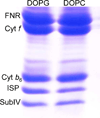
Lipid-dependence of stability of purified cyt b6f complex in lipids. Purified cyt b6f complex from spinach thylakoid membranes was reconstituted with lipids (DOPG or DOPC) at a molar ratio (protein:lipid) of 1:10. The molecular mass of the ISP subunit does not undergo a change in the presence of the anionic lipid DOPG thereby indicating that the polypeptide remains intact in the presence of the anionic lipid. The band for the enzyme FNR, which co-purifies with the spinach cyt b6f complex, is also shown.

References

    1. Schutz M, Brugna M, Lebrun E, Baymann F, Huber R, Stetter KO, Hauska G, Toci R, Lemesle-Meunier D, Tron P, Schmidt C, Nitschke W. Early evolution of cytochrome bc complexes. Journal of molecular biology. 2000;300:663–675. - PubMed
    1. Darrouzet E, Cooley JW, Daldal F. The Cytochrome bc (1) Complex and its Homologue the b (6) f Complex: Similarities and Differences. Photosynth Res. 2004;79:25–44. - PubMed
    1. Widger WR, Cramer WA, Herrmann RG, Trebst A. Sequence homology and structural similarity between cytochrome b of mitochondrial complex III and the chloroplast b6-f complex: position of the cytochrome b hemes in the membrane. Proceedings of the National Academy of Sciences of the United States of America. 1984;81:674–678. - PMC - PubMed
    1. Cramer WA, Hasan SS, Yamashita E. The Q cycle of cytochrome bc complexes: a structure perspective. Biochimica et biophysica acta. 2011;1807:788–802. - PMC - PubMed
    1. Berry EA, Guergova-Kuras M, Huang LS, Crofts AR. Structure and function of cytochrome bc complexes. Annual review of biochemistry. 2000;69:1005–1075. - PubMed

Publication types

MeSH terms

Associated data

LinkOut - more resources