. 2013 Mar 21;368(12):1101-10.
doi: 10.1056/NEJMoa1214271.

ERCC1 isoform expression and DNA repair in non-small-cell lung cancer

Affiliations

ERCC1 isoform expression and DNA repair in non-small-cell lung cancer

Luc Friboulet et al. N Engl J Med. .

Abstract

Background: The excision repair cross-complementation group 1 (ERCC1) protein is a potential prognostic biomarker of the efficacy of cisplatin-based chemotherapy in non-small-cell lung cancer (NSCLC). Although several ongoing trials are evaluating the level of expression of ERCC1, no consensus has been reached regarding a method for evaluation.

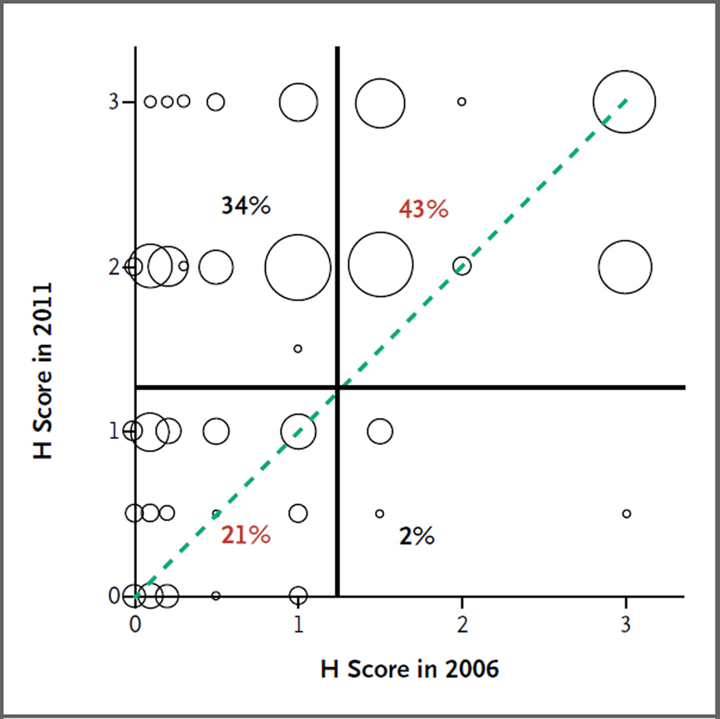
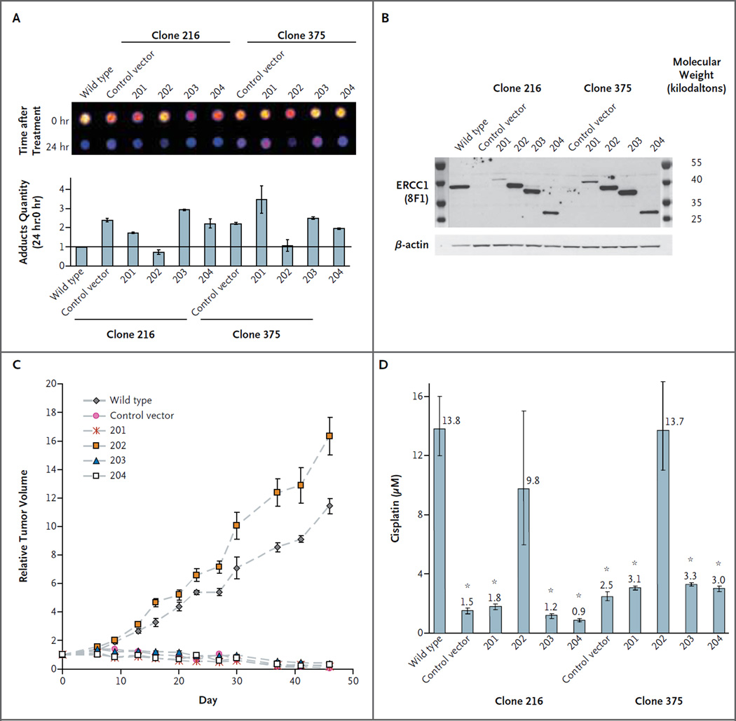
Methods: We used the 8F1 antibody to measure the level of expression of ERCC1 protein by means of immunohistochemical analysis in a validation set of samples obtained from 494 patients in two independent phase 3 trials (the National Cancer Institute of Canada Clinical Trials Group JBR.10 and the Cancer and Leukemia Group B 9633 trial from the Lung Adjuvant Cisplatin Evaluation Biology project). We compared the results of repeated staining of the entire original set of samples obtained from 589 patients in the International Adjuvant Lung Cancer Trial Biology study, which had led to the initial correlation between the absence of ERCC1 expression and platinum response, with our previous results in the same tumors. We mapped the epitope recognized by 16 commercially available ERCC1 antibodies and investigated the capacity of the different ERCC1 isoforms to repair platinum-induced DNA damage.

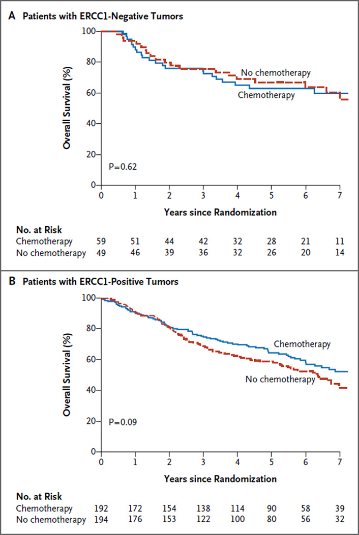
Results: We were unable to validate the predictive effect of immunostaining for ERCC1 protein. The discordance in the results of staining for ERCC1 suggested a change in the performance of the 8F1 antibody since 2006. We found that none of the 16 antibodies could distinguish among the four ERCC1 protein isoforms, whereas only one isoform produced a protein that had full capacities for nucleotide excision repair and cisplatin resistance.

Conclusions: Immunohistochemical analysis with the use of currently available ERCC1 antibodies did not specifically detect the unique functional ERCC1 isoform. As a result, its usefulness in guiding therapeutic decision making is limited. (Funded by Eli Lilly and others.).


Figures

Figure 1
Figure 1. Kaplan–Meier Estimates of Overall Survival in the Validation Cohort
Overall survival curves are shown according to treatment group for patients with ERCC1-negative tumors (Panel A) and those with ERCC1-positive tumors (Panel B). P values are based on multivariable Cox models. The numbers of events corresponding to the survival curves are shown in the forest plot in Figure S2 in the Supplementary Appendix.
Figure 2
Figure 2. Discrepancy in Results of Staining of Tumor Samples for ERCC1 between 2006 and 2011
A correlation plot shows the ERCC1 H scores from our previously reported results in 2006 and from repeated staining with the use of a different 8F1 antibody batch in samples obtained from the same 589 patients in 2011. Each circle is proportional to the number of patients with a particular combination of ERCC1 H scores in the two studies. The circles on the dotted line indicate patients with tumors that had the same H score in the two studies. The H score reflects the staining intensity of nuclei and the percentage of positive nuclei; ERCC1-positive tumors were defined as those with an H score higher than 1. The vertical and horizontal lines represent the cutoff values used to discriminate between patients with tumors that were positive and those with tumors that were negative for ERCC1. Percentages of patients with concordant scores (red) and discordant scores (black) are shown.
Figure 3
Figure 3. Mapping of Peptide Sequences Recognized by ERCC1 Antibodies across Different Isoforms
Recognition sites for 16 commercial antibodies (arrows) and corresponding amino acid sequences of the epitope are shown. XPA denotes xeroderma pigmentosum complementation group A protein, and XPF xeroderma pigmentosum complementation group F protein.
Figure 4
Figure 4. Capacity of ERCC1 Protein Isoforms to Allow Nucleotide Excision Repair and Cisplatin Resistance
Panel A shows the results of dot-blot analysis and quantification of removal of cisplatin–DNA adducts 24 hours after a 2-hour cisplatin treatment at 25 μM in A549 wild-type, ERCC1-deficient clones 216 and 375 (the control vector) and cells expressing single ERCC1 isoforms (201, 202, 203, or 204). The adduct quantity ratio (the ratio of the number of adducts at 24 hours to the number at 0 hours) in A549 wild-type cells was set at 1. Panel B shows control of isoform construct expression in established ERCC1 cell lines with 8F1 and β-actin antibodies. Panel C shows relative tumor volumes over the course of treatment after 106 ERCC1-deficient cells, with single isoform expression, were injected subcutaneously into nude mice. Twice weekly, tumors were measured, and cisplatin was injected intraperitoneally at a dose of 1 mg per kilogram of body weight. The I bars represent 95% confidence intervals. Panel D shows the results of a short-term growth assay for A549 wild-type, ERCC1-deficient, and isoform-expressing cells treated for 48 hours with increasing doses of cisplatin. The numbers above the bars represent the 50% inhibitory concentration for cisplatin. The stars indicate significant differences from wild-type cells (P<0.05). The I bars represent 95% confidence intervals.

References

    1. Pignon JP, Tribodet H, Scagliotti GV, et al. Lung adjuvant cisplatin evaluation: a pooled analysis by the LACE Collaborative Group. J Clin Oncol. 2008;26:3552–3559. - PubMed
    1. The NSCLC Meta-analyses Collaborative Group. Adjuvant chemotherapy, with or without postoperative radiotherapy, in operable non-small-cell lung cancer: two meta-analyses of individual patient data. Lancet. 2010;375:1267–1277. - PMC - PubMed
    1. Postel-Vinay S, Vanhecke E, Olaussen KA, Lord CJ, Ashworth A, Soria JC. The potential of exploiting DNA-repair defects for optimizing lung cancer treatment. Nat Rev Clin Oncol. 2012;9:144–155. - PubMed
    1. Bergstralh DT, Sekelsky J. Interstrand crosslink repair: can XPF-ERCC1 be let off the hook? Trends Genet. 2008;24:70–76. - PubMed
    1. Zheng Z, Chen T, Li X, Haura E, Sharma A, Bepler G. DNA synthesis and repair genes RRM1 and ERCC1 in lung cancer. N Engl J Med. 2007;356:800–808. - PubMed

Publication types

MeSH terms