. 2012 Apr;1(2):63-8.
doi: 10.1089/biores.2012.9902.

Human xenografts are not rejected in a naturally occurring immunodeficient porcine line: a human tumor model in pigs

Affiliations

Human xenografts are not rejected in a naturally occurring immunodeficient porcine line: a human tumor model in pigs

Matthew T Basel et al. Biores Open Access. 2012 Apr.

Abstract

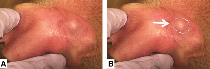
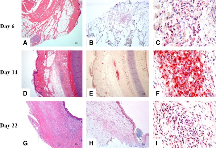
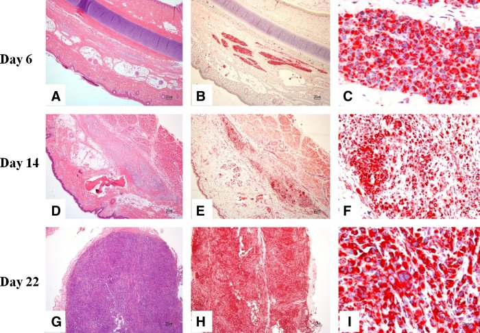
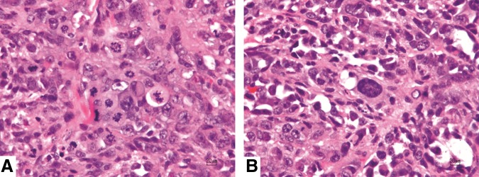
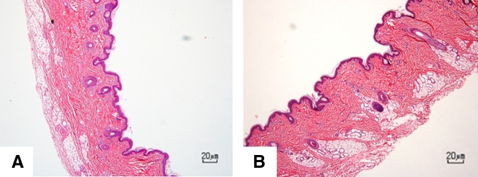
Animal models for cancer therapy are invaluable for preclinical testing of potential cancer treatments; however, therapies tested in such models often fail to translate into clinical settings. Therefore, a better preclinical model for cancer treatment testing is needed. Here we demonstrate that an immunodeficient line of pigs can host and support the growth of xenografted human tumors and has the potential to be an effective animal model for cancer therapy. Wild-type and immunodeficient pigs were injected subcutaneously in the left ear with human melanoma cells (A375SM cells) and in the right ear with human pancreatic carcinoma cells (PANC-1). All immunodeficient pigs developed tumors that were verified by histology and immunohistochemistry. Nonaffected littermates did not develop tumors. Immunodeficient pigs, which do not reject xenografted human tumors, have the potential to become an extremely useful animal model for cancer therapy because of their similarity in size, anatomy, and physiology to humans.

Keywords: immunodeficient swine; large-animal cancer model; melanoma; pancreatic carcinoma; xenografts.


Figures

FIG. 1.
FIG. 1.
Antemortem visual evidence of tumor growth in pig 3 (day 20). (A) Photograph indicates a large growth on the left ear. (B) Same photograph as in (A), with the growth outlined for visual reference.
FIG. 2.
FIG. 2.
Right ear with pancreatic carcinoma cells from pigs euthanized at day 6, 14, and 22 post-transplantation. (A) There is a focal, well-demarcated, unencapsulated neoplasm composed of nests and packets of neoplastic cells within the subcutis. (B, C) Strong positivity to anti-human mitochondrial antibody is evident within the cytoplasm of neoplastic cells. (D) Within the subcutis and skeletal muscle, there is an unencapsulated, moderately demarcated, and mildly infiltrative neoplasm composed of nests and packets of neoplastic cells. (E, F) Strong positivity to anti-human mitochondrial antibody is evident within the cytoplasm of neoplastic cells. (G) Within the dermis, subcutis, and skeletal muscle, there is an unencapsulated, poorly demarcated, and infiltrative neoplasm composed of nests and packets of neoplastic cells. (H, I) Strong positivity to anti-human mitochondrial antibody is evident within the cytoplasm of neoplastic cells. (A, D, G) H&E stain; (B, C, E, F, H, I) anti-human mitochondrial antibody immunohistochemistry. H&E, hematoxylin and eosin.
FIG. 3.
FIG. 3.
Left ear with melanoma cells from pigs euthanized at day 6, 14, and 22 post-transplantation. (A) Within the deep dermis, subcutis, and skeletal muscle, there is an unencapsulated and infiltrative neoplasm composed of nests and short streams of neoplastic cells. (B, C) Strong positivity to anti-human mitochondrial antibody is evident within the cytoplasm of neoplastic cells. (D) Within the deep dermis and subcutis, there is an unencapsulated and infiltrative neoplasm composed of nests and short streams of neoplastic cells within a moderate amount of fibrovascular stroma. (E, F) Strong positivity to anti-human mitochondrial antibody is evident within the cytoplasm of neoplastic cells. (G) Within the deep dermis and subcutis, there is an unencapsulated, moderately demarcated, and infiltrative neoplasm composed of nests and short streams of neoplastic cells within a moderate amount of fibrovascular stroma. (H, I) Strong positivity to anti-human mitochondrial antibody is evident within the cytoplasm of neoplastic cells. (A, D, G) H&E stain; (B, C, E, F, H, I) anti-human mitochondrial antibody immunohistochemistry.
FIG. 4.
FIG. 4.
Photomicrographs of the left ear of pig 3 demonstrating histologic features of neoplasia. (A) Note multiple mitotic figures. (B) Note significant cellular and nuclear pleomorphism. H&E stain.
FIG. 5.
FIG. 5.
Right ear (A) and left ear (B) from control pigs at day 22. No tumors were identified at the site of injection of human-origin neoplastic cells. H&E stain.

References

    1. Rothenberg ML. Carbone DP. Johnson DH. Improving the evaluation of new cancer treatments: challenges and opportunities. Nat Rev Cancer. 2003;3:303–309. - PubMed
    1. Richmand A. Su Y. Mouse xenograft models vs gem models for human cancer therapeutics. Dis Models Mech. 2008;1:78–82. - PMC - PubMed
    1. Kelland LR. “Of mice and men”: values and liabilities of the athymic nude mouse model in anticancer drug development. Eur J Cancer. 2004;40:827–836. - PubMed
    1. Liu M. Hicklin D. Human tumor xenograft efficacy models. In: Teicher BA, editor. Tumor Models in Cancer Research. Springer; New York: 2010. pp. 99–124.
    1. Sausville EA. Burger AM. Contributions of human tumor xenografts to anticancer drug development. Cancer Res. 2006;66:3351–3354. - PubMed