. 2013 May 10;288(19):13356-69.
doi: 10.1074/jbc.M113.455378. Epub 2013 Mar 20.

Structural insights into the regulation of foreign genes in Salmonella by the Hha/H-NS complex

Affiliations

Structural insights into the regulation of foreign genes in Salmonella by the Hha/H-NS complex

Sabrina S Ali et al. J Biol Chem. .

Abstract

Background: Hha facilitates H-NS-mediated silencing of foreign genes in bacteria.

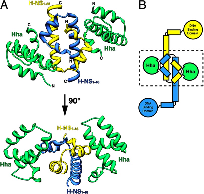
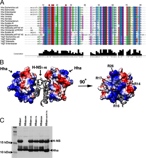
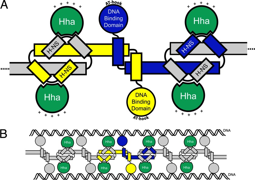
Results: Two Hha monomers bind opposing faces of the H-NS N-terminal dimerization domain.

Conclusion: Hha binds the dimerization domain of H-NS and may contact DNA via positively charged surface residues.

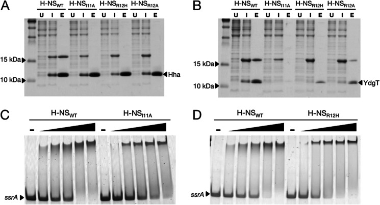
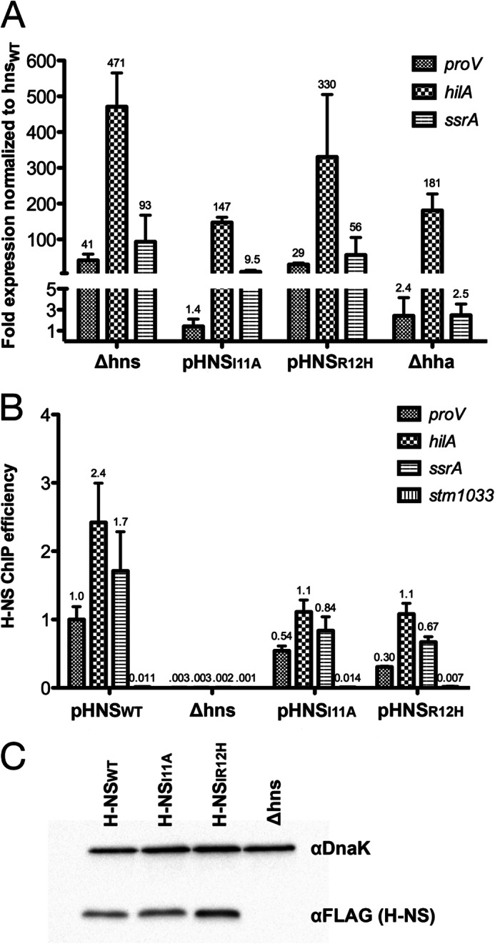
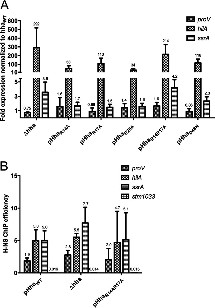
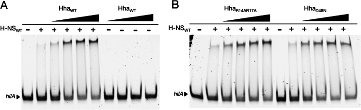
Significance: The structure of Hha and H-NS in complex provides a mechanistic model of how Hha may affect gene regulation. The bacterial nucleoid-associated proteins Hha and H-NS jointly repress horizontally acquired genes in Salmonella, including essential virulence loci encoded within Salmonella pathogenicity islands. Hha is known to interact with the N-terminal dimerization domain of H-NS; however, the manner in which this interaction enhances transcriptional silencing is not understood. To further understand this process, we solved the x-ray crystal structure of Hha in complex with the N-terminal dimerization domain of H-NS (H-NS(1-46)) to 3.2 Å resolution. Two monomers of Hha bind to symmetrical sites on either side of the H-NS(1-46) dimer. Disruption of the Hha/H-NS interaction by the H-NS site-specific mutation I11A results in increased expression of the Hha/H-NS co-regulated gene hilA without affecting the expression levels of proV, a target gene repressed by H-NS in an Hha-independent fashion. Examination of the structure revealed a cluster of conserved basic amino acids that protrude from the surface of Hha on the opposite side of the Hha/H-NS(1-46) interface. Hha mutants with a diminished positively charged surface maintain the ability to interact with H-NS but can no longer regulate hilA. Increased expression of the hilA locus did not correspond to significant depletion of H-NS at the promoter region in chromatin immunoprecipitation assays. However, in vitro, we find Hha improves H-NS binding to target DNA fragments. Taken together, our results show for the first time how Hha and H-NS interact to direct transcriptional repression and reveal that a positively charged surface of Hha enhances the silencing activity of H-NS nucleoprotein filaments.

Keywords: Bacterial Genetics; DNA-binding Protein; Gene Regulation; Gene Transfer; Microbial Pathogenesis; Microbiology.


Figures

FIGURE 1.
FIGURE 1.
Structure of Hha in complex with the N-terminal dimerization domain of H-NS (H-NS(1–46)). A, Hha and H-NS(1–46) crystallized in a 1:1 ratio with molecules of Hha binding either side of the H-NS(1–46) dimer. Hha is shown in green, and the H-NS(1–46) monomers are colored yellow and blue. The N and C termini are indicated in black. B, schematic representation of an H-NS dimer bound to two molecules of Hha. The dashed box indicates the region crystallized.
FIGURE 2.
FIGURE 2.
H-NS mutations at residues Ile-11 and Arg-12 disrupt the Hha/H-NS and YdgT/H-NS interactions without affecting DNA binding activity in vitro. A, Coomassie-stained SDS-PAGE of His6-tagged Hha co-expressed with FLAG-tagged H-NSWT, H-NSI11A, H-NSR12H, and H-NSR12A. Samples from co-expression cultures were taken prior to induction with isopropyl 1-thio-β-d-galactopyranoside (lanes marked U (for “uninduced”)), 16 h after the addition of 1 mm isopropyl 1-thio-β-d-galactopyranoside (lanes marked I (for “induced”)), and after purification by nickel chromatography (lanes marked E (for “eluate”)). His6-tagged Hha copurifies with H-NSWT but not the point mutants H-NSI11A, H-NSR12H, and H-NSR12A. B, nickel resin purification of His6-tagged YdgT after co-expression with the same H-NS constructs in A. C and D, purified H-NSWT, H-NSI11A, and H-NSR12H were added to a 300-bp PCR fragment from the ssrA promoter region at concentrations of 150, 200, 300, 400, and 500 nm. The dash above four of the lanes indicates that no protein was added to the samples. Protein-DNA binding reactions were separated on a 6% polyacrylamide gel by native gel electrophoresis and stained using SYBR Green nucleic acid stain. The H-NS point mutants H-NSI11A and H-NSR12H shift the ssrA promoter fragment at similar concentrations as H-NSWT.
FIGURE 3.
FIGURE 3.
H-NSI11A and H-NSR12H self-associate in a concentration-dependent manner. Nickel-purified H-NSWT, H-NSI11A, and H-NSR12H were applied to a Tricorn Superdex 200 10/300 GL column at concentrations of 25 μm (dashed lines), 75 μm (solid lines), and 100 μm (dotted lines). The molecular weight of the average protein complex in each sample was calculated according to prior calibration with molecular weight protein standards between 18.2 and 440 kDa (Table 3). As the sample concentration was increased, H-NSWT, H-NSI11A, and H-NSR12H eluted from the column as higher molecular weight oligomeric complexes.
FIGURE 4.
FIGURE 4.
H-NS mutants I11A and R12H differentially misregulate gene targets in S. Typhimurium. A, reverse transcription qPCR was performed on samples from S. Typhimurium Δhns strains harboring pHNSWT, pHNSI11A, pHNSR12H, an empty plasmid control (Δhns), and a Δhha strain. Total RNA was purified and reverse transcribed, and the resulting cDNA was quantified by qPCR with primers against proV, hilA, and ssrA. Transcript levels were graphed as -fold expression relative to the Δhns strain expressing pHNSWT, and the error bars represent the S.D. from three biological replicates. B, H-NS enrichment was measured by ChIP qPCR at the proV, hilA, ssrA, and stm1033 promoter regions with samples from the same strains used in A. stm1033 is a region previously shown to be unbound by H-NS. H-NS enrichment is expressed as the immunoprecipitation efficiencies (percentage recovery after immunoprecipitation compared with initial input). Error bars, S.D. on three biological replicates. C, Western blot analysis of H-NS expression levels from the complemented Δhns strains used in A and B. H-NS was probed with α-FLAG monoclonal antibody, and α-DnaK served as a loading control.
FIGURE 5.
FIGURE 5.
Highly conserved, positively charged residues protrude from the surface of Hha when in complex with H-NS(1–46). A, an alignment of diverse Hha-like and YdgT-like molecules from selected enteric bacterial species or their plasmids. Residues are colored according to conservation. The conserved, positively charged residues targeted for mutagenesis are indicated with red dots. Alignments were performed using the default settings on the COBALT server at NCBI (60), and results were displayed using Jalview (61). B, surface electrostatic representation of Hha in complex with H-NS(1–46). The 3.2 Å resolution of the Hha/H-NS(1–46) complex did not enable modeling of several surface-exposed side chains; therefore, the Hha solution structure (PDB code 1JW2) was aligned to Hha from the Hha/H-NS(1–46) complex (r.m.s. deviation = 1.9 Å over 67 Cα atoms). Positively charged residues are colored blue, and negatively charged residues are colored red. Surface-exposed basic residues of Hha that point away from H-NS(1–46) are labeled. C, Coomassie-stained SDS-PAGE after co-expression and Ni2+ purification of FLAG-tagged H-NSWT and His6-tagged HhaWT, HhaD48N, HhaR14A, HhaR17A, HhaR26A, and HhaR1A/R17A. All of the Hha mutants maintained an interaction with H-NSWT with the exception of the D48N point mutation, previously reported to disrupt the interaction.
FIGURE 6.
FIGURE 6.
Positively charged residues of the surface of Hha are critical for function. A, transcript analysis was performed on a Δhha strain of S. Typhimurium harboring an empty plasmid control (Δhha) or pHhaWT, pHhaR14A, pHhaR17A, pHhaR26A, or pHhaR14AR17A. Expression levels of proV, hilA, and ssrA were normalized to the Δhha strain harboring pHhaWT. All of the Hha variants tested resulted in significant up-regulation of hilA, whereas the expression profile of the HhaR14A/R17A double mutant most closely paralleled the Δhha strain. B, ChIP qPCR was performed with samples from the pHhaWT, Δhha, and pHhaR14A/R17A strains. H-NS enrichment at the proV, hilA, ssrA, and stm1033 promoter regions is expressed as ChIP efficiencies. Error bars, S.D. on three biological replicates. Significant differences between the ChIP efficiencies of H-NS from the pHhaWT, Δhha, and pHhaR14A/R17A strains were not noted.
FIGURE 7.
FIGURE 7.
Hha augments H-NS DNA binding in vitro. A, electrophoretic mobility shift assays performed with a 300-bp PCR product from the hilA promoter region. No protein was added to the samples marked with minus signs. The plus signs signify 300 nm H-NS in the binding reaction. The addition of HhaWT to the samples indicated at concentrations of 300, 600, 900, and 1200 nm resulted in increased shifting of the hilA fragment. When HhaWT is combined with the hilA fragment in the absence of H-NS, no shifting is observed. B, EMSAs performed with HhaR14R17A and HhaD48N also result in improved shifting of the hilA fragment. The same protein concentrations from A were applied.
FIGURE 8.
FIGURE 8.
Model of Hha interaction with H-NS. A, diagram of four Hha molecules (green) bound to two H-NS dimers. Monomers of the H-NS dimer are represented in yellow and blue. The AT-hook motif in the H-NS DNA binding domain interacts with the minor groove of target DNA sequences. The relative orientation of the positively charged surface of Hha is shown with plus signs. B, model for how Hha could be arranged in the H-NS nucleoprotein complex in “bridging” mode.

References

    1. Lawrence J. G., Ochman H. (1997) Amelioration of bacterial genomes. Rates of change and exchange. J. Mol. Evol. 44, 383–397 - PubMed
    1. Groisman E. A., Ochman H. (1996) Pathogenicity islands. Bacterial evolution in quantum leaps. Cell 87, 791–794 - PubMed
    1. Daubin V., Lerat E., Perrière G. (2003) The source of laterally transferred genes in bacterial genomes. Genome Biol. 4, R57. - PMC - PubMed
    1. Navarre W. W., Porwollik S., Wang Y., McClelland M., Rosen H., Libby S. J., Fang F. C. (2006) Selective silencing of foreign DNA with low GC content by the H-NS protein in Salmonella. Science 313, 236–238 - PubMed
    1. Lucchini S., Rowley G., Goldberg M. D., Hurd D., Harrison M., Hinton J. C. (2006) H-NS mediates the silencing of laterally acquired genes in bacteria. PLoS Pathog. 2, e81. - PMC - PubMed

Publication types

MeSH terms

LinkOut - more resources