. 2012 Dec;1(6):280-90.
doi: 10.1089/biores.2012.0271.

A standardized rat model of volumetric muscle loss injury for the development of tissue engineering therapies

Affiliations

A standardized rat model of volumetric muscle loss injury for the development of tissue engineering therapies

Xiaowu Wu et al. Biores Open Access. 2012 Dec.

Abstract

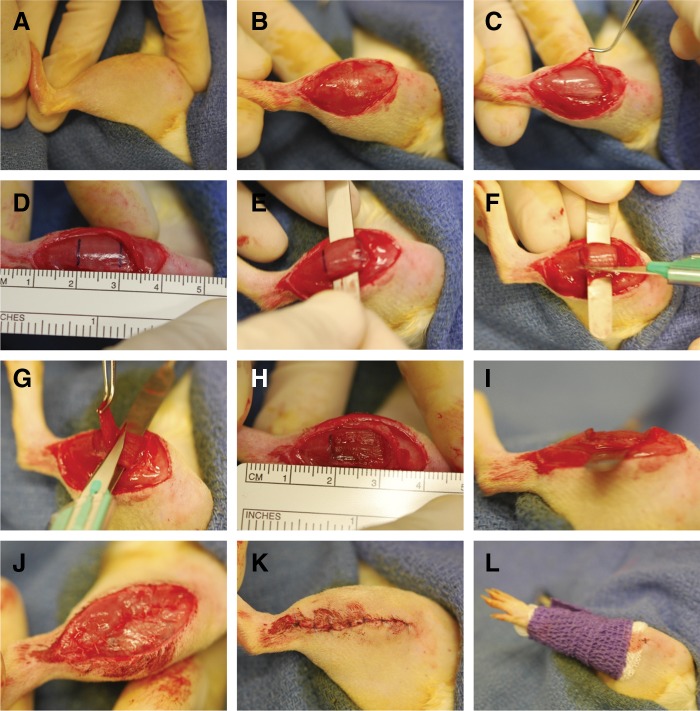
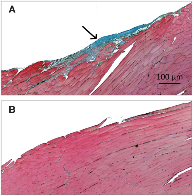
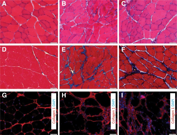
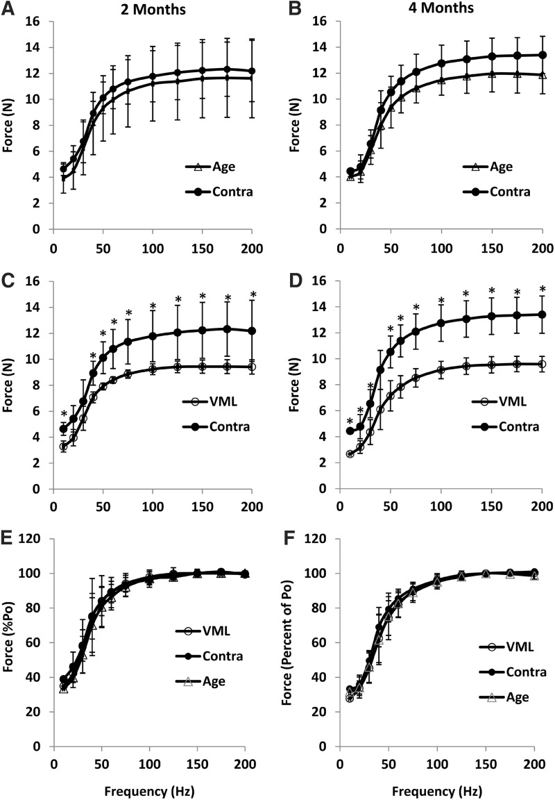
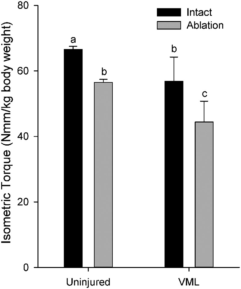
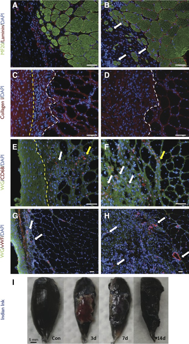
Soft tissue injuries involving volumetric muscle loss (VML) are defined as the traumatic or surgical loss of skeletal muscle with resultant functional impairment and represent a challenging clinical problem for both military and civilian medicine. In response, a variety of tissue engineering and regenerative medicine treatments are under preclinical development. A wide variety of animal models are being used, all with critical limitations. The objective of this study was to develop a model of VML that was reproducible and technically uncomplicated to provide a standardized platform for the development of tissue engineering and regenerative medicine solutions to VML repair. A rat model of VML involving excision of ∼20% of the muscle's mass from the superficial portion of the middle third of the tibialis anterior (TA) muscle was developed and was functionally characterized. The contralateral TA muscle served as the uninjured control. Additionally, uninjured age-matched control rats were also tested to determine the effect of VML on the contralateral limb. TA muscles were assessed at 2 and 4 months postinjury. VML muscles weighed 22.7% and 19.5% less than contralateral muscles at 2 and 4 months postinjury, respectively. These differences were accompanied by a reduction in peak isometric tetanic force (Po) of 28.4% and 32.5% at 2 and 4 months. Importantly, Po corrected for differences in body weight and muscle wet weights were similar between contralateral and age-matched control muscles, indicating that VML did not have a significant impact on the contralateral limb. Lastly, repair of the injury with a biological scaffold resulted in rapid vascularization and integration with the wound. The technical simplicity, reliability, and clinical relevance of the VML model developed in this study make it ideal as a standard model for the development of tissue engineering solutions for VML.

Keywords: extracellular matrix; fibrosis; muscle injury; muscle regeneration; regenerative medicine; tissue engineering.


Figures

FIG. 1.
FIG. 1.
Illustration of rat tibialis anterior (TA) muscle surgical procedure. Approximately 20% of the rat TA muscle was excised to create an endogenously irrecoverable VML injury. See Materials and Methods for a description of panels (A–L).
FIG. 2.
FIG. 2.
Longitudinal section of volumetric muscle loss (VML) injured (A) and uninjured contralateral control (B) (10×) with Masson's Trichrome stain at 2 months postinjury. A thin layer of connective tissue can be seen at the injury site indicative of fibrotic scarring. A number of disorganized muscle cells were present within or in the immediate vicinity of the wound site.
FIG. 3.
FIG. 3.
Histological evidence of muscle fiber regeneration, fibrosis, and remodeling during the months after VML injury. Uninjured (A, D, G) and VML-injured muscles 2 months (B, E, H) and 4 months (C, F, I) postinjury were stained with hematoxylin and eosin (HE) (A–C), Mason's Trichrome (D–F), or collagen I (nuclei with DAPI) (G–I). Cross sections were obtained from the area corresponding to the middle of the injury. Scale bar=50 μm.
FIG. 4.
FIG. 4.
The force–frequency relationships at 2 months (A, C, E) and 4 months (B, D, F). Age-matched cage control (Age) and uninjured contralateral control (Contra) were compared with VML-injured subjects at 2 and 4 months. At both time points, isometric force was significantly less at most stimulation frequencies compared to Contra (p>0.05). However, when force was expressed as percent of Po (E, F), there was no difference between groups at either time point. All values are mean±standard deviation.
FIG. 5.
FIG. 5.
In vivo isometric torque in the anterior crural muscles before (Intact) and after tenotomy of the extensor digitorum longus muscle (Ablation). abcLetters indicate that the value is significantly different from any different letter (p<0.05).
FIG. 6.
FIG. 6.
Surgical repair of VML with muscle-derived rat extracellular matrix (RAMM). Unrepaired (A, C, E, G) and RAMM-repaired (B, D, F, H) TA muscles harvested 2 months after injury were analyzed using immunohistochemistry. (A, B) Regenerating myosin-positive muscle fibers (white arrows) in isolation from the remaining muscle mass were only observed in the defect area of RAMM-repaired muscles. (C, D) Collagen I deposition was prominent in the defect area of both unrepaired and RAMM-repaired muscle. However, the extent of collagen 1 deposition was qualitatively greater in RAMM-repaired muscle [area to left of yellow line is fascia, between lines is scar tissue, and to right of white line is muscle remaining muscle mass; no fascia is depicted in (D)]. (E, F) Macrophages (CD68) were present in the remaining muscle mass (yellow arrows) or in the area of collagen 1 deposition in the defect area (white arrows). The area to the left of the yellow line in E is fascia. (G, H) Vascularization (white arrows) in the defect area was detected using von Willebrand (vWF) staining. Nuclei were stained with DAPI. Stains are identified for each slide with color coded text in the left margin of each row; WG, wheat germ agglutinin. Scale bars=50 μm. (I) Rats were perfused with Indian ink at 3, 7, or 14 days after implantation of RAMM to demonstrate an integrated vascular network with the host circulation.

References

    1. Grogan BF. Hsu JR. Volumetric muscle loss. J Am Acad Orthop Surg. 2011;19(Suppl 1):S35–37. - PubMed
    1. MacKenzie EJ. Bosse MJ. Kellam JF, et al. Characterization of patients with high-energy lower extremity trauma. J Orthop Trauma. 2000;14:455–466. - PubMed
    1. Grogan B. Hsu JR. Volumetric muscle loss. J Am Acad Orthop Surg. 2011;19(Suppl. 1):S35–S39. - PubMed
    1. Owens BD. Kragh JF., Jr Wenke JC, et al. Combat wounds in operation Iraqi Freedom and operation Enduring Freedom. J Trauma. 2008;64:295–299. - PubMed
    1. Lew TA. Walker JA. Wenke JC, et al. Characterization of craniomaxillofacial battle injuries sustained by United States service members in the current conflicts of Iraq and Afghanistan. J Oral Maxillofac Surg. 2010;68:3–7. - PubMed

LinkOut - more resources