. 2013 Mar 20;33(12):5085-94.
doi: 10.1523/JNEUROSCI.3560-12.2013.

Calpains, cleaved mini-dysferlinC72, and L-type channels underpin calcium-dependent muscle membrane repair

Affiliations

Calpains, cleaved mini-dysferlinC72, and L-type channels underpin calcium-dependent muscle membrane repair

Angela Lek et al. J Neurosci. .

Abstract

Dysferlin is proposed as a key mediator of calcium-dependent muscle membrane repair, although its precise role has remained elusive. Dysferlin interacts with a new membrane repair protein, mitsugumin 53 (MG53), an E3 ubiquitin ligase that shows rapid recruitment to injury sites. Using a novel ballistics assay in primary human myotubes, we show it is not full-length dysferlin recruited to sites of membrane injury but an injury-specific calpain-cleavage product, mini-dysferlinC72. Mini-dysferlinC72-rich vesicles are rapidly recruited to injury sites and fuse with plasma membrane compartments decorated by MG53 in a process coordinated by L-type calcium channels. Collective interplay between activated calpains, dysferlin, and L-type channels explains how muscle cells sense a membrane injury and mount a specialized response in the unique local environment of a membrane injury. Mini-dysferlinC72 and MG53 form an intricate lattice that intensely labels exposed phospholipids of injury sites, then infiltrates and stabilizes the membrane lesion during repair. Our results extend functional parallels between ferlins and synaptotagmins. Whereas otoferlin exists as long and short splice isoforms, dysferlin is subject to enzymatic cleavage releasing a synaptotagmin-like fragment with a specialized protein- or phospholipid-binding role for muscle membrane repair.


Figures

Figure 1.
Figure 1.
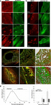
Dysferlin and MG53 show rapid, calcium-dependent accumulation at injury sites. a, 3D-SIM. Left, Dysferlin (green) and MG53 (red) form a concentrated lattice encircling injury sites in “+Ca,” interdigitating within the same xz plane (bottom rows). In low calcium conditions (middle: 10 s EGTA chelation, shot −Ca buffer) or −Ca conditions (right: 30 s EGTA chelation, shot −Ca buffer), dysferlin and MG53 remain as diffuse halos and occupy discrete spatial compartments in the xz plane (bottom rows), observed as clusters of dysferlin-rich vesicles positioned adjacent to plasma membrane compartments enriched with MG53. First row: scale bar, 2 μm. Zoomed images and xz slices: scale bar, 0.5 μm. b, MG53 and dysferlin transition from a diffuse halo in −Ca/low calcium, into concentrated rings at injury sites with >200 μm extracellular calcium. Scale bars, 10 μm. c, MG53 remains diffusely enriched at dysferlin-negative ballistics lesions. Scale bars, 4 μm. d, In the presence of calcium, MG53 and dysferlin colocalize at the wound site; a Pearson coefficient (Costes et al., 2004) of ∼0.7 is consistent with interdigitated and partially overlapping dysferlin and MG53 compartments. Distal to the wound site, or in the absence of calcium, dysferlin and MG53 do not colocalize (Pearson coefficient <0.4).
Figure 2.
Figure 2.
Temporal sequence of injury-activated recruitment of MG53, dysferlin, and annexin A1. Representative confocal images of MG53 and dysferlin (a) and MG53 and annexin A1 (b) recruitment to sites of injury in primary human myotubes fixed at 2, 10, 30, and 90 s after injury. Scale bar: 10 μm. c, 3D-SIM of two lesions at 90 s after injury; an unfilled lesion (top) with an expansive dysferlin (green) and MG53 (red) lattice surrounding the injury site, and a filled lesion (bottom) with the characteristic arc of strongly labeled dysferlin and MG53 among a tightly woven lattice. d, Ballistics lesions in human skeletal myotubes showed calcium-dependent expansion and contraction phases of membrane resealing. Points on the line graph (left) represent the average area (Leica SP2 ellipse formula) of lesions at 2 s (n = 44), 10 s (n = 40), 30 s (n = 47), and 60 s (n = 40); error bars indicate SE. The area at 90 s was set to 0 to reflect resealed patches. Histogram (right) shows the lack of expansion of ballistics lesions in −Ca conditions (2 s, n = 45; 10 s, n = 44; 30 s, n = 45). Later time points for −Ca conditions could not be calculated because of cellular lethality caused by injury in −Ca.
Figure 3.
Figure 3.
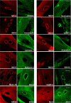
Screening for injury-activated recruitment of other muscular dystrophy proteins and endomembrane markers 10 s after ballistics injury of human skeletal myotubes. Rapid recruitment to injury sites is a specific feature of dysferlin and MG53, not observed for myoferlin, AHNAK, caveolin-3, or dystrophin. Moreover, we could find no evidence for specific recruitment of endomembrane compartments labeled for syntaxin-4, Munc-18c, VAMP-4, GM130, or annexin V, or molecular motor nonmuscle myosin 2A. Of note, we did not observe enrichment of LAMP-1 (or LAMP-2, data not shown) at sites of membrane injury in human myotubes, although we occasionally observed evidence for lysosomal exocytosis at sites distal to the membrane injury: the membrane bleb positively labeled for LAMP-1 directly adjacent to the site of membrane injury labeled by MG53.
Figure 4.
Figure 4.
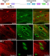
Injury-recruited dysferlin is only recognized by the C-terminal Hamlet-1 antibody. Dysferlin antibodies recognizing N-terminal epitopes do not detect dysferlin at injury sites at 10 s after injury (Hamlet-2, top row; anti-C2DE SAB2100636, middle row) or 90 s after injury (Romeo-1, bottom row).
Figure 5.
Figure 5.
Membrane injury triggers calpain cleavage of dysferlin to release a C-terminal mini-dysferlinC72 fragment with a specialized role in membrane repair. a, A mini-dysferlin band of 72 kDa is detected by Hamlet-1 with ballistics injury in +Ca (lane 1), but not when injured in calcium-free buffer (lane 2) or in uninjured cells (lane 3). b, Mini-dysferlinC72 is also produced with scrape injury in +Ca (lanes 3 and 4), but not when injured in calcium-free buffer (lanes 5 and 6), or in uninjured cells (lanes 1 and 2). c, Injury-induced production of mini-dysferlinC72 is attenuated or absent in myotubes from three patients with dysferlinopathy (D1-D3) but normal in myotubes from disease controls (α-sarcoglycanopathy S1, caveolinopathy C1). d, Production of mini-dysferlinC72 is calcium-dependent, activated by 200 μm extracellular calcium. e, Formation of mini-dysferlinC72 is inhibited by calpeptin treatment. Differentiating myoblasts were treated with 20 μm calpeptin or DMSO carrier 24 and 3 h before harvesting. D0-D4, days of differentiation. f, Maximal inhibition of dysferlin cleavage is achieved with ≥30 μm calpeptin using a 3 h preincubation treatment and refreshment of one-third media 30 min before injury. g, Dysferlin recruitment to sites of ballistics injury is attenuated in calpeptin-treated myotubes (top row, Hamlet-1), compared with untreated human myotubes. h, Maximum cleavage of dysferlin occurs at neutral pH. Cells were subjected to scrape injury in PBS buffered from pH 5.5–8.5.
Figure 6.
Figure 6.
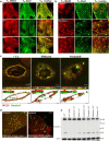
L-type calcium-channel signaling mediates coordinated fusion of mini-dysferlinC72 rich cytoplasmic vesicles with MG53 plasma membrane domains. a, Injury recruitment of dysferlin and MG53 is blocked by Cd2+, but not by Ni2+ or Gd3+. Scale bar, 10 μm. b, Specific L-type channel antagonists diltiazem, nifedipine, and verapamil attenuate dysferlin and MG53 injury recruitment. Scale bar, 10 μm. c, 3D-SIM of ballistics injuries performed in the presence or absence of specific L-type channel antagonists diltiazem and verapamil. Recruitment of dysferlin and MG53 is attenuated and appears uncoordinated, with clumps of dysferlin and MG53 at the edges of the lesions (xy, scale bar, 2 μm, MG53 red, Hamlet-1 green). Rotation of images in the xz or yz planes (scale bars, 1 μm) reveals spatial separation of dysferlin and MG53 compartments, similar to low calcium conditions (Fig. 1). d, 3D-SIM of large vesicles generated by verapamil treatment. The diameter of vesicles was 0.82 ± 0.4 μm (mean ± SD); n = 222. Vesicles positively label for MG53 and the C-terminal dysferlin antibody Hamlet-1 (left), but not the N-terminal dysferlin antibody Romeo-1, suggesting that they represent abnormal fusion of mini-dysferlinC72 and MG53 compartments. Romeo separately labels a population of smaller vesicles that are negative for Hamlet-1, suggesting separate subcellular localizations for each of the cleaved dysferlin fragments. e, Cadmium inhibits formation of mini-dysferlinC72 detected by Western blot, with normal production of mini-dysferlinC72 observed with Ni2+, Gd3+, and specific L-type VGCC antagonists.
Figure 7.
Figure 7.
Our proposed model for membrane repair. 0–5 s after injury: Membrane injury causes promiscuous influx of calcium at sites of membrane injury, local activation of calpains, and strong and persistent depolarization of L-type VGCCs. MG53 is mobilized and targeted to the injury site. Injury mobilization of MG53 is calcium-independent and may relate to its role as a ubiquitin ligase, perhaps targeting a receptor damaged by oxidation or calpain cleavage as a consequence of the membrane injury. Dysferlin is cleaved by activated calpains, releasing a C-terminal fragment, mini-dysferlinC72. Mini-dysferlinC72-rich cytoplasmic vesicles are rapidly recruited to sites of membrane injury and fuse with MG53-decorated plasma membrane compartments in a calcium-dependent process coordinated by L-type VGCCs. 2–10 s after injury: Mini-dysferlinC72 fuses into the plasma membrane and undergoes calcium-dependent phospholipid binding via its C2 domains, initiating a calcium-dependent phase of MG53 injury recruitment. Mini-dysferlinC72 and MG53 compartments interact to form an interdigitated lattice with strong affinity for exposed phospholipids surrounding the injury site. 10–30 s after injury: Annexin-A1 undergoes calcium-activated phospholipid binding. The role of annexin-A1 may be related to delivery of endomembrane compartments peripheral to the injury site to reduce plasma membrane tension and facilitate repair. 30–120 s after injury: Mini-dysferlinC72 and MG53 compartments infiltrate the plasma membrane surrounding the membrane injury, forming a lattice to stabilize the plasma membrane as it expands to reseal the membrane injury.

References

    1. Abreu-Blanco MT, Verboon JM, Parkhurst SM. Cell wound repair in Drosophila occurs through three distinct phases of membrane and cytoskeletal remodeling. J Cell Biol. 2011;193:455–464. - PMC - PubMed
    1. Bansal D, Miyake K, Vogel SS, Groh S, Chen CC, Williamson R, McNeil PL, Campbell KP. Defective membrane repair in dysferlin-deficient muscular dystrophy. Nature. 2003;423:168–172. - PubMed
    1. Bashir R, Britton S, Strachan T, Keers S, Vafiadaki E, Lako M, Richard I, Marchand S, Bourg N, Argov Z, Sadeh M, Mahjneh I, Marconi G, Passos-Bueno MR, Moreira Ede S, Zatz M, Beckmann JS, Bushby K. A gene related to Caenorhabditis elegans spermatogenesis factor fer-1 is mutated in limb-girdle muscular dystrophy type 2B. Nat Genet. 1998;20:37–42. - PubMed
    1. Bouter A, Gounou C, Berat R, Tan S, Gallois B, Granier T, d'Estaintot BL, Poschl E, Brachvogel B, Brisson AR. Annexin-A5 assembled into two-dimensional arrays promotes cell membrane repair. Nat Commun. 2011;2:272. - PMC - PubMed
    1. Cai C, Masumiya H, Weisleder N, Matsuda N, Nishi M, Hwang M, Ko JK, Lin P, Thornton A, Zhao X, Pan Z, Komazaki S, Brotto M, Takeshima H, Ma J. MG53 nucleates assembly of cell membrane repair machinery. Nat Cell Biol. 2009a;11:56–64. - PMC - PubMed

Publication types

MeSH terms

Supplementary concepts