. 2013 Mar 20;33(12):5275-84.
doi: 10.1523/JNEUROSCI.3200-12.2013.

Hypocretin/orexin neurons contribute to hippocampus-dependent social memory and synaptic plasticity in mice

Affiliations

Hypocretin/orexin neurons contribute to hippocampus-dependent social memory and synaptic plasticity in mice

Liya Yang et al. J Neurosci. .

Abstract

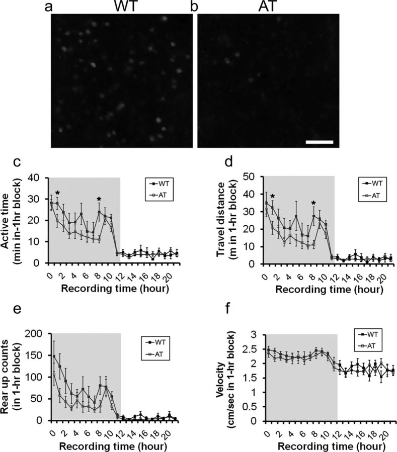
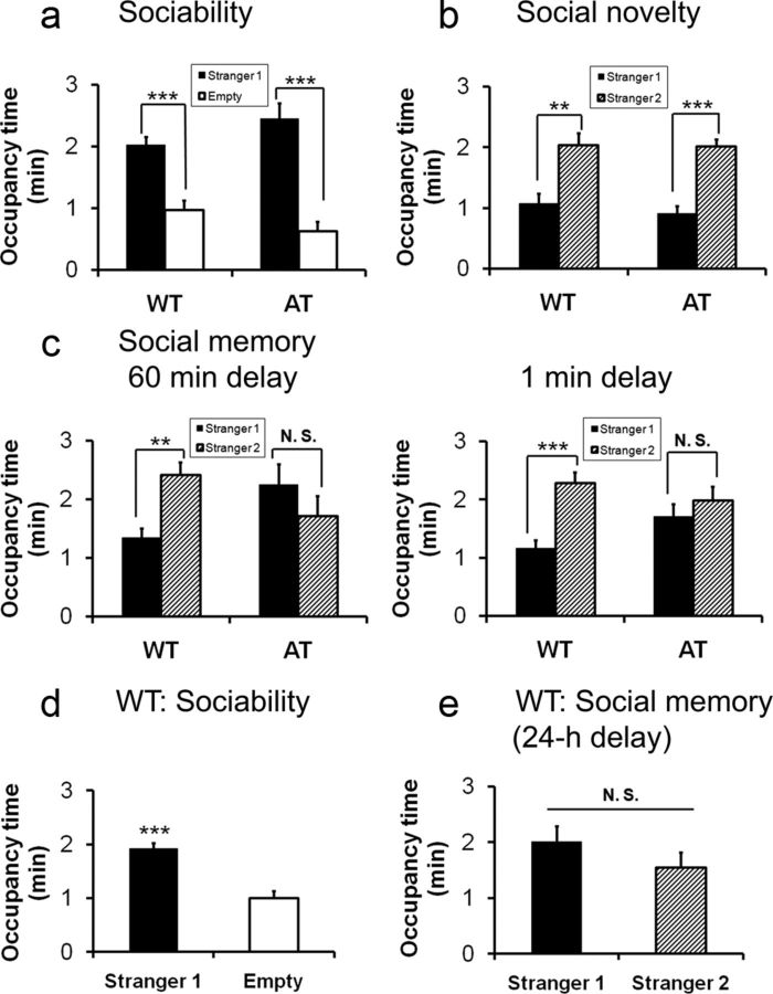
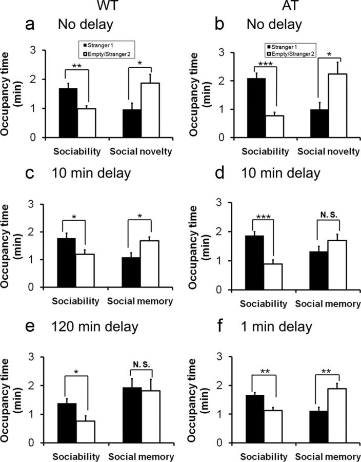
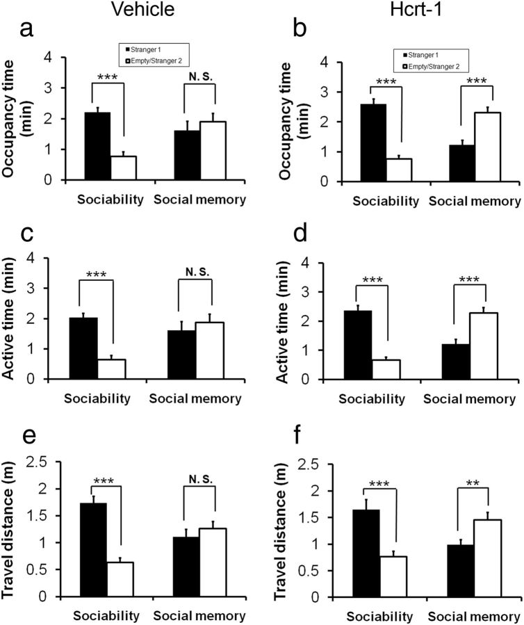
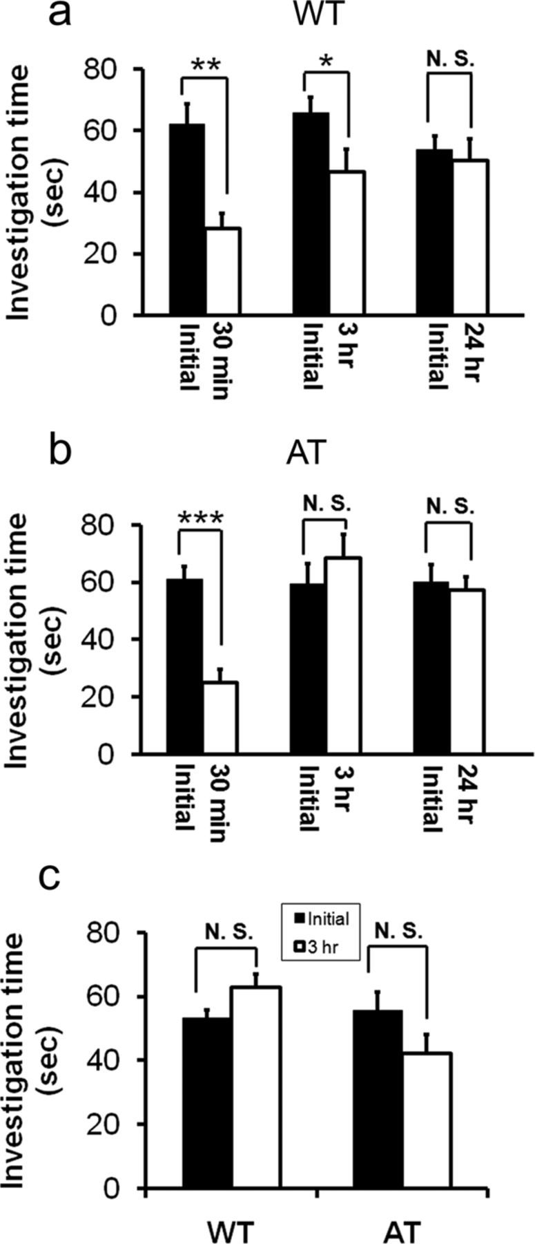
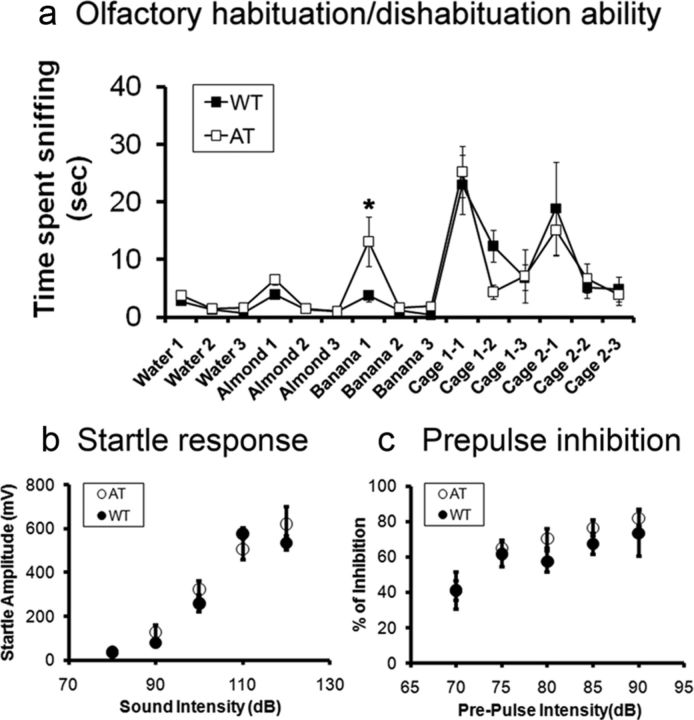
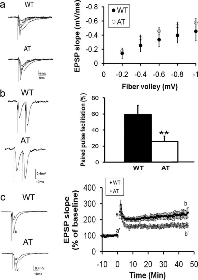
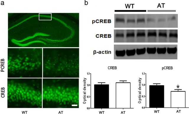
Hypocretin/orexin (Hcrt)-producing neurons in the lateral hypothalamus project throughout the brain, including to the hippocampus, where Hcrt receptors are widely expressed. Hcrt neurons activate these targets to orchestrate global arousal state, wake-sleep architecture, energy homeostasis, stress adaptation, and reward behaviors. Recently, Hcrt has been implicated in cognitive functions and social interaction. In the present study, we tested the hypothesis that Hcrt neurons are critical to social interaction, particularly social memory, using neurobehavioral assessment and electrophysiological approaches. The validated "two-enclosure homecage test" devices and procedure were used to test sociability, preference for social novelty (social novelty), and recognition memory. A conventional direct contact social test was conducted to corroborate the findings. We found that adult orexin/ataxin-3-transgenic (AT) mice, in which Hcrt neurons degenerate by 3 months of age, displayed normal sociability and social novelty with respect to their wild-type littermates. However, AT mice displayed deficits in long-term social memory. Nasal administration of exogenous Hcrt-1 restored social memory to an extent in AT mice. Hippocampal slices taken from AT mice exhibited decreases in degree of paired-pulse facilitation and magnitude of long-term potentiation, despite displaying normal basal synaptic neurotransmission in the CA1 area compared to wild-type hippocampal slices. AT hippocampi had lower levels of phosphorylated cAMP response element-binding protein (pCREB), an activity-dependent transcription factor important for synaptic plasticity and long-term memory storage. Our studies demonstrate that Hcrt neurons play an important role in the consolidation of social recognition memory, at least in part through enhancements of hippocampal synaptic plasticity and cAMP response element-binding protein phosphorylation.


Figures

Figure 1.
Figure 1.
Comparison of Hcrt-positive neurons and spontaneous homecage activity between AT mice and their WT littermates. Representative images of immunofluorescent staining of the lateral hypothalamus taken from WT mice (a) and AT mice (b) at 3 months of age are shown. Scale bar, 100 μm in b applied to a. Shown is a comparison of active time (c, recording hour 1,*p = 0.048, and recording hour 8, p = 0.015), traveling distance (d, recording hour 1, *p = 0.025, and recording hour 8, p = 0.014), rearing activity (e), and traveling speed (f) in AT and WT littermates in a 24 h record using the SmartCage system. Gray background in figures cf indicates dark phase during a 12:12 light:dark cycle (lights on at 7:00 A.M.; lights off at 7:00 P.M.). Data shown are mean ± SEM for each time point (1 h bin). Subjects were 5-month-old male mice (n = 8/genotype).
Figure 2.
Figure 2.
Comparisons of sociability, social novelty, and social recognition memory in AT mice and their WT littermates conducted during the light phase. a, The test AT mice displayed normal sociability similar to WT mice. b, AT mice displayed a normal degree of preference to a novel mouse (social novelty) compared with WT littermates when there was no delay between the end of the sociability session and the initiation of social novelty test. c, In two separate cohorts of animals, both genotypes exhibited normal sociability with Stranger 1 (data not shown). Stranger 2 was introduced 1 or 60 min after the sociability session. AT mice displayed deficits in social memory after a delay of 60 min (left) and 1 min (right), whereas WT mice displayed social memory in both delay time points. d, WT mice exhibited normal sociability toward Stranger 1. e, The same cohort of WT mice lost preference for social novelty after a 24 h delay to reexpose the same Stranger 1 and a novel Stranger 2. Data are shown as mean ± SEM. ac are the same cohorts of test mice, 3-month-old males (n = 8 per group); d and e are the same cohort of test WT 3-month-old males (n = 8). Paired Student's t test within genotype: *p < 0.05; **p < 0.01; ***p < 0.001.
Figure 3.
Figure 3.
Comparisons of social memory between AT mice and their WT littermates conducted during the first 3 h of the dark phase. a, b, Both WT and AT mice displayed normal sociability and social novelty without a delay between the two test sessions. c, WT mice displayed social recognition when the delay between sociability and social novelty tests was 10 min. d, AT mice displayed deficits in social memory with a delay of 10 min. e, WT mice displayed a loss of social memory with a delay of 120 min. f, AT mice displayed short-term social memory with a delay of 1 min. Data are mean ± SEM. Subjects were 3- to 6-month-old male mice (n = 8–12/group). Paired Student's t test: *p < 0.05; **p < 0.01; ***p < 0.001.
Figure 4.
Figure 4.
Nasal administration of Hcrt-1 improves social memory in AT mice. The AT subject mice received nasal administration of either vehicle (deionized water, n = 13, a) or Hcrt-1 (0.8 nmol, n = 14, b). The social memory test with a 10 min delay between the completion of sociability and the beginning of social novelty sessions. The AT mice that were treated with vehicle exhibited normal sociability and reproducible social memory deficits (a: for sociability, p < 0.001; for social memory, p = 0.362). The AT subjects that were treated with Hcrt-1 displayed normal sociability, but also showed a distinct shift in their investigation to Stranger 2 from familiarized Stranger 1 (b: for sociability, p < 0.001; for social memory, p < 0.001). Other two measureable social interaction parameters, active time (c and d, p < 0.001) and travel distance (e and f, p = 0.013), also indicated that the Hcrt-treated AT mice improved social recognition memory.
Figure 5.
Figure 5.
AT mice showed normal social interaction but with a social memory deficit compared with WT littermates in a direct contact social test. a, Test WT mice displayed social approach behavior toward the stimulus mouse (sociability) and reduced the approach behavior toward the reexposed social cue mouse 30 min and 3 h after initial contact as measured by a reduction in investigation time (n = 8 per genotype). Paired Student's t test: *p < 0.05; **p < 0.01. After a 24 h delay after the initial interaction, the test mouse spent the same amount of time investigating the same social cue mouse as in the initial trial. b, Test AT mice displayed social approach behavior toward the stimulus mouse (sociability) and reduced investigation time toward the same social cue 30 min after the initial interaction (n = 8 per genotype). Paired Student's t test: *p < 0.05; **p < 0.01. After a delay of 3 or 24 h after the initial interaction, the test mouse spent a similar amount of time investigating the reexposed social cue as in the initial interaction. c, AT and WT mice show similar investigation times toward two different, unfamiliar social cue mice between the initial trial and a second test after a 3 h delay. In the initial contact, the test subject displayed social approach behavior, as indicated by time investigating the stranger mouse. Three hours later, the same test subject was tested for its sociability toward a novel mouse (taken from another homecage). Subjects were 3-month-old male mice (n = 8/genotype). Paired Student's t test, p > 0.05).
Figure 6.
Figure 6.
Comparison of olfactory and auditory reflex functions in AT mice and their WT littermates. a, Olfactory habituation/dishabituation ability was measured as time spent sniffing a sequence of novel nonsocial (i.e., water, almond, and banana) and social odors (i.e., unfamiliar mouse cage odor) contained in cotton swab tips. Except for banana 1 (unpaired Student's t test, *p < 0.05), all data points were not significantly different between genotypes. b, Startle responses evoked by different sound intensities (80–120 dB). There were no significant differences between the genotypes (n = 8 per group). c, Prepulse inhibition induced by a constant startle stimulus (120 dB, 40 ms), which was preceded by a prepulse (70–90 dB, 20 ms) with an interval (100 ms) between the prepulse and startle stimuli. There were no significant differences between the genotypes. Subjects were 3- to 4-month-old male mice (n = 8/genotype).
Figure 7.
Figure 7.
AT mice exhibited normal basal synaptic neurotransmission but a decreased PPF ratio and attenuated LTP in the CA1 area of hippocampal slices compared with WT littermates. a, Left, A family of representative voltage traces of fEPSPs in the CA1 area of WT or AT hippocampal slices evoked by different stimulation intensities on the Schaffer collateral and commissural fibers. Right, I-O of the fEPSP. The fiber valley was binned with an interval of 0.2 mV. There were no significant differences in the I-O curves between the genotypes (WT: n = 14 slices from 5 mice; AT: n = 14 slices from 6 mice). b, Comparison of PPF ratio between AT and WT littermates. Left: Representative traces of paired pulse stimulation with 20 ms interval from WT and AT littermates, respectively. Right, The PPF ratio is significantly higher in WT mice than AT mice (WT: 59.3 ± 11.3%, n = 12 slices from 5 mice; AT: 25.7 ± 6.9%, n = 14 slices from 6 mice; unpaired Student's t test, p = 0.02). c, AT mice exhibited decreases in the amplitude of LTP in the CA1 area of hippocampal slices compared with WT littermates. Left, Representative voltage traces of fEPSP are superimposed before (a) and after (b) LTP induction by high-frequency stimulation of the Schaffer collateral and commissural fibers (100 Hz, 1 s, 4 trails, 20 s interval). Right, Each fEPSP slope was normalized to that just before the high-frequency stimulation and plotted as a function of recording time. The magnitude of LTP during the maintenance phase in WT slices is significantly larger than that in AT slices (WT: 234.6 ± 23.8%, n = 14 slices from 5 mice; AT: 164.3 ± 12.7%, n = 14 slices from 6 mice; 2-tailed unpaired Student's t test, p = 0.02).
Figure 8.
Figure 8.
Levels of pCREB in hippocampi of AT mice is lower compared with those in WT hippocampi. a, Representative images of immunochemistry staining shows heterogeneous CREB expression in the whole coronal section of the hippocampus taken from a WT mouse (top) and differences in expression of pCREB (middle) and CREB (bottom) in the CA1 area (indicated by white line box in the whole hippocampus image shown above) between WT and AT mice. Scale bar, 50 μm in the bottom panel applied to middle panel. b, Representative Western blot images of pCREB (top), CREB (middle), and β-actin (bottom) in hippocampi taken from AT or WT littermates (3 representative mice from a total n = 9 mice per genotype). Optical density of total CREB levels obtained from the Western blots are not significantly different between the genotypes (left). Optical density of pCREB (right) is significantly lower in the hippocampi taken from AT mice than that from WT littermates (n = 9 Western blot bands from n = 9 mice per genotype; unpaired Student's t test, p < 0.05). Optical densities of both CREB and pCREB were normalized to β-actin.

Similar articles

Cited by

References

    1. Aguirre M, Broughton R, Stuss D. Does memory impairment exist in narcolepsy-cataplexy? J Clin Exp Neuropsychol. 1985;7:14–24. - PubMed
    1. Akbari E, Naghdi N, Motamedi F. The selective orexin 1 receptor antagonist SB-334867-A impairs acquisition and consolidation but not retrieval of spatial memory in Morris water maze. Peptides. 2007;28:650–656. - PubMed
    1. Blouin AM, Fried F, Wilson C, Staba RJ, Behnke EJ, Lam HA, Maidment NT, Karlsson KA, Lapierre JL, Siegel JM. Human hypocretin and melanin concentrating hormone levels are linked to emotion and social interaction. Nat Communications. 2013 (in press) - PMC - PubMed
    1. Borgland SL, Taha SA, Sarti F, Fields HL, Bonci A. Orexin A in the VTA is critical for the induction of synaptic plasticity and behavioral sensitization to cocaine. Neuron. 2006;49:589–601. - PubMed
    1. Bourtchuladze R, Frenguelli B, Blendy J, Cioffi D, Schutz G, Silva AJ. Deficient long-term memory in mice with a targeted mutation of the cAMP-responsive element-binding protein. Cell. 1994;79:59–68. - PubMed

Publication types

MeSH terms