. 2013 Mar 20;33(12):5352-61.
doi: 10.1523/JNEUROSCI.6103-11.2013.

Dissociation of frontotemporal dementia-related deficits and neuroinflammation in progranulin haploinsufficient mice

Affiliations

Dissociation of frontotemporal dementia-related deficits and neuroinflammation in progranulin haploinsufficient mice

Anthony J Filiano et al. J Neurosci. .

Abstract

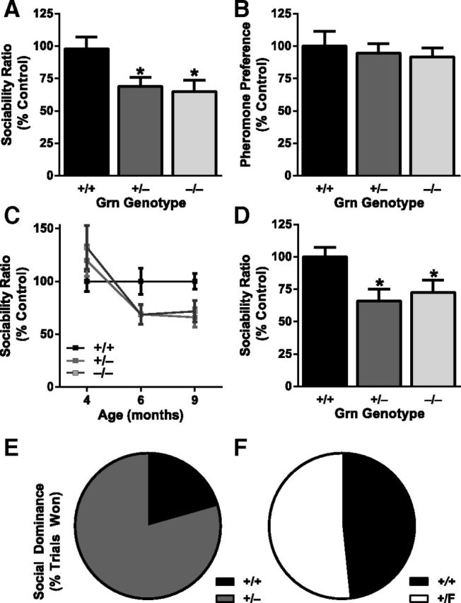
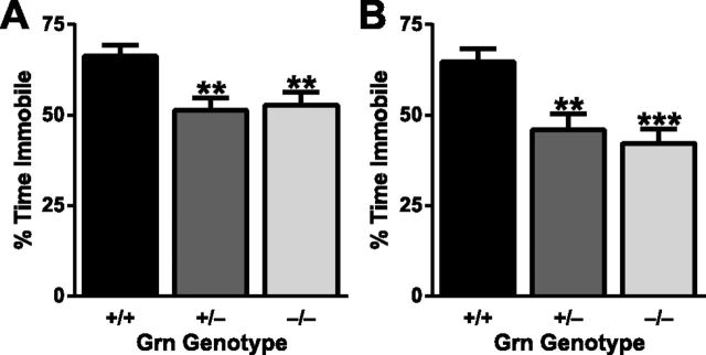
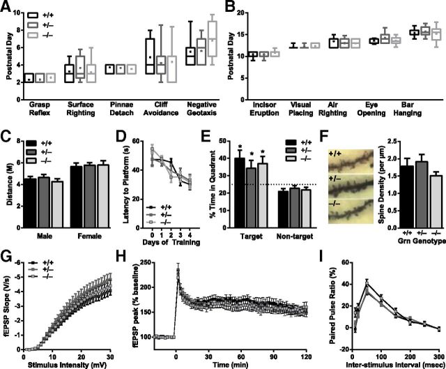
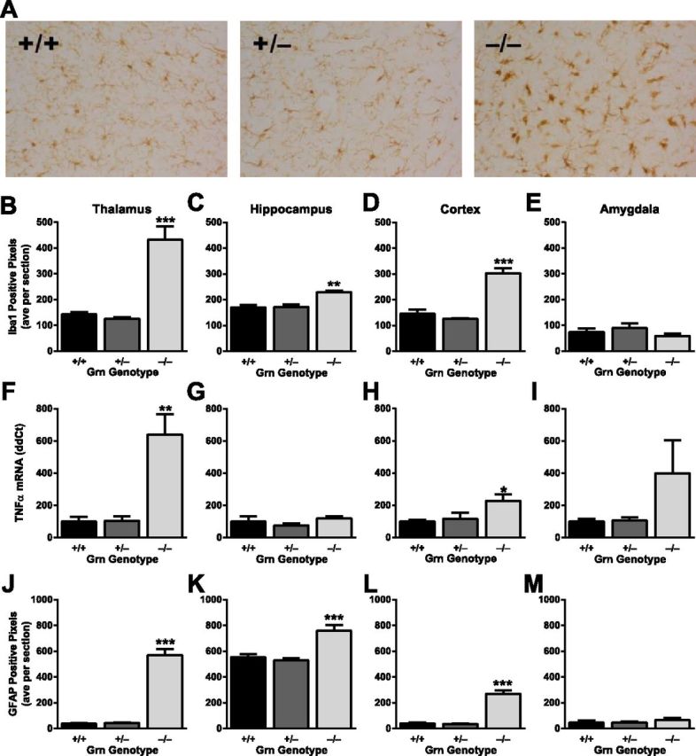
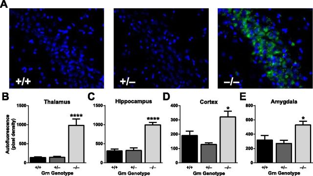
Frontotemporal dementia (FTD) is a neurodegenerative disease with hallmark deficits in social and emotional function. Heterozygous loss-of-function mutations in GRN, the progranulin gene, are a common genetic cause of the disorder, but the mechanisms by which progranulin haploinsufficiency causes neuronal dysfunction in FTD are unclear. Homozygous progranulin knock-out (Grn(-/-)) mice have been studied as a model of this disorder and show behavioral deficits and a neuroinflammatory phenotype with robust microglial activation. However, homozygous GRN mutations causing complete progranulin deficiency were recently shown to cause a different neurological disorder, neuronal ceroid lipofuscinosis, suggesting that the total absence of progranulin may have effects distinct from those of haploinsufficiency. Here, we studied progranulin heterozygous (Grn(+/-)) mice, which model progranulin haploinsufficiency. We found that Grn(+/-) mice developed age-dependent social and emotional deficits potentially relevant to FTD. However, unlike Grn(-/-) mice, behavioral deficits in Grn(+/-) mice occurred in the absence of gliosis or increased expression of tumor necrosis factor-α. Instead, we found neuronal abnormalities in the amygdala, an area of selective vulnerability in FTD, in Grn(+/-) mice. Our findings indicate that FTD-related deficits resulting from progranulin haploinsufficiency can develop in the absence of detectable gliosis and neuroinflammation, thereby dissociating microglial activation from functional deficits and suggesting an important effect of progranulin deficiency on neurons.


Figures

Figure 1.
Figure 1.
Progranulin levels in progranulin-deficient mice. A–D, Progranulin mRNA levels in cortex (A), hippocampus (B), thalamus (C), and amygdala (D). Relative expression levels quantified by quantitative PCR were calculated using the ΔΔCt method and are displayed as a percentage of the average level in wild-type mice (N = 5–6 mice per genotype; age 19 months). E, F, Progranulin protein levels in cortex (E; N = 6 mice per genotype) and plasma (F; N = 17 mice per genotype), quantified by ELISA (age 12 months).
Figure 2.
Figure 2.
Progranulin haploinsufficiency causes social deficits. A, C, D, Three-chamber sociability test, with sociability ratio (time spent investigating another mouse divided by time spent investigating an inanimate object) expressed as percentage of control. A, On a mixed background at 6–7 months, Grn+/− mice and Grn−/− mice had lower sociability than Grn+/+ mice (ANOVA, p < 0.01). *p < 0.05 (post hoc test). N = 51–58 mice per genotype. B, No deficit in pheromone preference was observed in progranulin-deficient mice. N = 17 mice per genotype; mixed background at 7–8 months. C, A cohort of mice on a congenic C57BL/6 background was tested longitudinally at mean ages of 4, 6, and 9 months. There were no sociability deficits at 4 months, but reduced sociability was apparent at older ages (age × Grn interaction, p < 0.05). N = 17–19 mice per genotype. D, On a congenic C57BL/6 background at 9–12 months, Grn+/− mice and Grn−/− mice had lower sociability than Grn+/+ mice (ANOVA, p < 0.05). *p < 0.05 (post hoc test). N = 17–21 mice per genotype. E, F, Social interactions were further tested with the tube test of social dominance at 4–6 months of age. E, Grn+/− mice won 79% (27 of 34) of trials against Grn+/+ mice (exact p = 0.0008). F, Grn+/F controls, which have normal progranulin levels, won 52% (16 of 31) of trials against Grn+/+ mice, no different from chance. Male and female mice were used for both social tests. No sex-dependent effects were observed, so the data from males and females were combined.
Figure 3.
Figure 3.
Progranulin haploinsufficiency impairs fear conditioning. Twelve-month-old mice from a mixed (A; N = 54–59 mice per genotype) or congenic C57BL/6 (B; N = 17–19 mice per genotype) background were tested for cued fear conditioning one day after training. A significant effect of progranulin deficiency was observed in mice on both backgrounds (ANOVA, p < 0.01). Both Grn+/− and Grn−/− mice spent significantly less time immobile than Grn+/+ mice: **p < 0.001 (post hoc test). ***p < 0.0001 (post hoc test).
Figure 4.
Figure 4.
Progranulin-deficient mice have normal hippocampal function and spine density. A, B, Box-and-whisker plots representing the age at which early (A) and late (B) milestones were attained for each genotype. Boxes indicate the 75th and 25th percentiles, with a horizontal line at the median and a dot at the mean; whiskers represent the 10th and 90th percentiles (N = 11–21 mice per genotype). C, No differences were observed in exploratory behavior in the open field. Data are represented as total distance traveled during 15 min (congenic C57BL/6 background; N = 12–15 mice per genotype; age 9 months). D, Learning curves on the Morris water maze hidden platform task were not affected by progranulin deficiency. E, Dwell time in the target quadrant on a probe trial conducted 24 h after completion of training was not affected by progranulin deficiency. *p < 0.05 versus nontarget quadrant for each genotype. N = 14 mice per genotype; age 7 months. F, Progranulin deficiency did not affect hippocampal spine density in second-order apical dendrites of Golgi-stained CA1 pyramidal neurons (N = 9 or 10 neurons from two mice per genotype; age 12 months). G–I, Extracellular field recordings in area CA1 of acute hippocampal slices (N = 31–40 slices from 5 or 6 mice per genotype; age 12 months). G, Progranulin deficiency did not affect baseline synaptic transmission, assessed by input-output curves. H, Progranulin deficiency did not affect LTP, either early or late stages. I, Progranulin deficiency did not affect paired-pulse facilitation.
Figure 5.
Figure 5.
Absence of neuroinflammation in Grn+/− mice with FTD-related behavioral abnormalities. A, Representative images of Iba1 immunohistochemistry in the thalamus of control and progranulin-deficient mice. B–E, Quantification of Iba1 immunoreactivity in various brain regions (N = 6 mice per genotype; age 12 months). B, Grn−/−, but not Grn+/−, mice had microgliosis in thalamus (ANOVA, p < 0.0001). On post hoc tests, only Grn−/− mice differ from other groups: ***p < 0.0001. C, Grn−/−, but not Grn+/−, mice had microgliosis in hippocampus (ANOVA, p < 0.005). On post hoc tests, only Grn−/− mice differ from other groups: **p < 0.001. D, Grn−/−, but not Grn+/−, mice had microgliosis in cortex (ANOVA, p < 0.0001). On post hoc tests, only Grn−/− mice differ from other groups: ***p < 0.0001. E, There were no significant differences in Iba1 immunoreactivity in amygdala. F–I, Quantitative PCR analysis of TNF-α mRNA levels in various brain regions (N = 5 or 6 mice per genotype; age 19 months). F, Grn−/−, but not Grn+/−, mice had increased TNF-α in thalamus (ANOVA, p < 0.005). On post hoc tests, only Grn−/− mice differ from other groups: **p < 0.001. G, There were no significant differences in TNF-α in hippocampus. H, Grn−/−, but not Grn+/−, mice had increased TNF-α in cortex (ANOVA, p < 0.05). On post hoc tests, only Grn−/− mice differ from other groups: *p < 0.05. I, Although trending, no significant differences in TNF-α in amygdala were detected (ANOVA, p = 0.12). J–M, Quantification of GFAP immunoreactivity in various brain regions (N = 6 mice per genotype; age 12 months). J, Grn−/−, but not Grn+/−, mice had astrocytosis in thalamus (ANOVA, p < 0.0001). On post hoc tests, only Grn−/− mice differ from other groups: ***p < 0.0001. K, Grn−/−, but not Grn+/−, mice had astrocytosis in hippocampus (ANOVA, p = 0.0001). On post hoc tests, only Grn−/− mice differ from other groups: ***p < 0.001. L, Grn−/−, but not Grn+/−, mice had astrocytosis in cortex (ANOVA, p < 0.0001). On post hoc tests, only Grn−/− mice differ from other groups: ***p < 0.0001. M, There were no significant differences in GFAP immunoreactivity in amygdala.
Figure 6.
Figure 6.
Absence of lipofuscinosis in Grn+/− mice. A, Representative images of autofluorescent lipofuscin granules in the CA3 region of the hippocampus. B–E, Quantification of autofluorescence in various brain regions (N = 6–8 mice per genotype; age 12 months). Increased autofluorescence was observed in Grn−/−, but not Grn+/− mice (ANOVA, p < 0.001; post hoc tests). Only Grn−/− mice differ from other groups: *p < 0.05; ****p < 0.0001.
Figure 7.
Figure 7.
Decreased neuronal activation in the amygdala of Grn+/− mice. A, Regions counted for c-Fos- and NeuN-positive neurons, including central amygdala (CeA), caudate/putamen (CPu), and dentate gyrus (DG). B–D, Active c-Fos-positive neurons were counted in various brain regions. Under resting, home-cage conditions, few neurons were c-Fos-positive. B, Fewer c-Fos-positive neurons were present in the amygdala of Grn+/− and Grn−/− mice after 2 h in a novel environment (ANOVA, p < 0.05). **p < 0.01 versus Grn+/+ mice (post hoc test). *p < 0.05 versus Grn+/+ mice (post hoc test). There was no difference in the number of c-Fos-positive neurons in the caudate/putamen (C) or dentate gyrus (D) of the hippocampus. n.s., Not significant. E, There were no differences between groups in the number of social interactions of various types during the 2 h in the novel environment. F, There were no differences in total NeuN-positive amygdala neuron counts of progranulin-deficit mice, so the decrease in c-Fos-positive neurons cannot be attributed to neuron loss, but rather to impaired activation.

References

    1. Adolphs R. What does the amygdala contribute to social cognition? Ann N Y Acad Sci. 2010;1191:42–61. - PMC - PubMed
    1. Ahmed Z, Sheng H, Xu YF, Lin WL, Innes AE, Gass J, Yu X, Wuertzer CA, Hou H, Chiba S, Yamanouchi K, Leissring M, Petrucelli L, Nishihara M, Hutton ML, McGowan E, Dickson DW, Lewis J. Accelerated lipofuscinosis and ubiquitination in granulin knockout mice suggest a role for progranulin in successful aging. Am J Pathol. 2010;177:311–324. - PMC - PubMed
    1. Arrasate M, Mitra S, Schweitzer ES, Segal MR, Finkbeiner S. Inclusion body formation reduces levels of mutant huntingtin and the risk of neuronal death. Nature. 2004;431:805–810. - PubMed
    1. Bickart KC, Wright CI, Dautoff RJ, Dickerson BC, Barrett LF. Amygdala volume and social network size in humans. Nat Neurosci. 2011;14:163–164. - PMC - PubMed
    1. Bosch OJ, Neumann ID. Vasopressin released within the central amygdala promotes maternal aggression. Eur J Neurosci. 2010;31:883–891. - PubMed

Publication types

MeSH terms