. 2013;8(3):e58535.
doi: 10.1371/journal.pone.0058535. Epub 2013 Mar 13.

Saccades during attempted fixation in parkinsonian disorders and recessive ataxia: from microsaccades to square-wave jerks

Affiliations

Saccades during attempted fixation in parkinsonian disorders and recessive ataxia: from microsaccades to square-wave jerks

Jorge Otero-Millan et al. PLoS One. 2013.

Abstract

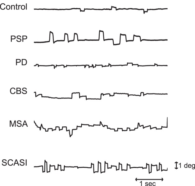
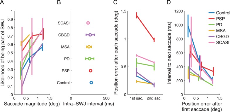
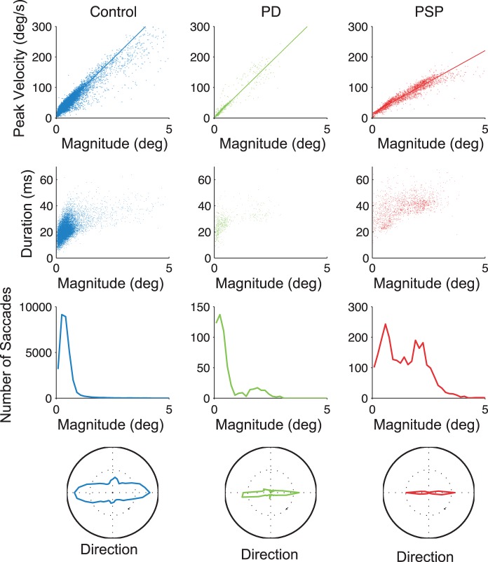
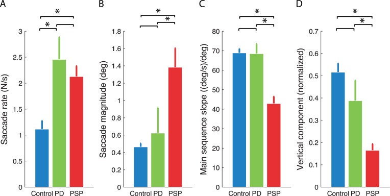
During attempted visual fixation, saccades of a range of sizes occur. These "fixational saccades" include microsaccades, which are not apparent in regular clinical tests, and "saccadic intrusions", predominantly horizontal saccades that interrupt accurate fixation. Square-wave jerks (SWJs), the most common type of saccadic intrusion, consist of an initial saccade away from the target followed, after a short delay, by a "return saccade" that brings the eye back onto target. SWJs are present in most human subjects, but are prominent by their increased frequency and size in certain parkinsonian disorders and in recessive, hereditary spinocerebellar ataxias. Here we asked whether fixational saccades showed distinctive features in various parkinsonian disorders and in recessive ataxia. Although some saccadic properties differed between patient groups, in all conditions larger saccades were more likely to form SWJs, and the intervals between the first and second saccade of SWJs were similar. These findings support the proposal of a common oculomotor mechanism that generates all fixational saccades, including microsaccades and SWJs. The same mechanism also explains how the return saccade in SWJs is triggered by the position error that occurs when the first saccadic component is large, both in the healthy brain and in neurological disease.


Conflict of interest statement

Competing Interests: Susana Martinez-Conde is a PLOS ONE Editorial Board member. This does not alter the authors’ adherence to all the PLOS ONE policies on sharing data and materials.

Figures

Figure 1
Figure 1. Examples of saccadic intrusions in a control subject, a PSP patient, a PD patient, a CBS patient, a MSA patient and a SCASI patient.
SWJs were present in all subject groups, although they were smaller and less frequent in healthy controls. Each trace represents a 5 s recording of horizontal eye positions containing SWJs. Horizontal position and timescales for all traces are as in the bottom trace.
Figure 2
Figure 2. A common square-wave coupling mechanism.
A) Correlation between saccade size and likelihood of being part of a SWJ. B) Intra-SWJ intervals across groups. C) Position error (see Methods for details) at the end of the first versus second saccade in a SWJ. D) Relationship between the estimated position error at the end of each saccade and the inter-saccadic interval to the next saccade. Error bars in all panels represent the standard error of the mean across subjects.
Figure 3
Figure 3. Characteristics of fixational saccades across subject groups.
First row, saccadic peak velocity/magnitude relationships. Second row, saccadic duration/magnitude relationships. Third row, saccade magnitude distributions. Fourth row, polar histograms of saccade directions. Each graph shows the combined data for all subjects in each group.
Figure 4
Figure 4. Saccadic parameters in PD patients, PSP patients and healthy controls.
Saccade rates, magnitudes, peak velocity-magnitude relationship slopes and vertical components (of saccade direction) are indicated. Bars represent the average value across subjects of each group and the error bars indicate the standard error of the mean. Asterisks show significance (p<0.05, t-test).
Figure 5
Figure 5. Microsaccade triggering model .
SC neurons present two gradients of connectivity, one that is strongest between rostral SC and OPNs, and one that is strongest between caudal SC and EBNs and IBNs – (longer lines represent stronger connections). The mutually inhibited OPNs and IBNs act as a trigger. During fixation, rostral SC activity drives the OPNs that inhibit the EBNs and IBNs. Directly preceding the launch of a microsaccade, activity in the rostral area shifts slightly caudally. At some point the balance of inhibition is broken, and the IBNs inhibit the OPNs more than the OPNs inhibit the IBNs. Then the EBNs start to burst initiating the microsaccade. Note that this representation is a one-dimension simplification of the circuit. The circuit functions in the same manner for vertical (up and down) BNs. Minus signs indicate inhibitory connections, plus signs excitatory ones.

References

    1. Abadi RV, Gowen E (2004) Characteristics of saccadic intrusions. Vision Res 44: 2675–2690. - PubMed
    1. Martinez-Conde S, Macknik SL, Hubel DH (2004) The role of fixational eye movements in visual perception. Nat Rev Neurosci 5: 229–240. - PubMed
    1. Leigh RJ, Zee DS (2006) The neurology of eye movements. Oxford Univ Press.
    1. Martinez-Conde S, Macknik SL, Troncoso XG, Dyar TA (2006) Microsaccades counteract visual fading during fixation. Neuron 49: 297–305. - PubMed
    1. Martinez-Conde S, Macknik SL, Troncoso XG, Hubel DH (2009) Microsaccades: a neurophysiological analysis. Trends Neurosci 32: 463–475. - PubMed

Publication types

LinkOut - more resources