. 2013;8(3):e58554.
doi: 10.1371/journal.pone.0058554. Epub 2013 Mar 13.

Myostatin suppression of Akirin1 mediates glucocorticoid-induced satellite cell dysfunction

Affiliations

Myostatin suppression of Akirin1 mediates glucocorticoid-induced satellite cell dysfunction

Yanjun Dong et al. PLoS One. 2013.

Abstract

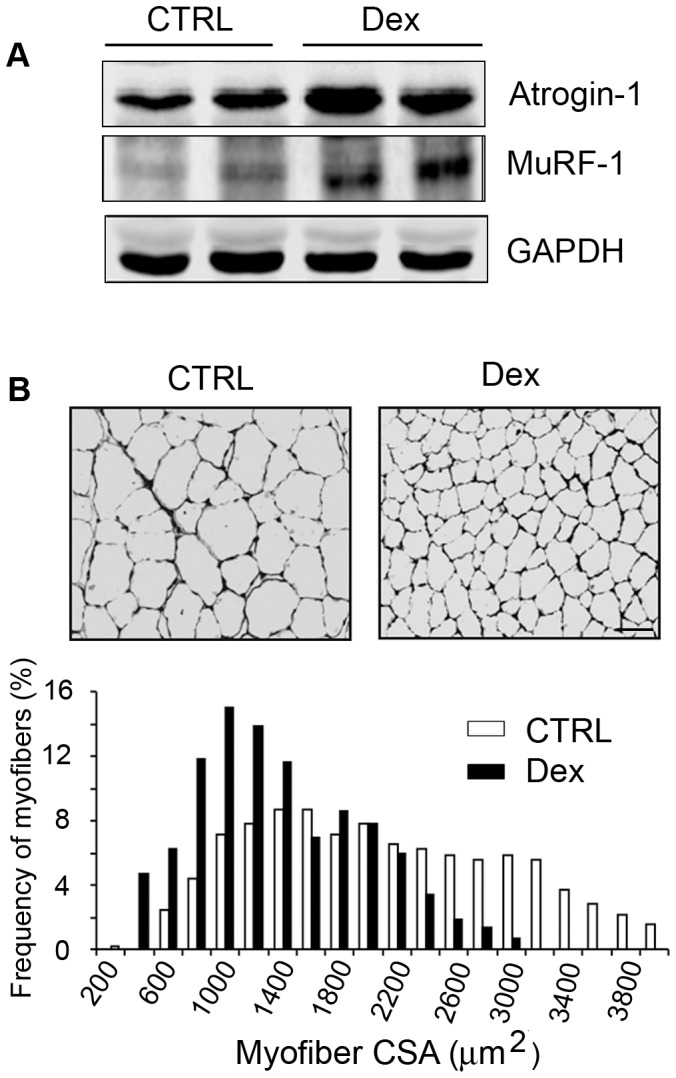
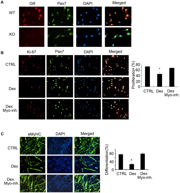
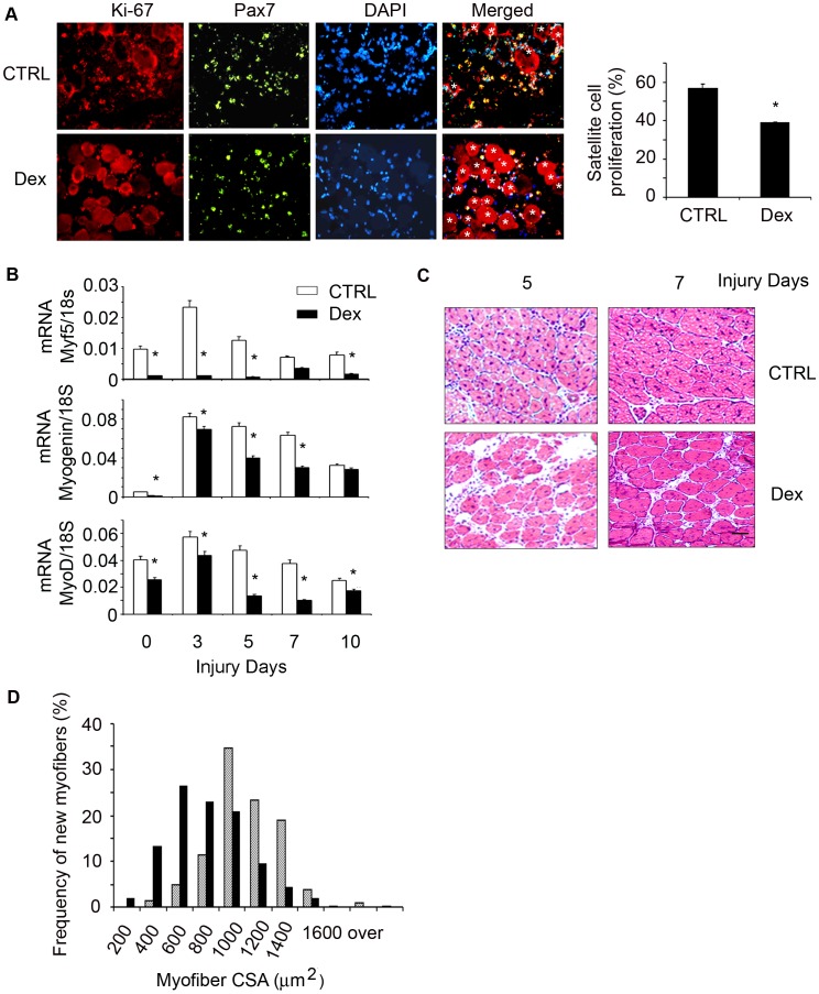
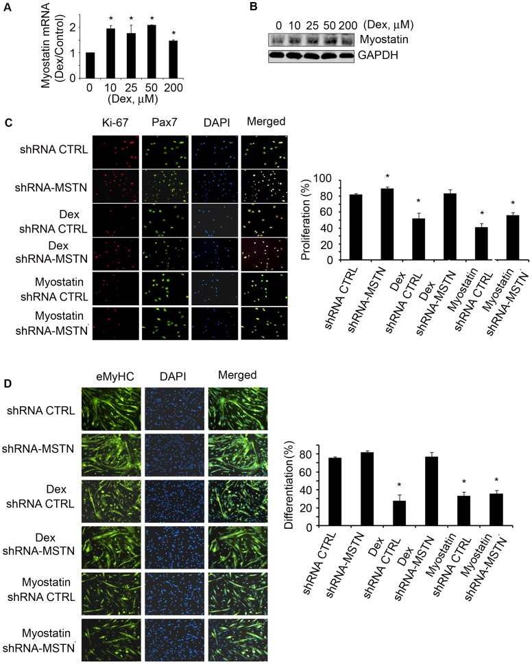
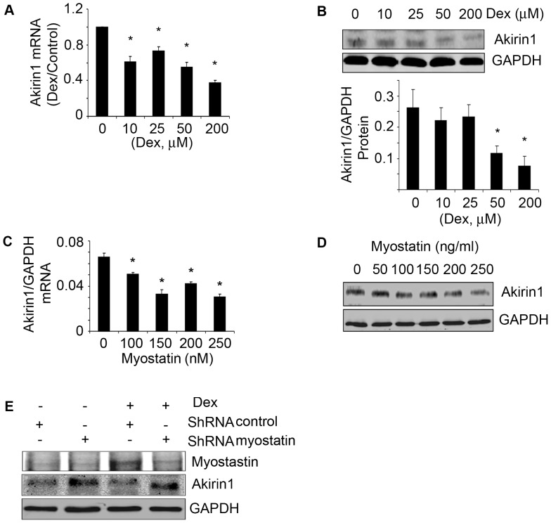
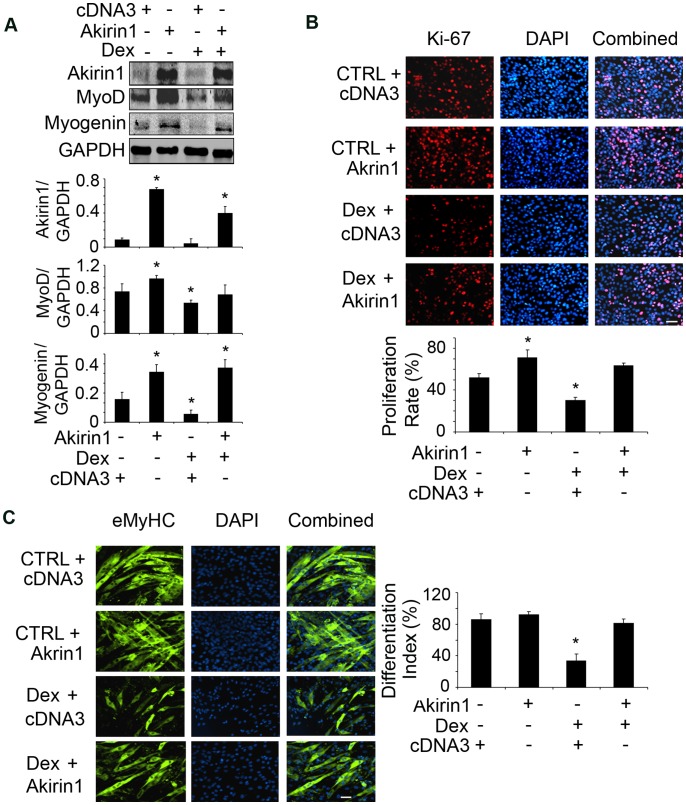
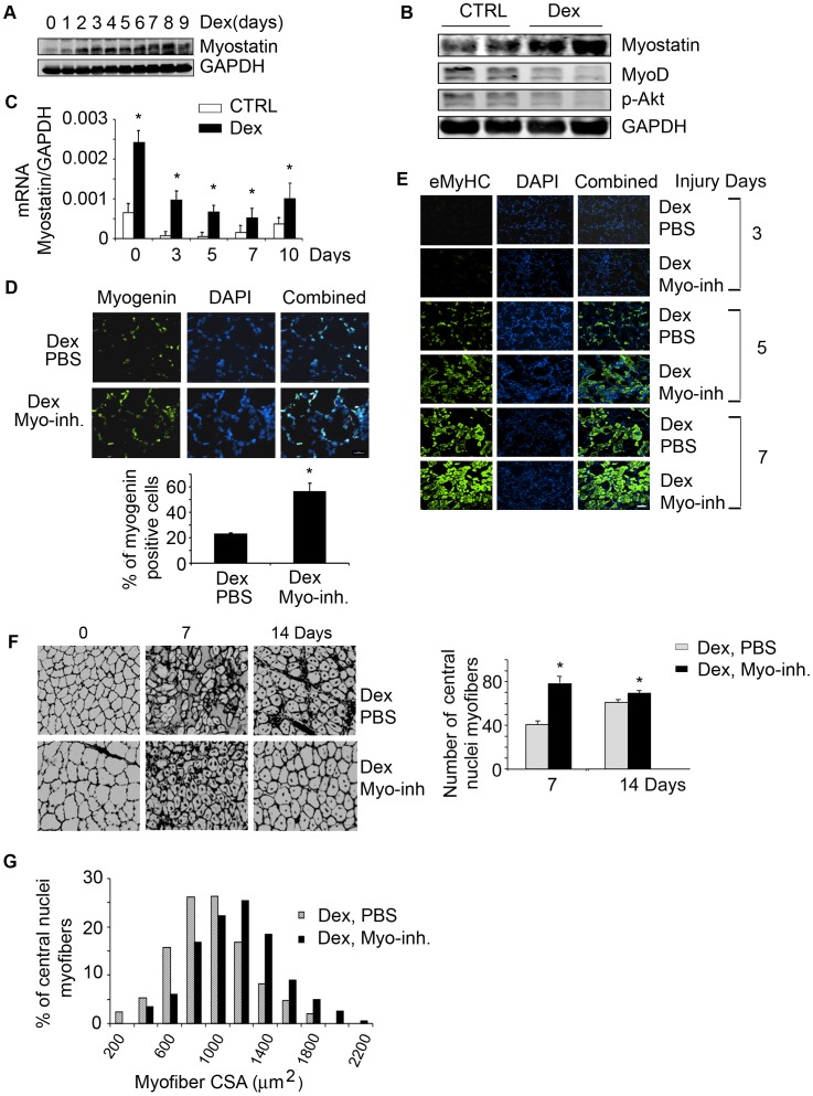
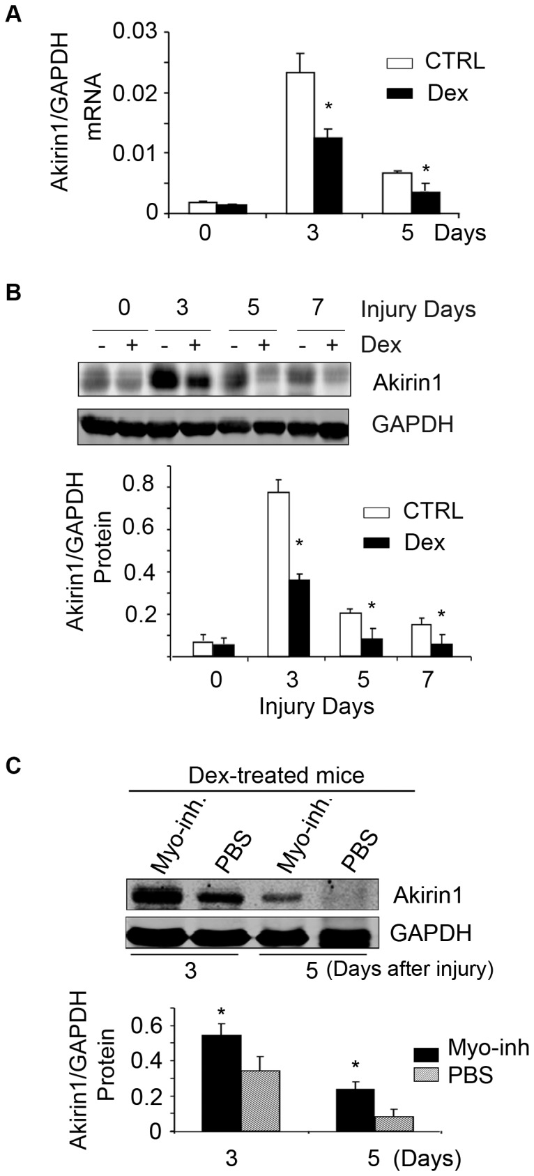
Glucocorticoids production is increased in many pathological conditions that are associated with muscle loss, but their role in causing muscle wasting is not fully understood. We have demonstrated a new mechanism of glucocorticoid-induced muscle atrophy: Dexamethasone (Dex) suppresses satellite cell function contributing to the development of muscle atrophy. Specifically, we found that Dex decreases satellite cell proliferation and differentiation in vitro and in vivo. The mechanism involved Dex-induced upregulation of myostatin and suppression of Akirin1, a promyogenic gene. When myostatin was inhibited in Dex-treated mice, Akirin1 expression increased as did satellite cell activity, muscle regeneration and muscle growth. In addition, silencing myostatin in myoblasts or satellite cells prevented Dex from suppressing Akirin1 expression and cellular proliferation and differentiation. Finally, overexpression of Akirin1 in myoblasts increased their expression of MyoD and myogenin and improved cellular proliferation and differentiation, theses improvements were no longer suppressed by Dex. We conclude that glucocorticoids stimulate myostatin which inhibits Akirin1 expression and the reparative functions of satellite cells. These responses attribute to muscle atrophy. Thus, inhibition of myostatin or increasing Akirin1 expression could lead to therapeutic strategies for improving satellite cell activation and enhancing muscle growth in diseases associated with increased glucocorticoid production.


Conflict of interest statement

Competing Interests: This study was partly funded by Satellite Health. There are no patents, products in development or marketed products to declare. This does not alter the authors' adherence to all the PLOS ONE policies on sharing data and materials.

Figures

Figure 1
Figure 1. Dex induces muscle proteolysis through the ubiquitin-proteasome system.
C57/BL6 mice were treated with Dex for 14 days. A. A representative western blot of Atrogin-1 or MuRF-1 in muscles. B. Cross-sections of TA muscles were immunostained with anti-laminin (upper panel, bar = 50 µm). Myofiber areas were measured and their distribution was calculated as the percentage of the number of myofibers in a designated area divided by the total number of myofibers assessed (lower panel).
Figure 2
Figure 2. Dex impairs satellite cell function in vitro.
A. Satellite cells were co-immunostained with anti-GR (Red) or Pax7 (green); nuclei were stained with DAPI (blue). In the merged picture (right column), satellite cells expressing glucocorticoid receptor (GR) are yellow (bar = 50 µm). B. Satellite cells were treated with Dex plus PBS or Dex plus myostatin inhibitor for 24 h, the percentage of Ki67 and Pax7 dual-positive (yellow) cells to total Pax-7 positive cells was calculated as the satellite cell proliferation rate (right panel) (*p<0.05; Dex vs. non-Dex-treatment, CTRL, bar = 50 µm). C. Satellite cells were cultured in differentiation media with Dex plus PBS or Dex plus myostatin inhibitor for 96 h, differentiated satellite cells were evaluated by immunostaining them with anti-eMyHC (green). The right panel summarizes the differentiation index (*p<0.05; Dex vs. non-Dex-treatment, CTRL, bar = 50 µm).
Figure 3
Figure 3. Dex suppresses satellite cell activation in vivo.
C57/BL6 control mice were treated with Dex and TA muscles were injured. A. At 1.5 day following injury, the cryo-cross-section of injured muscle were co-immunostained with anti-Pax7 (green) and Ki-67 (red); in the merged (right) column, dual positive (proliferating) cells are indicated by yellow color. *indicates injured myofibers. The proliferation rate was shown in the right column. B. The mRNAs of Myf5, MyoD and Myogenin in non-injured (designated as 0 injury days) or injured muscles (at different time points) were assessed by RT-PCR (*p<0.05; Dex vs. non-Dex-treated CTRL mice; n = 3 mice for each group). C. H/E staining of the cross-section of injured muscles (bar = 50 µm). D. At 7 days after injury, the newly formed myofiber sizes were measured and the myofiber size distribution was presented.
Figure 4
Figure 4. Dex stimulates myostatin expression in satellite cells suppressing their activation in vitro.
A. satellite cells were treated with different concentrations of Dex for 24 h. Myostatin mRNA was evaluated by RT-PCR (*p<0.05 vs. non-Dex; n = 3 independent experiments). B. Satellite cells were treated with different concentrations of Dex for 36 h; a representative western blot of myostatin is shown. C. Satellite cells were transducted with shRNA-myostatin or shRNA-control lentivirus and treated with/without Dex or myostatin for 24 h. The percentage of Ki67 and Pax7 dual positive cells (indicated by yellow color in the merged column) to total Pax7 positive cells (green) is shown in right panel. (*p<0.05 vs. shRNA-control bar = 50 µm). D. Satellite cells were transducted with shRNA-myostatin or shRNA-control lentivirus then exposed to differentiation media with/without Dex or myostatin for 96 h. The fixed cells were immunostained with anti-eMyHC (left panel). The differentiation index is shown in the right panel (*p<0.05 vs. shRNA-control bar = 50 µm).
Figure 5
Figure 5. Dex or myostatin can inhibit Akirin1 expression.
Satellite cells were treated with different concentrations of Dex for 24 h. A. Akirin-1 mRNA expression was examined by RT-PCR (corrected for GAPDH). The fold change vs. control (no Dex) are shown (*p<0.05; Dex vs. no-Dex; n = 3 independent experiments). B. A representative western blot of Akirin1 is in the upper panel. The band density of Akirin1 corrected for GAPDH are shown in lower panel. (*p<0.05; Dex vs. no-Dex; n = 3 independent experiments). C&D. Satellite cells were treated with different concentrations of myostatin. Akirin1 mRNA (C) and protein (D) was examined (*p<0.05 vs. no myostatin; n = 3 independent experiments). E. Stable cell lines were selected with puromycin from myoblasts transducted with lentivirus of shRNA-myostatin or shRNA-control and treated with or without Dex for 24 h. Representative Western blots of myostatin and Akirin1 are shown.
Figure 6
Figure 6. Overexpression of Akirin1 blocked Dex-induced suppression of myogenic gene expression and myoblast proliferation and differentiation.
A. C2C12 myoblasts were transfected with Akirin-1 or cDNA3 (control). After 24 h, cells were treated with 10 µM Dex in 2% horse serum for 24 h. Representative western blots of measured proteins are shown (upper panel) and band density corrected for GAPDH is shown in lower panel. (*p<0.05 vs. cells transfected with cDNA3 without Dex treatment; n = 3 repeats). B. Transfected cells were treated with 10 µM Dex and immunostained with anti-Ki67 (red). The percentage of Ki67 positive cells to the total number of cells in 10 areas was examined (lower panel) (*p<0.05 vs. cells transfected with cDNA3 without Dex treatment). C. Transfected cells were incubated in 2% horse serum with or without 20 µM Dex for 96 h to stimulate differentiation. Cells were immunostained with anti-eMyHC (green, left panel). The differentiation index is shown in right panel (*p<0.05 vs. cells transfected with cDNA3 without Dex treatment; n = 3 repeats).
Figure 7
Figure 7. Dex increases myostatin expression and impairs satellite cell activation in vivo.
A. Representative western blots of myostatin in gastrocnemius muscles of mice treated with Dex for different days. B. Representative western blots of indicated proteins from muscles of control or mice treated with Dex for 14 days. C. mRNA expression of myostatin was measured by RT-PCR in muscles of mice treated with or without Dex and injured with CTX (*p<0.05 vs. CTRL; n = 3 mice in each group). D. At 4 days after injury, cross-sections of muscle were immunostained with anti-myogenin (left panel) and the ratio of myogenin positive cells to DAPI expressed as a percentage is shown in right panel (*p<0.05 vs. Dex plus PBS). E. Cross-sections of injured TA muscles from mice injected with Dex plus PBS or Dex plus myostatin inhibitor were immunostained with anti-eMyHC (green). F. Sections in Fig. 7E were immunostained with laminin and DAPI to show the newly formed myofibers (left panel). The average number of central nuclei myofibers was calculated from 10 areas counted (*p<0.05 vs. Dex plus PBS, right panel). G. at 14 days after injury, newly formed myofiber cross-sectional areas were measured and the distribution is shown.
Figure 8
Figure 8. Dex decreases Akirin1 expression in activated satellite cells via myostatin expression in mice.
A. Akirin1 mRNA was evaluated in injured muscle of mice treated with/without Dex (*p<0.05 vs. no-Dex; n = 5 mice). B. Representative western blot of Akirin1 from injured muscle of mice treated with/without Dex (upper panel). The density of Akirin1 corrected for GAPDH is in lower panel (*p<0.05 vs. ctrl; n = 5 mice). C. A representative western blot of Akirin1 from injured TA muscles of mice treated with Dex plus PBS or Dex plus myostatin inhibitor (upper panel). The density of Akirin1 corrected for GAPDH is in lower panel (*p<0.05 vs. Dex plus PBS; n = 5 mice).

References

    1. Seale P, Sabourin LA, Girgis-Gabardo A, Mansouri A, Gruss P, et al. (2000) Pax7 is required for the specification of myogenic satellite cells. Cell 102: 777–786. - PubMed
    1. Mitchell PO, Pavlath GK (2004) Skeletal muscle atrophy leads to loss and dysfunction of muscle precursor cells. Am J Physiol Cell Physiol 287: C1753–C1762. - PubMed
    1. Jejurikar SS, Kuzon WM Jr (2003) Satellite cell depletion in degenerative skeletal muscle. Apoptosis 8: 573–578. - PubMed
    1. Zhang L, Wang XH, Wang H, Hu Z, Du J, et al. (2010) Satellite cell dysfunction and impaired IGF-1 signaling contribute to muscle atrophy in chronic kidney disease. J Am Soc Nephrol 21: 419–427. - PMC - PubMed
    1. Hasselgren PO (1999) Glucocorticoids and muscle catabolism. Curr Opin Clin Nutr Metab Care 2: 201–205. - PubMed

Publication types