. 2013 Aug:61:26-39.
doi: 10.1016/j.freeradbiomed.2013.03.012. Epub 2013 Mar 18.

The role of RhoA and cytoskeleton in myofibroblast transformation in hyperoxic lung fibrosis

Affiliations

The role of RhoA and cytoskeleton in myofibroblast transformation in hyperoxic lung fibrosis

Jixiang Ni et al. Free Radic Biol Med. 2013 Aug.

Abstract

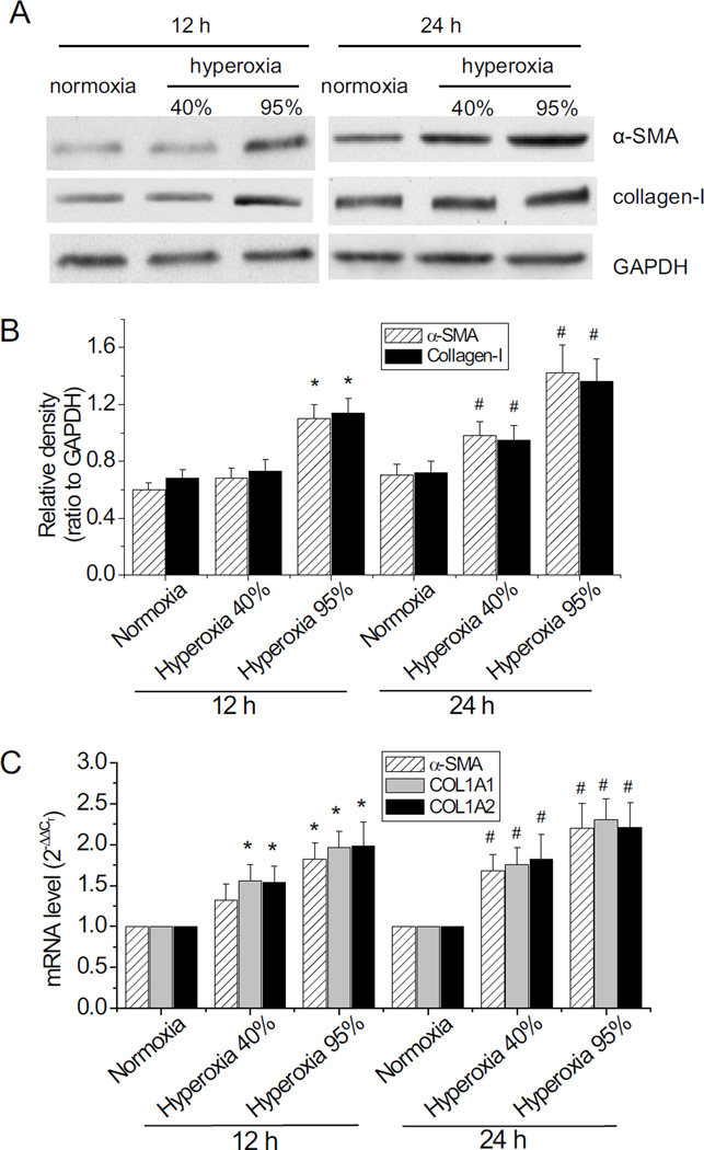
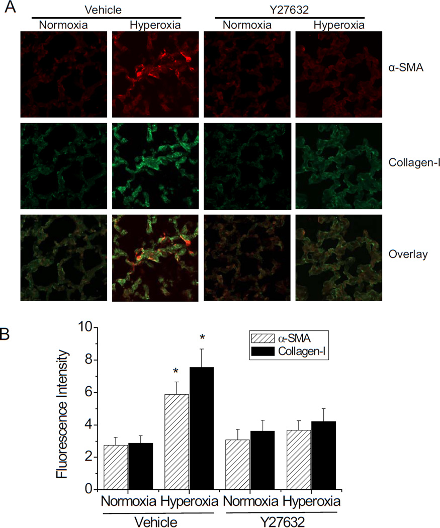
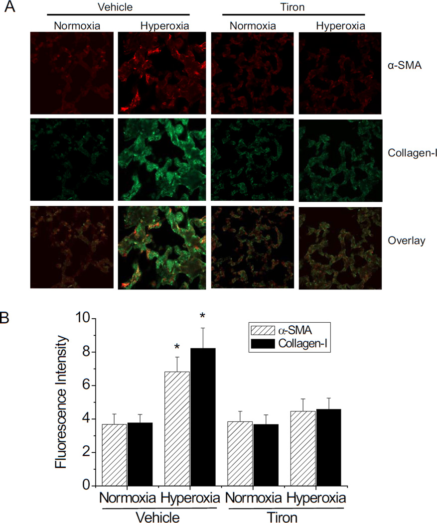
Myofibroblast transformation is a key process in the pathogenesis of lung fibrosis. We have previously reported that hyperoxia induces RhoA activation in HFL-1 lung fibroblasts and RhoA mediates collagen synthesis in hyperoxic lung fibrosis. In this study, we investigated the role of RhoA and actin cytoskeleton in hyperoxia-induced myofibroblast transformation. Exposure of HFL-1 lung fibroblasts to hyperoxia stimulated actin filament formation, shift of G-actin to F-actin, nuclear colocalization of myocardin-related transcription factor-A (MRTF-A), recruitment of MRTF-A to the α-smooth muscle actin (α-SMA) gene promoter, myofibroblast transformation, and collagen-I synthesis. Inhibition of RhoA by C3 transferase CT-04 or dominant-negative RhoA mutant T19N, and inhibition of ROCK by Y27632, prevented myofibroblast transformation and collagen-I synthesis. Moreover, inhibition of RhoA by CT-04 prevented hyperoxia-induced actin filament formation, shift of G-actin to F-actin, and nuclear colocalization of MRTF-A. In addition, disrupting actin filaments with cytochalasin D or scavenging reactive oxygen species (ROS) with tiron attenuated actin filament formation, nuclear colocalization of MRTF-A, myofibroblast transformation, and collagen-I synthesis. Furthermore, overexpression of constitutively active RhoA mutant Q63L or stabilization of actin filaments recapitulated the effects of hyperoxia on the actin cytoskeleton and nuclear colocalization of MRTF-A, myofibroblast transformation, and collagen-I synthesis. Interestingly, knocking down MRTF-A prevented hyperoxia-induced increase in the recruitment of MRTF-A to the serum response factor transcriptional complex on the α-SMA gene promoter, myofibroblast transformation, and collagen-I synthesis. Finally, Y27632 and tiron attenuated hyperoxia-induced increases in α-SMA and collagen-I in mouse lungs. Together, these results indicate that the actin cytoskeletal reorganization due to the ROS/RhoA-ROCK pathway mediates myofibroblast transformation and collagen synthesis in lung fibrosis of oxygen toxicity. MRTF-A contributes to the regulatory effect of the actin cytoskeleton on myofibroblast transformation during hyperoxia.

Keywords: Collagen; Fibroblasts; Free radicals; Lung; MRTF-A; Oxygen toxicity; Reactive oxygen species.


Figures

Figure 1
Figure 1
Exposure of lung fibroblasts to hyperoxia results in myofibroblast transformation and increased collagen synthesis. Human HFL-1 lung fibroblasts were exposed to normoxia or hyperoxia (40% and 95% oxygen) for 12–24 h after which the protein levels of α-SMA and collagen-I (A and B) and the mRNA levels of α-SMA, COL1A1 and COL1A2 (C) were determined as described in Materials and Methods. (A) is representative blot of four separate experiments. (B) is bar graph depicting the changes in α-SMA and collagen-I protein. (C) is bar graph depicting the changes in mRNA levels of α-SMA, COL1A1 and COL1A2. Results are expressed as mean ± SE; n=4. * P<0.05 vs normoxia 12 h; # P<0.05 vs normoxia 24 h.
Figure 2
Figure 2
RhoA inhibitor CT-04 and ROCK inhibitor Y27632 prevent hyperoxia-induced myofibroblast transformation and collagen synthesis. HFL-1 fibroblasts were exposed to normoxia or 95% oxygen in the presence and absence of the cell-permeative Rho inhibitor CT-04 (0.5 µg/ml) or ROCK inhibitor Y27632 (10 µM) for 24 h, after which the protein levels of α-SMA and collagen-I (A and B) and the mRNA levels of α-SMA, COL1A1 and COL1A2 (C) were determined as described in Materials and Methods. (A) is representative blot of four separate experiments. (B) is bar graph depicting the changes in α-SMA and collagen-I protein. (C) is bar graph depicting the changes in the mRNA levels of α-SMA, COL1A1 and COL1A2. Results are expressed as mean ± SE; n=4. * P<0.05 vs normoxia vehicle group in CT-04 experiment; # P<0.05 vs normoxia vehicle group in Y27632 experiment.
Figure 3
Figure 3
RhoA activation is responsible for hyperoxia-induced myofibroblast transformation and collagen synthesis. (A, B, and C): HFL-1 fibroblasts were transfected with or without plasmids containing the wild-type RhoA gene and constitutively active RhoA mutant Q63L. 48 h after transfection, the protein levels of α-SMA and collagen-I (A and B) and the mRNA levels of α-SMA, COL1A1 and COL1A2 (C) were determined as described in Materials and Methods. (D, E, and F): HFL-1 fibroblasts were transfected with or without plasmids containing the wild-type RhoA gene and constitutively dominant-negative RhoA mutant T19N. 48 h after transfection, the cells were exposed to normoxia or 95% oxygen for 24 h after which the protein levels of α-SMA and collagen-I (D and E) and the mRNA levels of α-SMA, COL1A1 and COL1A2 (F) were determined as described in Materials and Methods. Results are expressed as means± SE; n=4. * P < 0.05 vs the group of wild-type plasmids (WT plasmid); # P<0.05 vs normoxia WT plasmid group.
Figure 4
Figure 4
RhoA inhibitor CT-04 prevents hyperoxia-induced alterations in the actin cytoskeletal organization. HFL-1 fibroblasts were exposed to normoxia or 95% oxygen in the presence and absence of the cell-permeable Rho inhibitor CT-04 (0.5 µg/ml) for 24 h, after which the actin cytoskeleton was stained (A), and F-actin and G-actin were separated and measured using Western blot analysis (B and C). (A) is representative image of the actin cytoskeleton of four separate experiments. (B) is representative blot of four separate experiments. (C) is bar graph depicting the changes in F-actin/G-actin ratio. Results are expressed as mean ± SE; n=4. * P<0.05 vs normoxia vehicle group.
Figure 5
Figure 5
Jasplakinolide promotes myofibroblast transformation and collagen synthesis. HFL-1 fibroblasts were incubated with jasplakinolide (0–200 nM) for 24 h, after which the actin cytoskeleton was stained (A), F-actin and G-actin were separated and measured using Western blot (B and C), and the protein levels of α-SMA and collagen-I (D and E) and the mRNA levels of α-SMA, COL1A1 and COL1A2 (F) were determined as described in Materials and Methods. (A) is representative image of the actin cytoskeleton of four separate experiments. (B) and (D) are representative images of Western blot. (C) is bar graph depicting the changes in F-actin/G- actin ratio. (E) is bar graph depicting the changes in α-SMA and collagen-I protein. (F) is bar graph depicting the changes in the mRNA levels of α-SMA, COL1A1 and COL1A2. Results are expressed as mean ± SE; n=4. * P<0.05 vs control (0).
Figure 6
Figure 6
Cytochalasin D prevents hyperoxia-induced myofibroblast transformation and collagen synthesis. HFL-1 lung fibroblasts were treated with or without cytochalasin D (2 µM) for 1 h, a disrupter of the actin cytoskeleton, and then exposed to normoxia or hyperoxia (95% oxygen) for 24 h, after which the actin cytoskeleton was stained (A), the protein levels of α-SMA and collagen-I (B and C) and the mRNA levels of α-SMA, COL1A1 and COL1A2 (D) were determined as described in Materials and Methods. (A) is representative image of the actin cytoskeleton of four separate experiments. (B) is representative image of Western blot. (C) is bar graph depicting the changes in α-SMA and collagen-I protein. (D) is bar graph depicting the changes in mRNA levels of α-SMA, COL1A1 and COL1A2. Results are expressed as mean ± SE; n=4. * P<0.05 vs normoxia vehicle group.
Figure 7
Figure 7
Manipulating the actin cytoskeleton affects nuclear co-localization of MRTF-A in normoxic and hyperoxic HFL-1 fibroblasts. (A): HFL-1 lung fibroblasts were treated with or without cytochalasin D (2 µM) for 1 h and then exposed to normoxia or hyperoxia (95% oxygen) for 24 h. Some HFL-1 cells were exposed to normoxia or hyperoxia (95% oxygen) in the absence or presence of CT-04 (1 µg/ml) for 24 h. (B): HFL-1 fibroblasts were incubated with jasplakinolide (0–200 nM) for 24 h. Some HFL-1 fibroblasts were transfected with or without plasmids containing the wild-type RhoA gene and constitutively active RhoA mutant Q63L for 48 h. After these treatments, the cells were stained for the actin filaments and MRTF-A and counter-stained with DAPI. The images shown are representatives from four separate experiments.
Figure 8
Figure 8
Knocking-down MRTF-A prevents hyperoxia-induced myofibroblast transformation and collagen synthesis. HFL-1 cells were transfected with siRNA against MRTF-A or control siRNA. 48 h after the transfection, the cells were exposed to normoxia or hyperoxia (95% oxygen) for 24 h after which the protein levels of MRTF-A, α-SMA and collagen-I (A and B) and the mRNA levels of MRTF-A, α-SMA, COL1A1 and COL1A2 (C) were determined as described in Materials and Methods. Results are expressed as means ± SE; n=4. * P < 0.05 vs normoxia group without siRNA; # P<0.05 vs normoxia group with control siRNA.
Figure 9
Figure 9
Knocking-down MRTF-A prevents hyperoxia-induced recruitment of MRTF-A to α-SMA gene promoter. HFL-1 cells were transfected with siRNA against MRTF-A or control siRNA. 48 h after the transfection, the cells were exposed to normoxia or hyperoxia (95% oxygen) for 24 h after which a ChIP assay was performed as described in Materials and Methods. Results are expressed as means ± SE; n=4., *P < 0.05 vs normoxia with control siRNA.
Figure 10
Figure 10
ROS scavenger tiron attenuates hyperoxia-induced myofibroblast transformation and collagen synthesis. HFL-1 cells were exposed to normoxia or hyperoxia (95% oxygen) in the absence or presence of tiron (5mM) for 24 h after which the actin cytoskeleton was stained (A), the protein levels of MRTF-A, α-SMA and collagen-I (B and C) and the mRNA levels of MRTF-A, α-SMA, COL1A1 and COL1A2 (D) were determined as described in Materials and Methods. (A) is representative image of the actin cytoskeleton of four separate experiments. (B) is representative image of Western blot. (C) is bar graph depicting the changes in MRTF-A, α-SMA and collagen-I protein. (D) is bar graph depicting the changes in mRNA levels of MRTF-A, α-SMA, COL1A1 and COL1A2. Results are expressed as mean ± SE; n=4. * P<0.05 vs normoxia vehicle group.
Figure 11
Figure 11
Knocking-down Nox4 inhibits hyperoxia-induced ROS formation. HFL-1 cells were transfected with siRNA against Nox4 or control siRNA. 48 h after the transfection, the cells were exposed to normoxia or hyperoxia (95% oxygen) for 24 h after which ROS were assayed as described in Materials and Methods. Results are expressed as means ± SE; n=4. * P < 0.05 vs normoxia group.
Figure 12
Figure 12
ROCK inhibitor Y27632 attenuates hyperoxia-induced synthesis of α-SMA and collagen-I in mouse lungs. Male C57BL/6 mice were exposed to 80% oxygen for 5 days, and then exposed to 50% oxygen for another 10 days. Starting at the time when being changed to 50% oxygen, the hyperoxic mice were given daily injection of ROCK inhibitor Y27632 (5 mg/kg, daily by IP injection) or same volume of PBS. The normoxic mice were kept in room air and also received daily IP injection of same dose of Y27632 or PBS at same time. The lung sections were stained for α-SMA and collagen-I protein and subjected to fluorescence confocal microscopy. (A): representative images from eight separate experiments (magnification x400). (B): fluorescence intensity of α-SMA and collagen-I protein. Results are expressed as means ± SE; n=8. * P < 0.05 vs normoxia group.
Figure 13
Figure 13
ROS scavenger tiron attenuates hyperoxia-induced synthesis of α-SMA and collagen-I in mouse lungs. Male C57BL/6 mice were exposed to 80% oxygen for 5 days, and then exposed to 50% oxygen for another 10 days. Starting at the time when being changed to 50% oxygen, the hyperoxic mice were given daily injection of ROS scavenger tiron (1.5 g/kg, daily by IP injection) or same volume of PBS. The normoxic mice were kept in room air and also received daily IP injection of same dose of tiron or PBS at same time. The lung sections were stained for α-SMA and collagen-I protein and subjected to fluorescence confocal microscopy. (A): representative images from eight separate experiments (magnification x400). (B): fluorescence intensity of α-SMA and collagen-I protein. Results are expressed as means ± SE; n=8. * P < 0.05 vs normoxia group.

References

    1. Meyrick B. Pathology of the adult respiratory distress syndrome. Crit Care Clin. 1986;2:405–428. - PubMed
    1. Davis WB, Rennard SI, Bitterman PB, Crystal RG. Pulmonary oxygen toxicity. Early reversible changes in human alveolar structures induced by hyperoxia. N. Engl. J. Med. 1983;309:878–883. - PubMed
    1. Riley DJ, Kerr JS, Yu SY. Effect of proline analogs on oxygen toxicity-induced pulmonary fibrosis in the rat. Toxicol. Appl. Pharmacol. 1984;75:554–560. - PubMed
    1. Weinberger B, Laskin DL, Heck DE, Laskin JD. Oxygen toxicity in premature infants. Toxicol. Appl. Pharmacol. 2002;181:60–67. - PubMed
    1. Chen N, Li JJ, Xue XD. [Effect of losartan on lung fibrosis in neonatal rats with hyperoxia-induced chronic lung disease] Zhongguo Dang. Dai Er. Ke. Za Zhi. 2007;9:591–594. - PubMed

Publication types

MeSH terms