

A rapid, novel method of volumetric assessment of MRI-detected subchondral bone marrow lesions in knee osteoarthritis
- PMID: 23518154
- PMCID: PMC3701435
- DOI: 10.1016/j.joca.2013.03.007
A rapid, novel method of volumetric assessment of MRI-detected subchondral bone marrow lesions in knee osteoarthritis
Abstract
Purpose: To assess reliability and validity of a semi-automated quantitative method for osteoarthritis (OA)-related bone marrow lesion (BML) assessment in the femur and tibia.
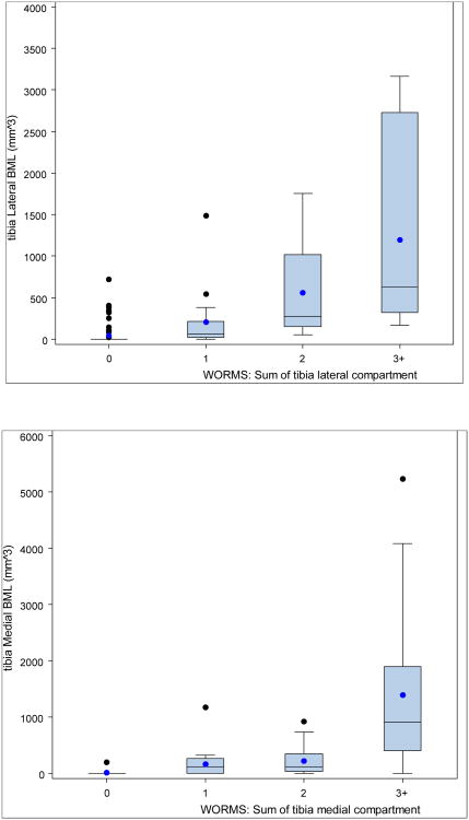
Methods: In a cross-sectional study of subjects with knee OA, we examined concurrent criterion and clinical validation of a novel method of semi-automated quantitative BML measurement. The primary outcome was total segmented BML volume in femoral and tibial medial and lateral knee compartments. Criterion validation was examined through comparison of BML volumes with Whole-Organ Magnetic Resonance Imaging Score (WORMS) scoring. Clinical validation was examined via associations of tibial and femoral BML volume with the Western Ontario and McMaster University OA Index weight-bearing pain questions.
Results: Among the 115 subjects, mean age was 62 years, mean BMI 30.4 (kg/m(2)), 84% were white and 52% male. The intra-class correlation coefficients (ICC) for intra-reader reliability was 0.96 and 0.97 for inter-reader reliability. Significant Spearman's correlations were found between segmented BML volume and WORMS BML scoring for tibial medial (0.75) and lateral (0.73) compartments, and for femoral medial (0.72) and lateral (0.88) compartments. Significant positive associations were found between weight-bearing pain and total femoral BML volume (P < 0.003), but not total tibial BML (P < 0.101).
Conclusion: We have documented a moderately strong correlation between a novel measurement method of femoral and tibial BML volume and semi-quantitative WORMS scores, providing evidence of criterion validity. The hypothesis that weight-bearing pain was associated with BML volume was confirmed for total femoral BML volume but not total tibial BML volume. The lack of association between tibial BML volume and pain requires further investigation.
Copyright © 2013 Osteoarthritis Research Society International. Published by Elsevier Ltd. All rights reserved.
Conflict of interest statement
Figures
Similar articles
-
Quantitative bone marrow lesion size in osteoarthritic knees correlates with cartilage damage and predicts longitudinal cartilage loss.BMC Musculoskelet Disord. 2011 Sep 30;12:217. doi: 10.1186/1471-2474-12-217. BMC Musculoskelet Disord. 2011. PMID: 21961433 Free PMC article. Clinical Trial.
-
Association of subchondral bone marrow lesion localization with weight-bearing pain in people with knee osteoarthritis: data from the Osteoarthritis Initiative.Arthritis Res Ther. 2021 Jan 19;23(1):35. doi: 10.1186/s13075-021-02422-0. Arthritis Res Ther. 2021. PMID: 33468243 Free PMC article.
-
Bone marrow lesion volume reduction is not associated with improvement of other periarticular bone measures: data from the Osteoarthritis Initiative.Arthritis Res Ther. 2013;15(5):R153. doi: 10.1186/ar4336. Arthritis Res Ther. 2013. PMID: 24432365 Free PMC article.
-
The reliability of a new scoring system for knee osteoarthritis MRI and the validity of bone marrow lesion assessment: BLOKS (Boston Leeds Osteoarthritis Knee Score).Ann Rheum Dis. 2008 Feb;67(2):206-11. doi: 10.1136/ard.2006.066183. Epub 2007 May 1. Ann Rheum Dis. 2008. PMID: 17472995 Review.
-
Degree of Preoperative Subchondral Bone Marrow Lesion Is Associated With Postoperative Outcome After Medial Opening Wedge High Tibial Osteotomy.Am J Sports Med. 2019 Aug;47(10):2454-2463. doi: 10.1177/0363546519858996. Epub 2019 Jul 9. Am J Sports Med. 2019. PMID: 31287714
Cited by
-
Association of quantitative measures of effusion-synovitis and hoffa-synovitis with radiographic and pain progression: Data from the FNIH OA biomarkers consortium.Osteoarthr Cartil Open. 2021 Jan 11;3(1):100138. doi: 10.1016/j.ocarto.2021.100138. eCollection 2021 Mar. Osteoarthr Cartil Open. 2021. PMID: 36475070 Free PMC article.
-
Bone marrow lesions in osteoarthritis: biomarker or treatment target? A narrative review.Skeletal Radiol. 2025 Feb;54(2):175-191. doi: 10.1007/s00256-024-04725-0. Epub 2024 Jun 14. Skeletal Radiol. 2025. PMID: 38877110 Review.
-
Measurement of bone marrow lesions by MR imaging in knee osteoarthritis using quantitative segmentation methods--a reliability and sensitivity to change analysis.BMC Musculoskelet Disord. 2014 Dec 20;15:447. doi: 10.1186/1471-2474-15-447. BMC Musculoskelet Disord. 2014. PMID: 25528153 Free PMC article. Clinical Trial.
-
Clinical and translational potential of MRI evaluation in knee osteoarthritis.Curr Rheumatol Rep. 2014 Jan;16(1):391. doi: 10.1007/s11926-013-0391-6. Curr Rheumatol Rep. 2014. PMID: 24318386 Free PMC article. Review.
-
The relation of oral bisphosphonates to bone marrow lesion volume among women with osteoarthritis.Osteoarthritis Cartilage. 2020 Oct;28(10):1325-1329. doi: 10.1016/j.joca.2020.07.006. Epub 2020 Aug 5. Osteoarthritis Cartilage. 2020. PMID: 32768598 Free PMC article.
References
Publication types
MeSH terms
Grants and funding
- R01 AR056664/AR/NIAMS NIH HHS/United States
- N01-AR-2-2261/AR/NIAMS NIH HHS/United States
- N01-AR-2-2260/AR/NIAMS NIH HHS/United States
- CAPMC/ CIHR/Canada
- K24 AR 057827/AR/NIAMS NIH HHS/United States
- P60 AR047782/AR/NIAMS NIH HHS/United States
- N01-AR-2-2259/AR/NIAMS NIH HHS/United States
- P60 AR 47782/AR/NIAMS NIH HHS/United States
- T32 AR 055885/AR/NIAMS NIH HHS/United States
- N01-AR-2-2258/AR/NIAMS NIH HHS/United States
- K24 AR057827/AR/NIAMS NIH HHS/United States
- R01AR056664/AR/NIAMS NIH HHS/United States
- N01-AR-2-2262/AR/NIAMS NIH HHS/United States
- T32 AR055885/AR/NIAMS NIH HHS/United States
LinkOut - more resources
Full Text Sources
Other Literature Sources
Medical
