Review
. 2013 Mar 21;5(3):954-80.
doi: 10.3390/v5030954.

Hepatitis C virus-induced mitochondrial dysfunctions

Affiliations
Review

Hepatitis C virus-induced mitochondrial dysfunctions

Charlène Brault et al. Viruses. .

Abstract

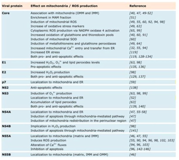
Chronic hepatitis C is characterized by metabolic disorders and a microenvironment in the liver dominated by oxidative stress, inflammation and regeneration processes that lead in the long term to hepatocellular carcinoma. Many lines of evidence suggest that mitochondrial dysfunctions, including modification of metabolic fluxes, generation and elimination of oxidative stress, Ca2+ signaling and apoptosis, play a central role in these processes. However, how these dysfunctions are induced by the virus and whether they play a role in disease progression and neoplastic transformation remains to be determined. Most in vitro studies performed so far have shown that several of the hepatitis C virus (HCV) proteins localize to mitochondria, but the consequences of these interactions on mitochondrial functions remain contradictory, probably due to the use of artificial expression and replication systems. In vivo studies are hampered by the fact that innate and adaptive immune responses will overlay mitochondrial dysfunctions induced directly in the hepatocyte by HCV. Thus, the molecular aspects underlying HCV-induced mitochondrial dysfunctions and their roles in viral replication and the associated pathology need yet to be confirmed in the context of productively replicating virus and physiologically relevant in vitro and in vivo model systems.


Figures

Figure 1
Figure 1
Structure and functions of mitochondria. Mitochondrial membranes and compartments comprise the matrix, inner mitochondrial membrane (IMM), outer mitochondrial membrane (OMM), cristae and mitochondria-associated membrane (MAM). MAMs are points of contact between mitochondria and smooth endoplasmic reticulum (ER), enriched in certain factors, including the inositol triphosphate receptor (IP3R), voltage-dependent anion channel (VDAC) and sarco/endoplasmic reticulum calcium ATPase (SERCA). The main functions of mitochondria are represented in color codes. In red: fatty acids and acetyl coenzyme A (acetyl-CoA) are oxidized by β-oxidation (also called Lynen helix) and the tricarboxylic acid cycle (TCA), respectively, and energy is transferred onto redox coenzymes. In green: the electron transport chain (ETC) is composed of five respiratory complexes (numbered I to V) and uses the electrons of the coenzymes generated by β-oxidation and the TCA cycle as substrates to generate a proton gradient (mtΔΨ) across the inner membrane. The flux of protons from the intermembrane space through complex V (ATP synthase) back into the mitochondrial matrix finally leads to ATP generation. ATP is exported to cytosol via adenine nucleotide translocase (ANT). In brown: electrons that leak from the ETC react with O2, forming superoxide anion O2•- and hydrogen peroxide H2O2, also referred to as reactive oxygen species (ROS). In blue: a number of caspase activators are retained in the intermitochondrial membrane space, including, e.g., cytochrome c, endonuclease G and apoptosis-inducing factor (AIF). These are released into the cytosol by pro-apoptotic signaling, which leads to opening of the mitochondrial permeability transition pore (mPTP). In violet: mitochondria constantly exchange calcium with the cytosol via the mitochondrial calcium uniporter (MCU) and Na+Ca2+ exchanger (NaCaE) and with the ER via the mPTP, which comprises, e.g., IP3R and VDAC.
Figure 2
Figure 2
ROS generation and scavenging in HCV infected cells. The main source of mt reactive oxygen species (ROS) is the electron transport chain (ETC); electrons leak from complexes I and IV and produce superoxide anion (O2•-) and hydrogen peroxide (H2O2), which is membrane permeable. Non-mt sources of O2•- and H2O2 are cytosolic xanthine oxidase (XO) and plasma membrane NAD(P)H oxidases. The cellular detoxification system scavenges ROS and converts it into H2O and O2: Mitochondrial (Mn) or cytosolic (Cu/Zn) superoxide dismutases (SOD) degrade O2•- into H2O2, which is then converted into H2O and O2 by either catalase (CAT), glutathione-peroxidases (GPx) or peroxiredoxin (PRX), which oxidize glutathione (GSH) in the process. Recycling of oxidized glutathione (GSSG) into GSH requires NADPH coenzyme. In addition to GSH, metallothionein (MT) and thioredoxins (Trx) are also used to scavenge ROS peroxidation products. If ROS are considerably augmented, hydroxyl radicals (HO), the most damaging form of ROS, can be produced by the Fenton reaction. ROS producing or scavenging factors whose action is modified by HCV are indicated by red stars and include: 1) oxidation of the glutathione pool [40,60]; 2) decrease in NADPH content [61]; 3) increased ROS production from ETC complex I [40,61]; 4) induction of mitochondrial SOD expression [60]; 5) catalase activation [60]; 6) induction of glutathione peroxidase [49,62]; 7) oxidation of the thioredoxin pool [60]; 8) lipid peroxidation [49,63]; 9) induction of metallothionein [49,64]; and 10) cytoplasmic ROS production by NADPH oxidase activation [65,66].
Figure 3
Figure 3
Metabolism in HCV infected cells. The major metabolic pathways that use glucose (Glc) to either drive nucleotide synthesis and NADPH production via the pentose phosphate shunt (PPP) or to drive the TCA cycle and respiration in mitochondria are depicted. TCA reactions catalyze the reduction of NAD+ and FAD into NADH and FADH2, which in turn feed the electron transport chain (ETC). The ETC maintains the mitochondrial membrane potential, mtΔΨ, required for ATP synthesis. Electrons that leak from complex I and IV of the ETC form reactive oxygen species (ROS). ROS scavenging requires reduced glutathione (GSH), which is oxidized in the process. Recycling of the GSH pool requires NADPH, which is mainly produced by the PP shunt. Glucose-6-phosphate (G6P), pyruvate (Pyr), lactate (Lac), acetyl coenzyme A (AcCoA), pyruvate dehydrogenase complex (PDC), citrate synthase (CS), aconitase (ACO), isocitrate dehydrogenase (ICDH), α-ketoglutarate dehydrogenase (αKGDH), succinyl coenzyme A ligase (SuCLA), succinate dehydrogenase (SDH), fumarate hydratase (FH), malate dehydrogenase (MDH). Factors and events targeted by HCV are indicated by red stars and include: activation of glycolytic enzymes (1) with potential re-routing of the flux into the pentose phosphate shunt, (2) induction of TCA cycle (3) and lipogenic enzymes (4) and complex I of the ETC (5).
Figure 4
Figure 4
Calcium signaling in HCV infected cells. Mitochondria take up Ca2+ via the mtΔΨ-dependent calcium uniporter (MCU), the voltage-dependent anion channel (VDAC), the Na+/Ca2+ exchanger (NaCaE) and the mitochondrial permeability transition pore (mPTP). ER Ca2+ uptake is regulated by the sarco/endoplasmic reticulum calcium ATP-ase (SERCA). Ca2+ fluxes are represented by dotted violet arrows. Mt Ca2+ levels control energy metabolism by activating several enzymes of the TCA cycle, such as isocitrate dehydrogenase (ICDH) and α-ketoglutarate dehydrogenase (αKGDH), as well as adenosine nucleotide translocase (ANT), which is a component of the mPTP. Mt Ca2+ increase (violet arrows) inhibits ETC function by decreasing mtΔΨ, as well as complex V activity and induces ATP depletion by mPTP opening, leading to the release of pro-apoptotic factors. Kick off effects of Ca2+ increase are marked by black arrows and comprise changes in mtΔΨ, mPTP opening, amplification of ETC inhibition and concomitant ROS generation and ATP depletion. Factors and events targeted by HCV are indicated by red stars and include 1) increase of BIP/Grp78, an endoplasmic reticulum chaperone [35,119], 2) Ca2+ transfer from the ER into mitochondria, mediated by NS5A-induced ER stress [96] and by Core expression [55], 3) Core-induced increase of MCU activity [32] and 4) Core-induced SERCA expression [119].
Figure 5
Figure 5
Effect on mitochondria.

References

    1. Lemon S.M., Walker C.M., Alter M.J., Yi M.–K. Hepatitis c virus. In: Knipe D.M., Howley P.M., editors. Fields Virology. 5th. 2007. pp. 1253–1304.
    1. Ge D., Fellay J., Thompson A.J., Simon J.S., Shianna K.V., Urban T.J., Heinzen E.L., Qiu P., Bertelsen A.H., Muir A.J., et al. Genetic variation in il28b predicts hepatitis c treatment–induced viral clearance. Nature. 2009;461:399–401. - PubMed
    1. Alaei M., Negro F. Hepatitis c virus and glucose and lipid metabolism. Diabetes Metab. 2008;34:692–700. doi: 10.1016/S1262-3636(08)74606-8. - DOI - PubMed
    1. Bartosch B. Hepatitis c virus and its complex interplay with hepatic glucose and lipid metabolism. J. Hepatol. 2009;50:845–847. doi: 10.1016/j.jhep.2009.02.009. - DOI - PubMed
    1. Moradpour D., Penin F., Rice C.M. Replication of hepatitis c virus. Nat. Rev. Microbiol. 2007;5:453–463. doi: 10.1038/nrmicro1645. - DOI - PubMed

Publication types

LinkOut - more resources