. 2013 May;27(5):754-68.
doi: 10.1210/me.2012-1331. Epub 2013 Mar 21.

Dicer deficiency reveals microRNAs predicted to control gene expression in the developing adrenal cortex

Affiliations

Dicer deficiency reveals microRNAs predicted to control gene expression in the developing adrenal cortex

Kenneth T Krill et al. Mol Endocrinol. 2013 May.

Abstract

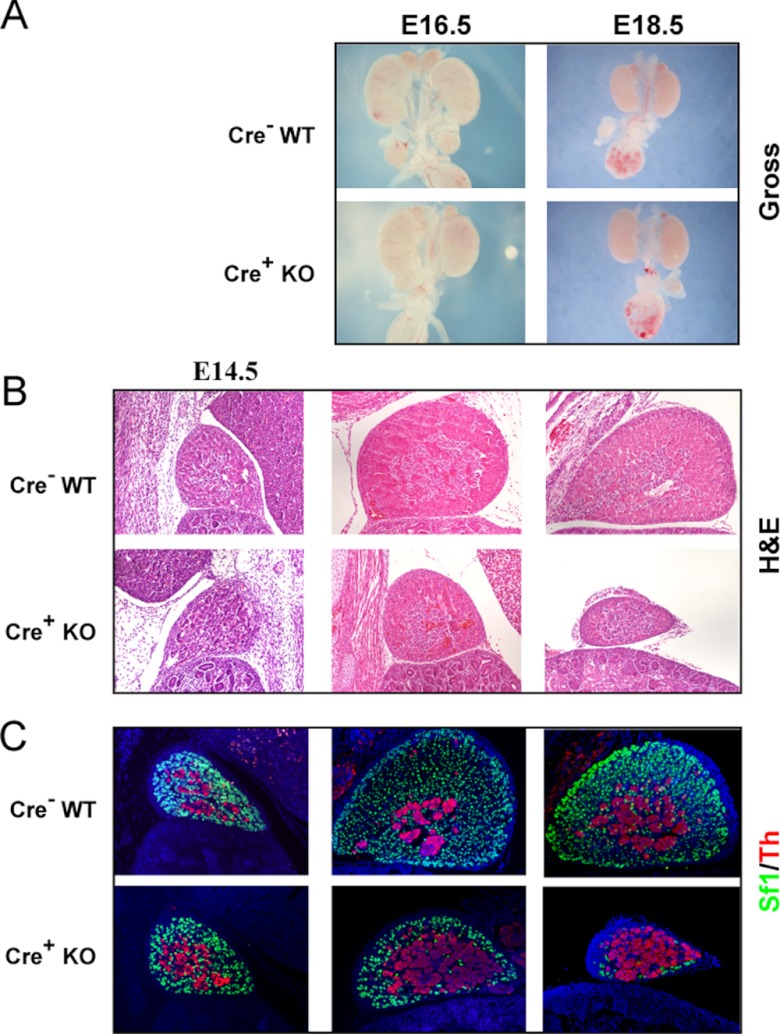
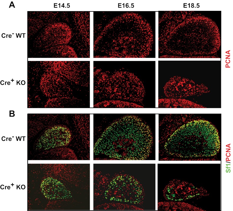
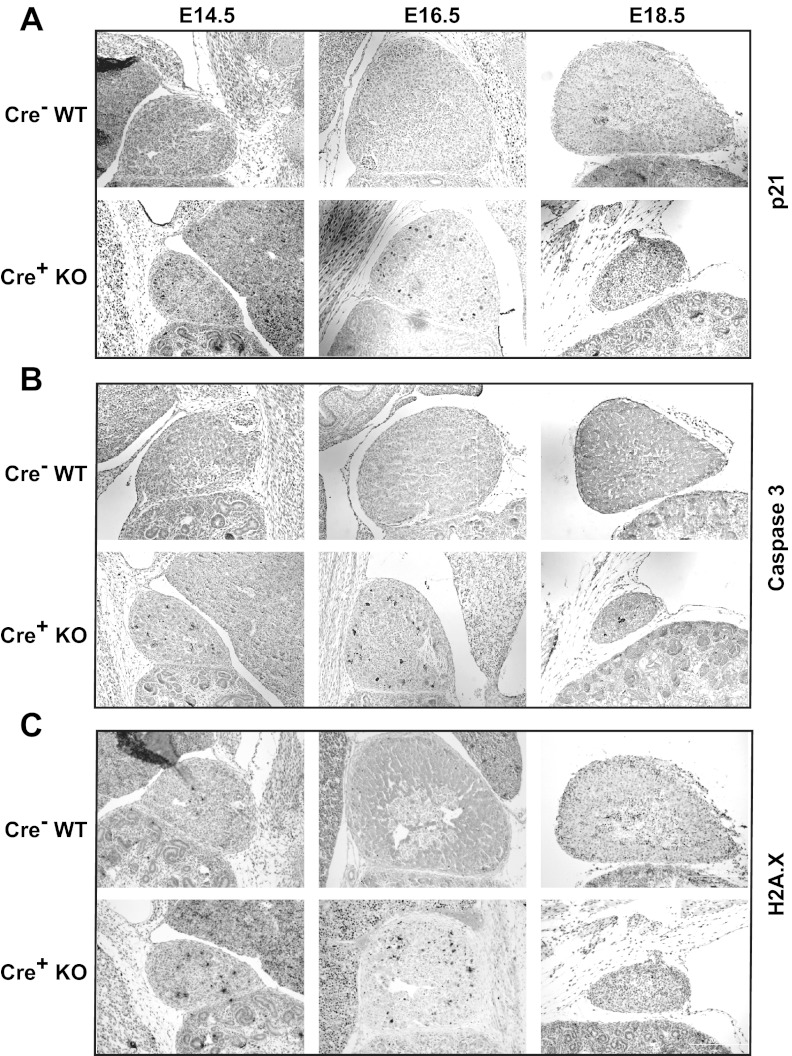
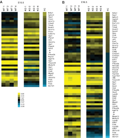
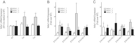
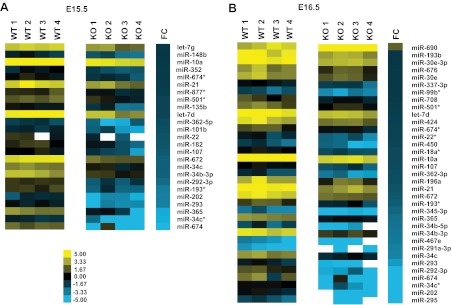
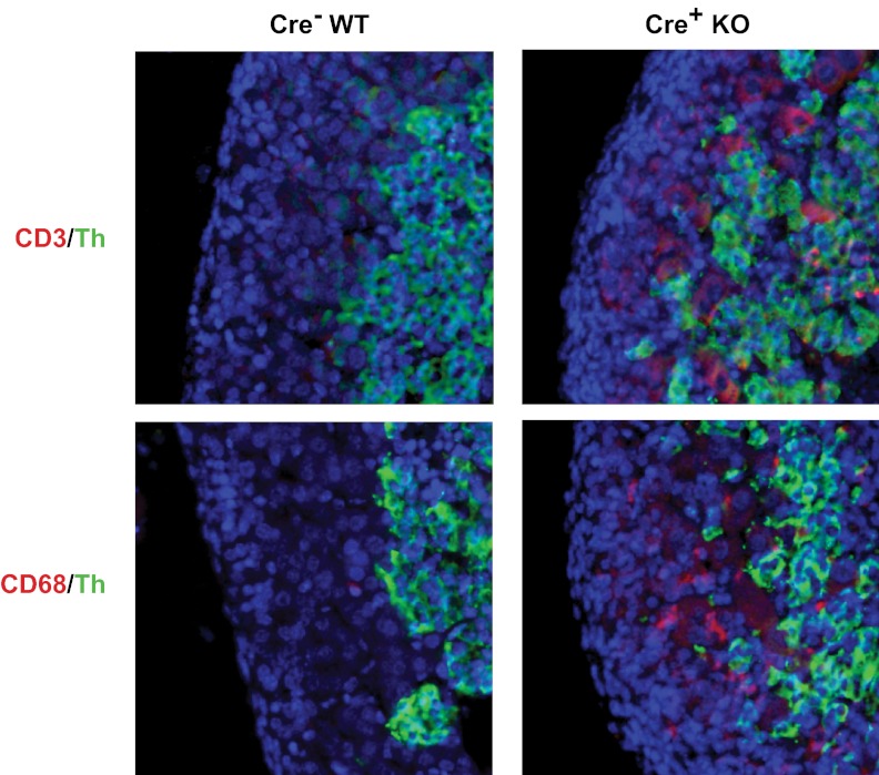
MicroRNAs (miRNAs) are small, endogenous, non-protein-coding RNAs that are an important means of posttranscriptional gene regulation. Deletion of Dicer, a key miRNA processing enzyme, is embryonic lethal in mice, and tissue-specific Dicer deletion results in developmental defects. Using a conditional knockout model, we generated mice lacking Dicer in the adrenal cortex. These Dicer-knockout (KO) mice exhibited perinatal mortality and failure of the adrenal cortex during late gestation between embryonic day 16.5 (E16.5) and E18.5. Further study of Dicer-KO adrenals demonstrated a significant loss of steroidogenic factor 1-expressing cortical cells that was histologically evident as early as E16.5 coincident with an increase in p21 and cleaved-caspase 3 staining in the cortex. However, peripheral cortical proliferation persisted in KO adrenals as assessed by staining of proliferating cell nuclear antigen. To further characterize the embryonic adrenals from Dicer-KO mice, we performed microarray analyses for both gene and miRNA expression on purified RNA isolated from control and KO adrenals of E15.5 and E16.5 embryos. Consistent with the absence of Dicer and the associated loss of miRNA-mediated mRNA degradation, we observed an up-regulation of a small subset of adrenal transcripts in Dicer-KO mice, most notably the transcripts coded by the genes Nr6a1 and Acvr1c. Indeed, several miRNAs, including let-7, miR-34c, and miR-21, that are predicted to target these genes for degradation, were also markedly down-regulated in Dicer-KO adrenals. Together these data suggest a role for miRNA-mediated regulation of a subset of genes that are essential for normal adrenal growth and homeostasis.


Figures

Figure 1.
Figure 1.
Analysis of adrenals from Sf1-Cre/Dicerlox/lox mice. A, Gross photos of adrenals and kidneys from E16.5 and E18.5 Dicer-KO (cre+ KO) embryos and cre littermate controls (WT). Photos of the E16.5 time point were taken at a 2× higher magnification than the E18.5 photos. B and C, Histological comparison of adrenal sections from Dicer-KO (cre+ KO) embryos and cre littermate controls (WT) at E14.5, E16.5, and E18.5 by hematoxylin and eosin (H&E) (B) and immunofluorescent straining (C) for Sf1 and tyrosine hydroxylase (Th). In C, sections were counterstained with DAPI (blue) before visualization and images were merged to show colocalization. Images in B and C are taken at ×100 magnification.
Figure 2.
Figure 2.
Assessment of proliferation in adrenals from Sf1-Cre/Dicerlox/lox mice. Immunofluorescent staining of adrenals from Dicer-KO (cre+ KO) embryos and cre littermate controls (WT) at E14.5, E16.5, and E18.5. A, PCNA alone. B, Sf1 (green) and PCNA (red). Images are taken at ×100 magnification.
Figure 3.
Figure 3.
Assessment of cell cycle arrest/apoptosis and DNA damage in adrenals from Sf1-Cre/Dicerlox/lox mice. DAB immunohistochemistry of adrenals Dicer-KO (cre+ KO) embryos and cre littermate controls (WT) at E14.5, E16.5, and E18.5. A, p21 (Cdkn1a), a cell cycle inhibitor. B, Cleaved-caspase 3, an apoptotic marker. C, Phospho-γ-H2A.X, an indicator of DNA damage repair. Tissues were counterstained with hematoxylin or eosin. Images are taken at ×100 magnification.
Figure 4.
Figure 4.
Gene expression differences between adrenals from control and Sf1-Cre/Dicerlox/lox embryos at E15.5 and E16.5. Heatmaps generated from Affymetrix gene expression arrays illustrate differentially expressed genes in adrenals from Dicer-KO and control (WT, cre littermates) embryos at E15.5 (A) and E16.5 (B). Data were filtered by excluding probe sets with an FDR of ≥.05 and a log2-fold change of ≤1.5 or ≥−1.5. Yellow bars indicate an increase over the mean chip intensity, and blue indicates a decrease over mean intensity. FC represents log2-fold change between the 4 Dicer-KO litters/replicates compared with the 4 WT litters/replicates.
Figure 5.
Figure 5.
Quantitative real-time PCR to confirm Nr6a1 expression in adrenals from Sf1-Cre/Dicerlox/lox and Sf1-Cre–only tissues. Total RNA was isolated from adrenals and reverse transcribed and quantitative PCR performed as described in Materials and Methods. A, Comparison of Nr6a1 expression in adrenals from adult mice expressing only the Sf1-Cre transgene (Cre), relative to WT animals. B and C, Expression of the three Nr6a1 transcript isoforms in 4 individual samples of E15.5 and E16.5 Sf1-Cre/Dicerlox/lox adrenals relative to their corresponding WT control samples. Statistical significance was determined using unpaired t tests. *, P < .05; **, P ≤ .01; ***, P ≤ .001.
Figure 6.
Figure 6.
Differentially expressed miRNAs in adrenals from Sf1-Cre/Dicerlox/lox embryos. Heatmaps were generated from OpenArray rodent miRNA analysis as described in Materials and Methods. A, Differentially expressed miRNAs at both E15.5 and E16.5 in Dicer-KO adrenals relative to control (WT, cre littermates) adrenals were filtered to select only those whose expression was changed significantly with a P value ≤ .05. Only miRNAs with a log2-fold change of ≥1.5 or ≤−1.5 were included. FC represents log2-fold change between the 4 Dicer-KO litters/replicates/samples compared with the 4 WT samples.
Figure 7.
Figure 7.
Expression of CD3 and CD68 in adrenals from E18.5 Sf1-Cre/Dicerlox/lox embryos. Immunohistochemistry of adrenals from E18.5 Dicer-KO (cre+ KO) embryos and cre littermate controls (WT). Top panel shows immunofluorescence staining for CD3 (red) and Th (green). Bottom panel shows immunofluorescence staining form CD68 (red) and Th (green). Sections were counterstained with DAPI (blue) before visualization, and images were merged to show colocalization.

References

    1. Hammer GD, Parker KL, Schimmer BP. Minireview: transcriptional regulation of adrenocortical development. Endocrinology. 2005;146:1018–1024 - PubMed
    1. Kim AC, Barlaskar FM, Heaton JH, et al. In search of adrenocortical stem and progenitor cells. Endo Rev. 2009;30:241–263 - PMC - PubMed
    1. Lalli E. Adrenal cortex ontogenesis. Best Pract Res Clin Endocrinol Metab. 2010;24:853–864 - PubMed
    1. Parker KL, Schimmer BP. The roles of the nuclear receptor steroidogenic factor 1 in endocrine differentiation and development. Trends Endocrinol Metab. 1996;7:203–207 - PubMed
    1. Wood MA, Hammer GD. Adrenocortical stem and progenitor cells: unifying model of two proposed origins. Mol Cell Endocrinol. 2011;336:206–212 - PMC - PubMed

Publication types

MeSH terms