. 2013 Jul;6(4):1043-8.
doi: 10.1242/dmm.011601. Epub 2013 Mar 15.

Central and haematopoietic interleukin-1 both contribute to ischaemic brain injury in mice

Affiliations

Central and haematopoietic interleukin-1 both contribute to ischaemic brain injury in mice

Adam Denes et al. Dis Model Mech. 2013 Jul.

Abstract

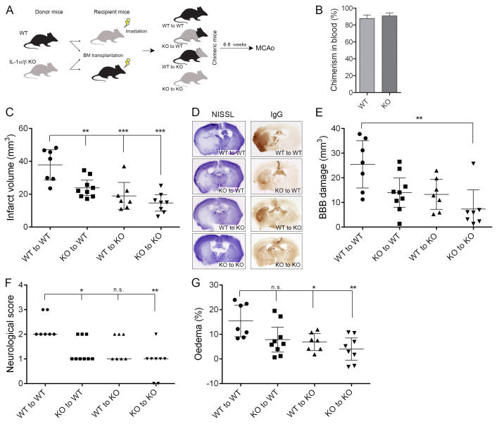
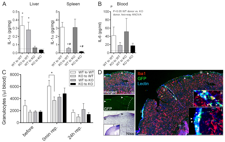
Interleukin-1 (IL-1) is a key regulator of inflammation and ischaemic brain injury, but the contribution of central and peripheral sources of IL-1 to brain injury is not well understood. Here we show that haematopoietic-derived IL-1 is a key driver of ischaemic brain injury. Wild type (WT) mice transplanted with IL-1αβ-deficient bone marrow displayed a significant (40%) reduction in brain injury induced by focal cerebral ischaemia compared with WT mice transplanted with WT bone marrow. This was paralleled by improved neurological outcome and the almost complete absence of splenic-derived, but not liver-derived, IL-1α after stroke in WT mice lacking haematopoietic-derived IL-1. IL-1αβ knockout (KO) mice transplanted with IL-1αβ-deficient bone marrow showed a 60% reduction in brain injury compared with WT mice receiving WT bone marrow. Transplantation of WT bone marrow in IL-1αβ KO mice resulted in a similar level of blood-brain-barrier injury to that observed in WT mice receiving IL-1αβ-deficient bone marrow. Cerebral oedema after brain injury was reduced in IL-1αβ KO recipients irrespective of donor-derived IL-1, but a lack of haematopoetic IL-1 has also been associated with smaller brain oedema independently of recipient status. Thus, both central and haematopoietic-derived IL-1 are important contributors to brain injury after cerebral ischaemia. Identification of the cellular sources of IL-1 in the periphery could allow targeted interventions at these sites.


Figures

Fig. 1.
Fig. 1.
Both central and haematopoietic-derived IL-1 are important contributors to brain injury after cerebral ischaemia. (A) Wild-type (WT) and IL-1αβ-deficient (KO) mice were subjected to whole body irradiation followed by transplantation of WT or IL-1αβ KO bone marrow. After 6–8 weeks of recovery, MCAo was performed. (B) Chimerism in blood as assessed by relative proportion of donor-derived (GFP+) leukocytes within the circulating CD45-positive cell population was uniform in WT and KO mice. (C) Infarct volume is significantly reduced in mice receiving KO bone marrow or in KO mice transplanted with WT bone marrow compared with WT mice reconstituted with WT bone marrow. (D) Representative images showing cresyl violet (Nissl)-stained brain sections or BBB injury (IgG infiltration). (E) Quantification of BBB injury based on IgG infiltration to the brain parenchyma. (F) Neurological deficit scores showing improved outcome in mice receiving IL-1αβ KO bone marrow. (G) Brain oedema is significantly reduced in mice lacking central IL-1. *P<0.05, **P<0.01, ***P<0.01, one-way ANOVA followed by Tukey’s multiple post-hoc comparison (C,E,G) or Kruskal-Wallis test followed by Dunn’s multiple comparison (F). Data are expressed as mean ± s.e.m. (B; n=9), mean and 95% confidence interval (C–E) or median (F). n.s., non-significant.
Fig. 2.
Fig. 2.
Lack of haematopoietic-derived IL-1 results in altered systemic inflammatory responses after cerebral ischaemia. (A) IL-1α production in the liver is dependent on host-derived cells with slow turnover, whereas splenic leukocytes expressing IL-1α are readily replaced by bone-marrow-derived cells within 6-8 weeks after bone marrow transplantation. IL-1α levels in liver and spleen homogenates at 24 hours reperfusion are shown. (B) Plasma IL-6 levels are reduced 24 hours after MCAo in mice receiving IL-1αβ KO bone marrow. Dashed lines in panels A and B indicate the detection limit of the assay. (C) Stroke results in a rapid increase in circulating granulocytes (P<0.01, 45 minutes after the onset of ischaemia compared with prior to occlusion), which is blunted in WT mice transplanted with IL-1αβ KO bone marrow. (D) Recruitment of donor-derived (GFP+, green) leukocytes (arrowheads) to the brain 24 hours after MCAo is most pronounced in the meninges (lectin, blue) and in large cortical blood vessels (lectin, blue). Tissue infiltration of blood-borne cells is seen only in superficial cortical layers (green, arrows) and is minimal deep in the brain parenchyma, with very minor contribution of donor-derived (GFP+) macrophages (Iba1, red). Inserts show a higher magnification of the areas labelled with i and ii. (iii) Recruitment of blood-borne cells into the brain (GFP, arrowheads) is associated with neuronal loss (Nissl, asterisk on insert is showing the area of interest in the ipsilateral hemisphere). Dashed line indicates the boundary of the infarct. Cc, corpus callosum. Scale bar: 200 μm. *P<0.05 versus KO to WT, and KO to KO, respectively (A, left), *P<0.05 versus WT to WT, and #P<0.05 versus WT to KO (A, right), oneway ANOVA followed by Tukey’s multiple post-hoc comparison (A), two-way ANOVA (B), and two-way ANOVA followed by Bonferroni’s multiple comparison (C). n=7–9, data are expressed as mean ± s.e.m.

References

    1. Ajami B., Bennett J. L., Krieger C., Tetzlaff W., Rossi F. M. (2007). Local self-renewal can sustain CNS microglia maintenance and function throughout adult life. Nat. Neurosci. 10, 1538–1543 - PubMed
    1. Allan S. M., Tyrrell P. J., Rothwell N. J. (2005). Interleukin-1 and neuronal injury. Nat. Rev. Immunol. 5, 629–640 - PubMed
    1. Bigger B. W., Siapati E. K., Mistry A., Waddington S. N., Nivsarkar M. S., Jacobs L., Perrett R., Holder M. V., Ridler C., Kemball-Cook G. (2006). Permanent partial phenotypic correction and tolerance in a mouse model of hemophilia B by stem cell gene delivery of human factor IX. Gene Ther. 13, 117–126 - PubMed
    1. Clausen B. H., Lambertsen K. L., Babcock A. A., Holm T. H., Dagnaes-Hansen F., Finsen B. (2008). Interleukin-1beta and tumor necrosis factor-alpha are expressed by different subsets of microglia and macrophages after ischemic stroke in mice. J. Neuroinflammation 5, 46. - PMC - PubMed
    1. Clausen F., Hånell A., Israelsson C., Hedin J., Ebendal T., Mir A. K., Gram H., Marklund N. (2011). Neutralization of interleukin-1β reduces cerebral edema and tissue loss and improves late cognitive outcome following traumatic brain injury in mice. Eur. J. Neurosci. 34, 110–123 - PubMed

Publication types