. 2013 Jul;20(7):920-30.
doi: 10.1038/cdd.2013.19. Epub 2013 Mar 22.

PINK1 protects against cell death induced by mitochondrial depolarization, by phosphorylating Bcl-xL and impairing its pro-apoptotic cleavage

Affiliations

PINK1 protects against cell death induced by mitochondrial depolarization, by phosphorylating Bcl-xL and impairing its pro-apoptotic cleavage

G Arena et al. Cell Death Differ. 2013 Jul.

Abstract

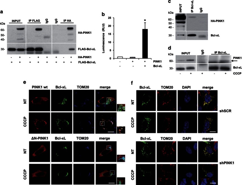
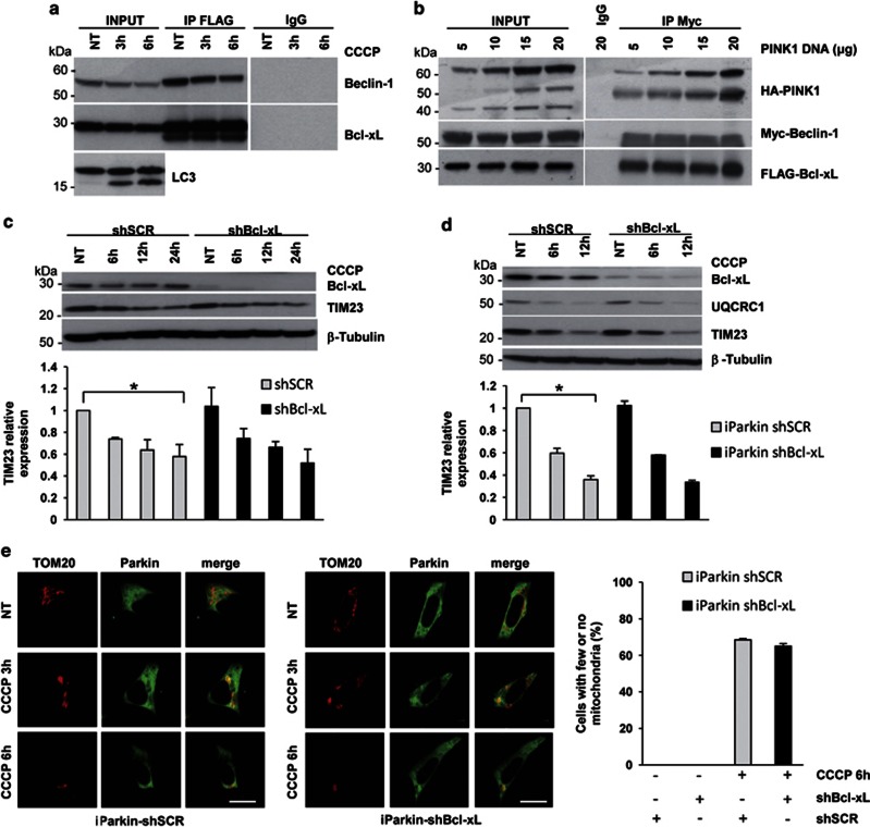
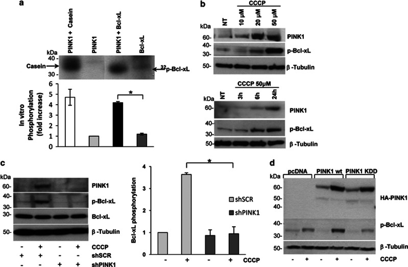
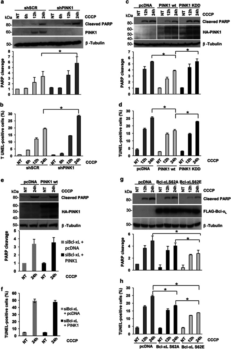
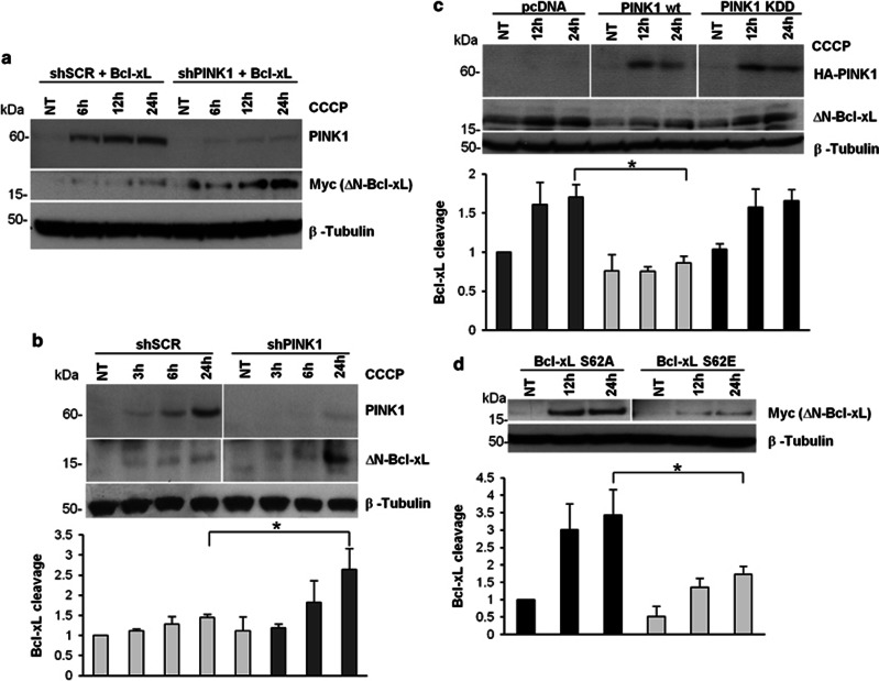
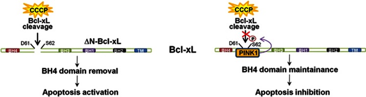
Mutations in the PINK1 gene are a frequent cause of autosomal recessive Parkinson's disease (PD). PINK1 encodes a mitochondrial kinase with neuroprotective activity, implicated in maintaining mitochondrial homeostasis and function. In concurrence with Parkin, PINK1 regulates mitochondrial trafficking and degradation of damaged mitochondria through mitophagy. Moreover, PINK1 can activate autophagy by interacting with the pro-autophagic protein Beclin-1. Here, we report that, upon mitochondrial depolarization, PINK1 interacts with and phosphorylates Bcl-xL, an anti-apoptotic protein also known to inhibit autophagy through its binding to Beclin-1. PINK1-Bcl-xL interaction does not interfere either with Beclin-1 release from Bcl-xL or the mitophagy pathway; rather it protects against cell death by hindering the pro-apoptotic cleavage of Bcl-xL. Our data provide a functional link between PINK1, Bcl-xL and apoptosis, suggesting a novel mechanism through which PINK1 regulates cell survival. This pathway could be relevant for the pathogenesis of PD as well as other diseases including cancer.


Figures

Figure 1
Figure 1
PINK1 interacts with Bcl-xL on depolarized mitochondria. (a) Reciprocal co-immunoprecipitations (co-IPs) of overexpressed PINK1 and Bcl-xL in HEK293 cells. PINK1 and Bcl-xL were immunoprecipitated with HA and FLAG antibodies, respectively. (b) Two-hybrid luciferase assay in HEK293 cells overexpressing PINK1 and Bcl-xL. HEK293 cells were transfected and processed as described in the Method section. In cells overexpressing both PINK1 and Bcl-xL, we observed a significant increase of luminescence compared with negative controls (Relative Light Units (RLU): 17.94±4.03, P=0.026). (c) co-IP of endogenous Bcl-xL and overexpressed PINK1 in SH-SY5Y cells. IP was performed with Bcl-xL antibody, followed by western blotting with HA antibody to detect PINK1. (d) co-IP of endogenous PINK1 and Bcl-xL. Lysates from SH-SY5Y cells, treated with CCCP or vehicle, were subjected to IP with Bcl-xL antibody, followed by immunoblotting with PINK1 antibody. (e) Colocalization of PINK1 and Bcl-xL at mitochondria. SH-SY5Y cells co-transfected with HA-PINK1 wt or ΔN and Bcl-xL–EGFP were treated with 6 h CCCP and subjected to confocal microscopy analysis. PINK1 was immunostained with HA antibody (red); TOM20 antibody was used to label the outer mitochondrial membrane (blue). Bcl-xL strongly colocalized with TOM20 in all experimental settings; conversely, PINK1 wt, but not PINK1-ΔN, colocalized with both Bcl-xL and TOM20 only in the presence of CCCP (see Supplementary Table S1 for overlap coefficients). (f) Colocalization of Bcl-xL and TOM20 upon PINK1 silencing. SH-SY5Y cells stably infected with scramble-shRNA (shSCR) or shPINK1 were transfected with Bcl-xL–EGFP and immunostained with TOM20 antibody (red). Merge pictures reveal colocalization. Bcl-xL strongly colocalized with TOM20 in all settings, regardless of PINK1 silencing (see Supplementary Table S2 for overlap coefficients). Scale bars: 10 μM
Figure 2
Figure 2
The interaction between PINK1 and Bcl-xL does not regulate mitophagy. (a) Bcl-xL–Beclin-1 co-IP in CCCP-treated SH-SY5Y cells. Bcl-xL was immunoprecipitated with FLAG antibody, followed by anti-Myc immunoblotting to detect Beclin-1. Autophagy activation was confirmed by LC3 western blotting. (b) Bcl-xL–Beclin-1 co-IP in HEK293 cells transfected with increasing amount of PINK1. Lysates were subjected to IP with Myc antibody, followed by immunoblotting with anti-FLAG. (c and d) Western blotting of mitochondrial markers in CCCP-treated wt and Parkin-inducible SH-SY5Y cells upon Bcl-xL silencing. Cells infected with either shSCR or Bcl-xL-shRNA (shBcl-xL) were treated with CCCP at the indicated times. Mitophagy was evaluated by assessing the decrease of the mitochondrial markers TIM23 and UQCRC1 by western blotting. Densitometric analysis revealed a significant decrease of TIM23 in both shSCR wt and Parkin-inducible SH-SY5Y cells treated with CCCP for 24 and 12 h, respectively (0.57±0.11, P=0.033 and 0.35±0.03, P=0.0013); however, we observed no significant differences in TIM23 levels related to Bcl-xL silencing (0.57±0.11 versus 0.51±0.12, P=0.67 and 0.35±0.03 versus 0.33±0.01, P=0.47).(e) Confocal microscopy analysis in CCCP-treated iParkin SH-SY5Y cells upon Bcl-xL silencing. Colocalization between Parkin (green) and TOM20 (red) was used as measure of mitophagy induction. Mitochondrial degradation was analyzed by counting the percentage of cells with few or no mitochondria after 6 h CCCP. Differences between shSCR and shBcl-xL cells were not significant (68.5±0.70 versus 65.0±1.41, P=0.09). Scale bars: 10 μM. *P-values<0.05
Figure 3
Figure 3
PINK1 phosphorylates Bcl-xL. (a) Mix beads in vitro kinase assay. Immunopurified PINK1 and Bcl-xL were processed as described in the Methods section. Casein was used as a positive control of the PINK1 kinase activity. PINK1 and Bcl-xL alone were used as negative controls. Bcl-xL phosphorylation significantly increased in the presence of PINK1 (4.12±0.10, P=0.001). (b) In vivo phosphorylation of endogenous Bcl-xL. SH-SY5Y cells were either subjected to increasing concentration of CCCP (upper panel) or treated with 50 μM CCCP up to 24 h (lower panel). Lysates were processed for western blotting using p-Bcl-xL and PINK1 antibodies. (c) Bcl-xL phosphorylation upon PINK1 silencing. Lysates from SH-SY5Y cells infected with either shSCR or shPINK1 were treated with 50 μM CCCP for 24 h. PINK1 silencing and p-Bcl-xL levels were assessed by immunoblotting. Densitometric analysis revealed a significant reduction of p-Bcl-xL in CCCP-treated shPINK1 cells compared with control (0.95±0.31 versus 3.63±0.08, P=0.007). (d) Bcl-xL phosphorylation after overexpression of PINK1. Lysates from CCCP-treated SH-SY5Y cells transfected with vector alone (pcDNA), PINK1 wt or KDD were processed for western blotting using HA and p-Bcl-xL antibodies. *P-values<0.05
Figure 4
Figure 4
The interaction between PINK1 and Bcl-xL protects against CCCP-induced apoptosis. (a and b) Apoptosis evaluation upon PINK1 silencing. Either shSCR or shPINK1 cells were treated with CCCP followed by cleaved PARP immunoblotting (a) or FACS analysis (b). Densitometric analysis on cleaved PARP revealed, at 24 h CCCP, a significant increase of cell death in shPINK1 cells compared with control (5.78±1.21 versus 3.34±0.92, P=0.03). The percentage of TUNEL-positive cells at 24 h was also significantly higher in shPINK1 compared with shSCR cells (28.47±0.66 versus 19.38±0.55, P=0.04).(c and d) Apoptosis rescue by PINK1. shPINK1 cells were transfected with PINK1 wt or KDD or vector alone (pcDNA). After 24 h CCCP, cleaved PARP levels were significantly lower in presence of PINK1 wt compared with both empty vector (3.86±0.08 versus 5.34±0.07, P=0.003), and KDD (5.37±0.45, P=0.042). The proportion of TUNEL-positive cells also significantly decreased in presence of PINK1 wt compared with both control (16.79±0.81 versus 25.40±0.68, P=0.009), and KDD (22.77±0.27, P=0.02). (e and f) Apoptosis evaluation in Bcl-xL-silenced cells after PINK1 overexpression. SH-SY5Y cells subjected to Bcl-xL knockdown were transfected with PINK1 wt or empty vector (pcDNA) and then treated with CCCP for 24 h. Upon mitochondrial depolarization, the increase of cleaved PARP levels in PINK1 overexpressing cells was similar to that observed in cells transfected with the empty vector (3.54±0.61 versus 3.37±0.57, P=0.85); the same result was obtained analyzing the percentage of TUNEL-positive cells by FACS (47.56±2.51 versus 49.28±2.47, P=0.82). (g and h) Apoptosis rescue by Bcl-xL S62E. shPINK1 cells were transfected with Bcl-xL S62E or S62A, or with the empty vector. At 24 h CCCP, cleaved PARP levels were significantly lower in presence of Bcl-xL S62E compared with both Bcl-xL S62A (2.76±0.55 versus 4.03±0.50, P=0.042) and empty vector (4.87±0.81, P=0.02). Similarly, the percentage of TUNEL-positive cells was significantly lower in shPINK1 cells transfected with Bcl-xL S62E compared with both Bcl-xL S62A (13.76±0.13 versus 18.41±0.35, P=0.003) and vector alone (24.48±0.73, P=0.0008). *P-values<0.05
Figure 5
Figure 5
PINK1-dependent Bcl-xL phosphorylation impairs Bcl-xL cleavage. (a and b) ΔN-Bcl-xL formation upon PINK1 silencing. Lysates from shSCR or shPINK1 cells, either transfected with Myc-Bcl-xL (a) or untransfected (b), were treated with CCCP and processed for western blotting. The cleavage of overexpressed Bcl-xL was well evident upon mitochondrial depolarization in PINK1-silenced cells (a). The endogenous levels of ΔN-Bcl-xL also significantly increased at 24 h CCCP in shPINK1 compared with shSCR cells (2.63±0.52 versus 1.44±0.07, P=0.017). (c) Rescue of Bcl-xL cleavage by PINK1. shPINK1 cells were transfected with PINK1 wt or KDD or vector alone (pcDNA). After 24 h CCCP, cleaved endogenous Bcl-xL was significantly reduced in presence of PINK1 wt compared with both control (0.85±0.09 versus 1.70±0.15, P=0.022) and KDD (1.65±0.14, P=0.022). (d) Cleavage of Bcl-xL S62E and S62A. Lysates from CCCP-treated shPINK1 cells transfected with the indicated Bcl-xL constructs were processed for immunoblotting. Cleaved Bcl-xL was detected using the Myc antibody. At 24 h CCCP, cleavage was significantly reduced in presence of Bcl-xL S62E compared with S62A (1.72±0.23 versus 3.43±0.72, P=0.018). *P-values<0.05
Figure 6
Figure 6
Schematic model of the mechanism through which PINK1–Bcl-xL interaction could protect against cell death induced by mitochondrial depolarization. Mitochondrial depolarization induces cleavage of Bcl-xL at the aspartate 61 (D61). The resulting C-terminal fragment (ΔN-Bcl-xL) lacks the anti-apoptotic N-terminal BH4 domain and is a potent inducer of cell death. PINK1 phosphorylates the serine 62 (S62) of Bcl-xL, impairing cleavage at the adjacent D61 site. In this way, the BH4 domain of Bcl-xL is maintained and the full-length protein can exert its anti-apoptotic function

Similar articles

Cited by

References

    1. Siderowf A, Stern M. Update on Parkinson disease. Ann Intern Med. 2003;138:651–658. - PubMed
    1. Schapira AH. Mitochondria in the etiology and pathogenesis of Parkinson's disease. Lancet Neurol. 2008;7:97–109. - PubMed
    1. Belin AC, Westerlund M. Parkinson's disease: a genetic perspective. FEBS J. 2008;275:1377–1383. - PubMed
    1. Deas E, Plun-Favreau H, Gandhi S, Desmond H, Kjaer S, Loh S, et al. PINK1 cleavage at position A103 by the mitochondrial protease PARL. Hum Mol Genet. 2011;20:867–879. - PMC - PubMed
    1. Jin SM, Lazarou M, Wang C, Kane LA, Narendra DP, Youle RJ. Mitochondrial membrane potential regulates PINK1 import and proteolytic destabilization by PARL. J Cell Biol. 2010;191:933–942. - PMC - PubMed

Publication types