. 2013 Jun;159(Pt 6):1120-1135.
doi: 10.1099/mic.0.063495-0. Epub 2013 Mar 21.

Evaluation of the roles played by Hcp and VgrG type 6 secretion system effectors in Aeromonas hydrophila SSU pathogenesis

Affiliations

Evaluation of the roles played by Hcp and VgrG type 6 secretion system effectors in Aeromonas hydrophila SSU pathogenesis

Jian Sha et al. Microbiology (Reading). 2013 Jun.

Abstract

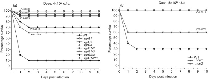
Aeromonas hydrophila, a Gram-negative bacterium, is an emerging human pathogen equipped with both a type 3 and a type 6 secretion system (T6SS). In this study, we evaluated the roles played by paralogous T6SS effector proteins, hemolysin co-regulated proteins (Hcp-1 and -2) and valine glycine repeat G (VgrG-1, -2 and -3) protein family members in A. hydrophila SSU pathogenesis by generating various combinations of deletion mutants of the their genes. In addition to their predicted roles as structural components and effector proteins of the T6SS, our data clearly demonstrated that paralogues of Hcp and VgrG also influenced bacterial motility, protease production and biofilm formation. Surprisingly, there was limited to no observed functional redundancy among and/or between the aforementioned T6SS effector paralogues in multiple assays. Our data indicated that Hcp and VgrG paralogues located within the T6SS cluster were more involved in forming T6SS structures, while the primary roles of Hcp-1 and VgrG-1, located outside of the T6SS cluster, were as T6SS effectors. In terms of influence on bacterial physiology, Hcp-1, but not Hcp-2, influenced bacterial motility and protease production, and in its absence, increases in both of the aforementioned activities were observed. Likewise, VgrG-1 played a major role in regulating bacterial protease production, while VgrG-2 and VgrG-3 were critical in regulating bacterial motility and biofilm formation. In an intraperitoneal murine model of infection, all Hcp and VgrG paralogues were required for optimal bacterial virulence and dissemination to mouse peripheral organs. Importantly, the observed phenotypic alterations of the T6SS mutants could be fully complemented. Taking these results together, we have further established the roles played by the two known T6SS effectors of A. hydrophila by defining their contributions to T6SS function and virulence in both in vitro and in vivo models of infection.


Figures

Fig. 1.
Fig. 1.
Amino acid sequence comparison between Hcp-1 and Hcp-2 (a) and among VgrG-1, VgrG-2 and VgrG-3 (b) of A. hydrophila SSU using online clustal 2.1 Multiple Sequence Alignments software. *Conserved amino acid residues; :, amino acid residues with strongly similar amino acid groups; ., amino acid residues with weakly similar amino acid groups.
Fig. 2.
Fig. 2.
Secretion of Hcp by the A. hydrophila SSU T6SS mutants. Immunoblot analysis was performed using polyclonal anti-Hcp antibodies to measure the production of Hcp in both the supernatants (a) and pellets (b) of WT A. hydrophila and its various Hcp and VgrG mutant strains.
Fig. 3.
Fig. 3.
Swimming motility assay of the A. hydrophila SSU T6SS mutants. Semi-solid media plates were inoculated with WT and various Hcp (a) and VgrG (b) mutant strains of A. hydrophila. The plates were incubated at 37 °C overnight, and the distances of bacterial migration (cm) through the agar from the centre towards the periphery of the plate were measured. Student’s t-test was used for data analysis, and a single asterisk represents a statistically significant difference relative to the WT strain with a P-value of <0.05, while a double asterisk represents a statistically significant difference relative to the WT strain with a P-value of <0.001. Both images of the motility on plates and quantification of the migration are shown.
Fig. 4.
Fig. 4.
Protease activity of the A. hydrophila SSU T6SS mutants. The overnight culture filtrates from WT and various Hcp (a) and VgrG (b) mutant strains of A. hydrophila were mixed with 5 mg Hide powder azure substrate and incubated in a shaker incubator at 37 °C for 1–3 h. As the protease in the culture filtrates degraded the substrate, blue colour was released and quantified at OD595. The protease activity was calculated per ml of culture filtrate per 108 c.f.u. Student’s t-test was used for the data analysis, and a single asterisk represents a statistically significant difference relative to the WT strain with a P-value of <0.05, while a double asterisk represents a statistically significant difference relative to the WT strain with a P-value of <0.001.
Fig. 5.
Fig. 5.
Biofilm formation by A. hydrophila SSU T6SS mutants. A crystal violet tube-based assay was used to detect biofilm production of WT and various Hcp (a) and VgrG (b) mutant strains of A. hydrophila SSU. The bar graph of optical quantification (OD570) of biofilm formation and an image of the actual crystal violet (CV) staining tubes of WT and various VgrG mutants are shown. Student’s t-test was used to analyse the data, and an asterisk represents a statistically significant difference relative to the WT strain with a P-value of ≤0.05.
Fig. 6.
Fig. 6.
Survival curves of mice infected with A. hydrophila SSU T6SS mutants. Groups of 20 female, Swiss Webster mice were i.p. challenged with A. hydrophila SSU WT or various VgrG mutant strains at the dose of 4×10⁷ c.f.u. (a) or A. hydrophila SSU WT and various Hcp mutant strains at the dose of 8×10⁶ c.f.u. (b). Mouse survival was recorded during the course of the experiments, and the Kaplan–Meier survival estimate was used to analyse the survival of mice. The P-values represented statistically significant differences relative to the WT-infected group.
Fig. 7.
Fig. 7.
Bacterial dissemination of A. hydrophila SSU T6SS mutants in infected mice. In vivo dissemination of the ΔvgrG1/2/3 mutant relative to the WT strain following a 2×10⁷ c.f.u. i.p. challenge was measured by viable c.f.u. plate counts of homogenized mice livers and spleens on day 1 (a) and day 2 (b) post-infection (p.i.). Similarly, in vivo dissemination of the Δhcp1 mutant relative to the WT strain following an 8×10⁶ c.f.u. i.p. challenge was measured by viable c.f.u. plate counts of homogenized livers and spleens on day 2 p.i. (c). Average counts are represented as a horizontal bar with individual counts represented by circles. ‘No survivors’ denotes mice that had expired prior to sampling.
Fig. 8.
Fig. 8.
Competitive growth of WT A. hydrophila SSU and the T6SS mutant ΔvgrG1/2/3 in mice. Mice were i.p. infected with a mixed culture of WT bacteria (streptomycin-resistant, Smr) and its ΔvgrG1/2/3 mutant (rifampicin resistant, Rifr) (Table 1) at the ratio of 1 : 1 with doses of 2×10⁷ c.f.u. (a) and 4×10⁷ c.f.u. (b). At 48 h p.i., the bacterial loads of WT and those of ΔvgrG1/2/3 mutant in livers and spleens from five mice were determined according to their antibiotic resistance patterns. The CI in the liver and spleen [calculated as (mutant output/competitor output)/(mutant input/competitor input)] for each animal is shown. The horizontal lines represent mean CI values. Student’s t-test was used to analyse the bacterial loads, and an asterisk represents a statistically significant difference relative to the WT strain (Smr) with P<0.05.
Fig. 9.
Fig. 9.
Complementation of the phenotypic alterations associated with various A. hydrophila SSU T6SS mutants. The T6SS genes (i.e. hcps and vgrGs) with their putative promoter regions were cloned into plasmid pBR322 for complementing the corresponding T6SS mutants (Table 1). The complemented (comp) strains, along with the mutants, the WT bacterium and the ΔvasH as well as ΔvasK mutants (with inactive T6SS) were evaluated for biofilm formation (a), swimming motility (b), protease production (c), and virulence in mice (d). The Kaplan–Meier survival estimate was used to analyse the survival of mice. The P-values represented statistically significant differences relative to the WT-infected group. Student’s t-test was used to analyse the data in other assays, and an asterisk represents a statistically significant difference relative to the WT bacterium with a P-value of <0.05.

References

    1. Altwegg M., Martinetti Lucchini G., Lüthy-Hottenstein J., Rohrbach M. (1991). Aeromonas-associated gastroenteritis after consumption of contaminated shrimp. Eur J Clin Microbiol Infect Dis 10, 44–45. - PubMed
    1. Aschtgen M. S., Bernard C. S., De Bentzmann S., Lloubès R., Cascales E. (2008). SciN is an outer membrane lipoprotein required for type VI secretion in enteroaggregative Escherichia coli. J Bacteriol 190, 7523–7531. 10.1128/JB.00945-08 - DOI - PMC - PubMed
    1. Ballister E. R., Lai A. H., Zuckermann R. N., Cheng Y., Mougous J. D. (2008). In vitro self-assembly of tailorable nanotubes from a simple protein building block. Proc Natl Acad Sci U S A 105, 3733–3738. 10.1073/pnas.0712247105 - DOI - PMC - PubMed
    1. Basler M., Pilhofer M., Henderson G. P., Jensen G. J., Mekalanos J. J. (2012). Type VI secretion requires a dynamic contractile phage tail-like structure. Nature 483, 182–186. 10.1038/nature10846 - DOI - PMC - PubMed
    1. Bröms J. E., Meyer L., Lavander M., Larsson P., Sjöstedt A. (2012). DotU and VgrG, core components of type VI secretion systems, are essential for Francisella LVS pathogenicity. PLoS ONE 7, e34639. 10.1371/journal.pone.0034639 - DOI - PMC - PubMed

Publication types

MeSH terms

Associated data

LinkOut - more resources