Comparative Study
. 2013 May 3;340(6132):615-9.
doi: 10.1126/science.1232808. Epub 2013 Mar 21.

Structural features for functional selectivity at serotonin receptors

Affiliations
Comparative Study

Structural features for functional selectivity at serotonin receptors

Daniel Wacker et al. Science. .

Abstract

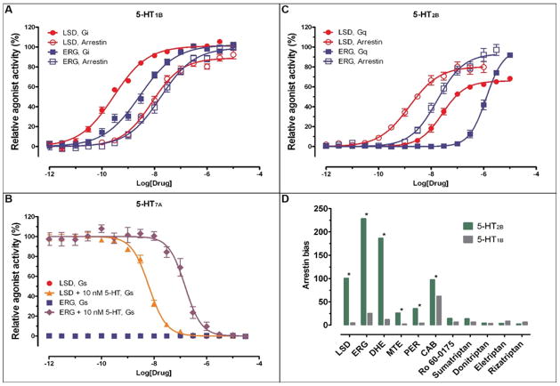
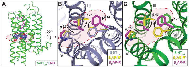
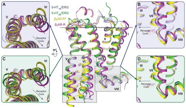
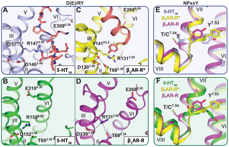
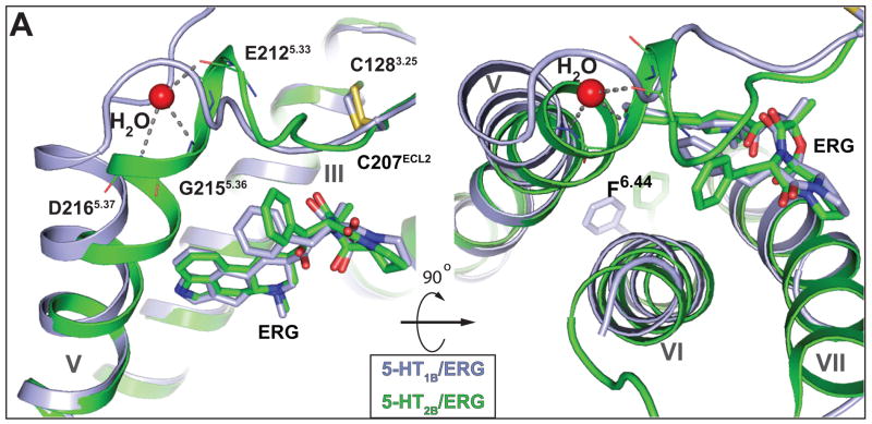
Drugs active at G protein-coupled receptors (GPCRs) can differentially modulate either canonical or noncanonical signaling pathways via a phenomenon known as functional selectivity or biased signaling. We report biochemical studies showing that the hallucinogen lysergic acid diethylamide, its precursor ergotamine (ERG), and related ergolines display strong functional selectivity for β-arrestin signaling at the 5-HT2B 5-hydroxytryptamine (5-HT) receptor, whereas they are relatively unbiased at the 5-HT1B receptor. To investigate the structural basis for biased signaling, we determined the crystal structure of the human 5-HT2B receptor bound to ERG and compared it with the 5-HT1B/ERG structure. Given the relatively poor understanding of GPCR structure and function to date, insight into different GPCR signaling pathways is important to better understand both adverse and favorable therapeutic activities.


Figures

Fig. 1
Fig. 1
Distinct signaling properties of lysergic acid diethylamide (LSD) and ergotamine (ERG) at 5-HT1B, 5-HT7A and 5-HT2B receptors. We used luminescence-based assays to measure 5-HT1B receptor mediated Gi activation and cAMP production; fluorescence-based calcium mobilization assays to measure 5-HT2B receptor mediated Gq activation and β-arrestin translocation-dependent luciferase reporter assays to measure 5-HT1B and 5-HT2B receptors mediated β-arrestin recruitment, all in HEK293 derived cells. (A) Normalized concentration-response studies for LSD and ERG at human cloned 5-HT1B receptor-mediated activation of Gi and non-canonical (Arrestin) signaling. (B) Normalized concentration-response studies for LSD and ERG at human cloned 5-HT7A receptor-mediated activation of Gs signaling in the presence and absence of 5-Hydroxytryptamine (5-HT). (C) Normalized concentration-response studies for LSD and ERG at human cloned 5-HT2B receptor-mediated activation of Gq and non-canonical (Arrestin) signaling. The solid circles for LSD-Gs signals are superimposed by the solid squares for ERG-Gs signals and thus, not visible. (D) Mean β-arrestin bias factors were calculated for serotonergic agonists (dihydroergotamine (DHE), methylergonovine (MTE), pergolide (PER) and cabergoline (CAB)), at 5-HT2B and 5-HT1B receptors. Concentration-responses curves were fit to the Black and Leff operational model to obtain transduction coefficients [Log(τ/KA)] for each ligand at each corresponding pathway. The ΔLog(τ/KA) was then calculated with 5-HT as a reference agonist for each pathway, and the ΔΔLog(τ/KA) was calculated between two pathways for each ligand. The bias factor is unit-less and defined as 10ΔΔLog(τ/KA) (28). Compounds with values close to one represent unbiased agonists while compounds with large numerical values, typically >100, represent extremely biased agonists. *p<0.0001 via two-way ANOVA comparing 5-HT2B vs 5-HT1B bias factors; N=3–6 separate experiments. ERG, DHE and, to a lesser extent, LSD, MTE and PER show strong β-arrestin bias at 5-HT2B receptor, but not 5-HT1B receptor.
Fig. 2
Fig. 2
Trigger motif P-I-F displays active- and intermediate active-state for 5-HT1B and 5-HT2B receptors, respectively. Residues of the P-I-F motif are highlighted in a dashed red circle. (A) Overall architecture of the 5-HT2B receptor (green) bound to ERG (magenta); residues of the P5.50 I3.40 F6.44 motif are illustrated in space-filling representation. (B) Alignment between 5-HT1B receptor (grey), β2AR-R (magenta; PDB ID: 2RH1) and β2AR-R*(yellow; PDB ID: 3SN6) indicates an activated P-I-F motif in the 5-HT1B/ERG structure. Black arrows indicate likely rearrangements upon 5-HT1B receptor activation according to analysis of β2AR. (C) Alignment between 5-HT2B receptor (green), β2AR-R (magenta) and β2AR-R* (yellow) suggests intermediate active-state of the P-I-F motif in the 5-HT2B/ERG structure, with F6.44 in an inactive conformation. Black arrows indicate likely rearrangements upon 5-HT2B receptor activation according to analysis of β2AR.
Fig. 3
Fig. 3
Structural alignment with β2AR-R and β2AR-R* reveals distinct active-state 7TM conformations of the 5-HT1B and 5-HT2B receptor structures. All structures were analyzed based on the last membrane embedded residue to minimize the effect of G proteins and fusion partners on the relative helix positions (see supplementary section). Center panel shows overall 7TM configuration of β2AR-R (magenta; PDB ID: 2RH1), β2AR-R*(yellow; PDB ID: 3SN6), 5-HT1B (grey) and 5-HT2B (green) receptors aligned through helices I–IV. Membrane boundaries are indicated by grey dots according to the OPM database (29). (A, C) Intracellular view of helix V and helix VI in β2AR-R, β2AR-R* compared to (A) the 5-HT1B, or (C) the 5-HT2B receptor. Residues on the intracellular side of the membrane have been removed for better comparison of ligand-induced helical rearrangements (see main text). Helix VI of the 5-HT1B and 5-HT2B receptors is in an intermediate active-state compared to β2AR. (B, D) Side view of helix VII and helix VIII in β2AR-R, β2AR-R* and (B) 5-HT1B receptor, or (D) 5-HT2B receptor. Helix VII of the 5-HT1B and 5-HT2B receptors is in an intermediate active-state compared to β2AR, with more pronounced activation features for the 5-HT2B receptor.
Fig. 4
Fig. 4
Activation state of the D(E)/RY and NPxxY motifs in 5-HT1B and 5-HT2B receptors compared to β2AR-R and β2AR-R*. Configuration of the D(E)/RY motif in (A) the 5-HT1B receptor (grey), (B) the 5-HT2B receptor (green), (C) β2AR-R* (yellow; PDB ID: 3SN6) and (D) β2AR-R (magenta; PDB ID: 2RH1). Residues of the G protein in β2AR-R* (C) and G protein mimicking BRIL loop (A) are highlighted in orange. The conformation of the D(E)/RY motif in the 5-HT1B receptor is similar to that observed in β2AR-R*, whereas the configuration of the 5-HT2B receptor compares to that of β2AR-R. (E, F) Conformational states of Y7.53 of the NPxxY motif and the proceeding residue 7.54 in β2AR-R, β2AR-R* and (E) the 5-HT1B, or (F) 5-HT2B receptor. When compared to β2AR, the conformation of the NPxxY motif in the 5-HT1B receptor is in an intermediate active-state, whereas the configuration of the 5-HT2B receptor is similar to β2AR-R*.
Fig. 5
Fig. 5
Structural differences in extracellular configuration of helix V between 5-HT1B and 5-HT2B receptors likely explain β-arrestin functional selectivity at the 5-HT2B receptor. Side view (left) and top view (right) of 5-HT1B (grey) and 5-HT2B (green) receptors shows a kink in the extracellular end of helix V in the 5-HT2B receptor. A water molecule (red sphere) was found to stabilize the kink through hydrogen bonds (grey dashed lines) with the E2125.33 main-chain carbonyl oxygen and the main-chain nitrogens of D2165.37 and G2155.36. Main-chain atoms of both residues are shown as lines.

Comment in

  • Biochemistry. As good as chocolate.
    Palczewski K, Kiser PD. Palczewski K, et al. Science. 2013 May 3;340(6132):562-3. doi: 10.1126/science.1238521. Science. 2013. PMID: 23641106 Free PMC article. No abstract available.

References

    1. Luttrell LM, et al. Beta-arrestin-dependent formation of beta2 adrenergic receptor-Src protein kinase complexes. Science. 1999;283:655. - PubMed
    1. Reiter E, Ahn S, Shukla AK, Lefkowitz RJ. Molecular mechanism of beta-arrestin-biased agonism at seven-transmembrane receptors. Annu Rev Pharmacol Toxicol. 2012;52:179. - PMC - PubMed
    1. Kenakin T. Functional selectivity and biased receptor signaling. J Pharmacol Exp Ther. 2011;336:296. - PubMed
    1. Allen JA, Roth BL. Strategies to discover unexpected targets for drugs active at G protein-coupled receptors. Annu Rev Pharmacol Toxicol. 2011;51:117. - PubMed
    1. Urban JD, et al. Functional selectivity and classical concepts of quantitative pharmacology. J Pharmacol Exp Ther. 2007;320:1. - PubMed

Publication types

MeSH terms

Associated data