

Endoscopic submucosal dissection as a treatment for gastric subepithelial tumors that originate from the muscularis propria layer: a preliminary analysis of appropriate indications
- PMID: 23519491
- PMCID: PMC3751271
- DOI: 10.1007/s00464-013-2904-9
Endoscopic submucosal dissection as a treatment for gastric subepithelial tumors that originate from the muscularis propria layer: a preliminary analysis of appropriate indications
Abstract
Background: Endoscopic submucosal dissection (ESD) is a well-established method for the treatment of gastrointestinal epithelial tumors. However, the treatment of gastric subepithelial tumors (SETs) that originate from the muscularis propria layer still depends primarily on surgical techniques. We evaluated the appropriate indications for ESD in the treatment of SETs that originate from the muscularis propria layer.
Methods: Thirty-five patients with gastric SETs that originate from the muscularis propria layer who underwent ESD were enrolled, and the charts were retrospectively reviewed to investigate the parameters predictive complete resection and complications.
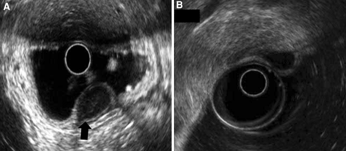
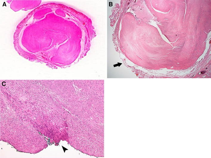
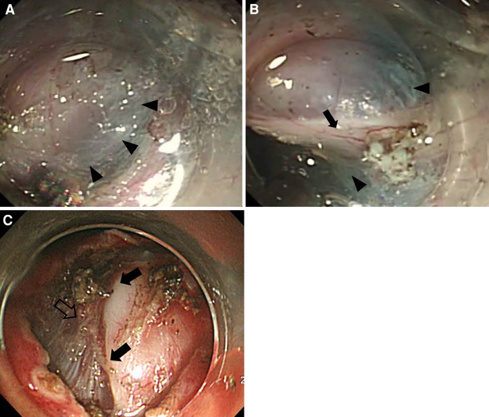
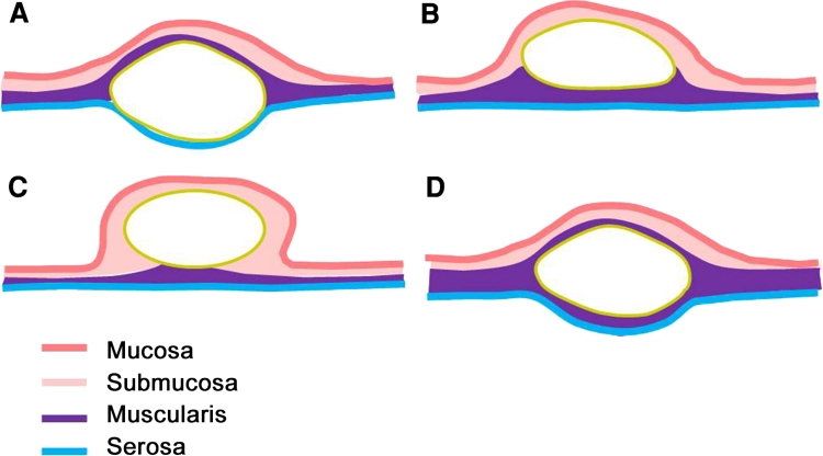
Results: The mean age of the patients was 54.15 ± 9.3 years, and the male/female ratio was 2:3. Twenty-eight of the 35 SETs (85.7%) were movable, and 15 (45.7%) had a positive rolling sign. The most frequent location of the SETs was high body (n = 14). The most common pathological diagnoses were leiomyoma (60%) and gastrointestinal stromal tumor (28.6%). The complete resection rate was 74.3%. A positive rolling sign (p = 0.022) and small tumor size (≤20 mm; p = 0.038) were significantly associated with complete resection. Two patients (6.1 %) developed perforations that required surgical treatment; their SMTs were neurogenic tumors with fixed lesion. Tumor mobility was significantly associated with perforation (p = 0.017).
Conclusions: The ESD method appears to be relatively safe for use in the complete resection of SETs that originate from the muscularis propria layer. Small tumor size (≤20 mm) and a positive rolling sign are appropriate indications for ESD.
Figures
References
-
- Kawamoto K, Yamada Y, Utsunomiya T, Okamura H, Mizuguchi M, Motooka M, Hirata N, Watanabe H, Sakai K, Kitaqawa S, Kinukawa N, Masuda K. Gastrointestinal submucosal tumors: evaluation with endoscopic US. Radiology. 1997;205:733–740. - PubMed
MeSH terms
LinkOut - more resources
Full Text Sources
Other Literature Sources
Medical
Miscellaneous
