. 2013 May;41(9):5062-74.
doi: 10.1093/nar/gkt189. Epub 2013 Mar 21.

Characterizing TDP-43 interaction with its RNA targets

Affiliations

Characterizing TDP-43 interaction with its RNA targets

Amit Bhardwaj et al. Nucleic Acids Res. 2013 May.

Abstract

One of the most important functional features of nuclear factor TDP-43 is its ability to bind UG-repeats with high efficiency. Several cross-linking and immunoprecipitation (CLIP) and RNA immunoprecipitation-sequencing (RIP-seq) analyses have indicated that TDP-43 in vivo can also specifically bind loosely conserved UG/GU-rich repeats interspersed by other nucleotides. These sequences are predominantly localized within long introns and in the 3'UTR of various genes. Most importantly, some of these sequences have been found to exist in the 3'UTR region of TDP-43 itself. In the TDP-43 3'UTR context, the presence of these UG-like sequences is essential for TDP-43 to autoregulate its own levels through a negative feedback loop. In this work, we have compared the binding of TDP-43 with these types of sequences as opposed to perfect UG-stretches. We show that the binding affinity to the UG-like sequences has a dissociation constant (Kd) of ∼110 nM compared with a Kd of 8 nM for straight UGs, and have mapped the region of contact between protein and RNA. In addition, our results indicate that the local concentration of UG dinucleotides in the CLIP sequences is one of the major factors influencing the interaction of these RNA sequences with TDP-43.


Figures

Figure 1.
Figure 1.
(A) Illustrates a schematic representation of TDP-43 gene showing the coding exons (black boxes), untranslated region (grey box), TDPBR (shaded box) and polyadenylation sites (black lines). The CLIP sequences analyzed in this study are shown in bold and underlined. Sequence of mutated CLIP34nt (CLIP34nt_UG6) is also shown below. (B) Shows an EMSA binding analysis of CLIP sequences: γ-P32 ATP labelled RNA oligonucleotides of CLIP sequences were mixed with GST tagged TDP-43 (101–261) and subjected to EMSA analysis. (C) Shows the binding profile of (B) following UV-cross-linking.
Figure 2.
Figure 2.
Quantitative EMSA analysis of various TDP–RNA complexes. Panel (A–C) shows the gel profiles and binding curves plotted using quantitative EMSA analysis for CLIP34nt_UG6, CLIP34nt and CLIP6 with GST-TDP (101–261). Panel (D–F) show the gel profile and binding curves plotted using quantitative EMSA analysis for CLIP34nt_UG6, CLIP34nt and CLIP6 with GST-TDPmut2 (101–261 F229L and F231L). The concentration (nM) of each probe used to determine the Kd is mentioned on the top of each gel. Each experiment was repeated at least three times to plot the binding curves.
Figure 3.
Figure 3.
Extracted ion Chromatograms of acetylated His-TDP (101–261). The extracted ion chromatograms for the acetylated chymotryptic peptide 132MVQKKDLKTGHSKGF147 (Chym132–147) were generated for samples in the absence and presence of RNA. Importantly, the light acetylation was used to label TDP-43 in the absence of RNA (red traces, Peak ‘L’), while the heavy reagent was used to label TDP-43 in the presence of RNA (blue traces, Peak ‘H’). For each binary comparison, the samples were mixed before chymotryptic digestion and analyzed in the same LC-MS/MS run. The production of the acetylated Chym132–147 peptides from no RNA controls are shown compared with TDP-43 + (UG)6 (A), TDP-43 + CLIP34nt (B) and TDP-43 + CLIP6 (C). The x-axis shows the intensity of the eluting peptides and, in all cases, the presence of RNA inhibited the acetylation of Lysine145 (underlined) as indicated by the greater intensity of the blue traces relative to the red traces. y-axis shows the elution time of these peptides.
Figure 4.
Figure 4.
Titration experiments for various TDP–RNA complexes using circular dichroism. Starting from 0 μM (blue), 5 μM (violet), 10 μM (green) and 15 μM (red) of GST-TDP (101–261) were used to perform titration experiments against a fixed concentration of various RNAs. Panels A, B, C and D shows the subtracted CD spectra of (UG)6 (18 μM), CLIP34nt (10 μM), CLIP6 (9 μM) and CLIP34nt_UG6 (8 μM) in the presence of various concentration of GST-TDP (101–261), respectively. Panel E and F show the subtracted CD spectra of CLIP34nt_UG6 (8 μM) in the presence of various concentrations of GST-TDPmut2 (101–261) and GST-TDPmut1 (101–261), respectively.
Figure 5.
Figure 5.
Circular dichroism analysis of GST-TDP (101–261) in the presence of RNA: (A) lane 1 of coomassie-stained EMSA gel shows the mobility of free GST-TDP (101–261) (arrow 1). In lane 2, a faster-moving band appeared in the presence of (UG)6 RNA (arrow 2), indicating the formation of a stable protein–RNA complex. Mobility of GST-TDP (101–261) did not change in the presence of CLIP1 RNA (negative control, lane3). Lane (4–5) shows that the addition of CLIP34nt and CLIP6 resulted in the appearance of a smear indicating the formation of weak complex. (B) All the samples from A were subjected to circular dichroism, and no significant changes were observed. (C) Appearance of a faster-moving band showing the stable complex between GST-TDP (101–261) and CLIP34nt_UG. (D) Far-UV CD spectra of GST-TDP (101–261) in the presence of CLIP34nt_UG.
Figure 6.
Figure 6.
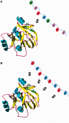
Proposed binding mechanism of TDP-43 (via RRM1) and multi-site RNA targets containing UG-repeats. In this model, long UG-repeats maintain a high local concentration of UGs, which in turn increases the probability of binding other shorter or single UGs stretches that prevent the protein to diffuse away before rebinding can occur.

References

    1. Buratti E, Baralle FE. TDP-43: gumming up neurons through protein-protein and protein-RNA interactions. Trends Biochem. Sci. 2012;37:237–247. - PubMed
    1. Neumann M, Sampathu DM, Kwong LK, Truax AC, Micsenyi MC, Chou TT, Bruce J, Schuck T, Grossman M, Clark CM, et al. Ubiquitinated TDP-43 in frontotemporal lobar degeneration and amyotrophic lateral sclerosis. Science. 2006;314:130–133. - PubMed
    1. Rademakers R, Neumann M, Mackenzie IR. Advances in understanding the molecular basis of frontotemporal dementia. Nat. Rev. Neurol. 2012;8:423–434. - PMC - PubMed
    1. Barmada SJ, Finkbeiner S. Pathogenic TARDBP mutations in amyotrophic lateral sclerosis and frontotemporal dementia: disease-associated pathways. Rev. Neurosci. 2010;21:251–272. - PubMed
    1. Mackenzie IR, Rademakers R, Neumann M. TDP-43 and FUS in amyotrophic lateral sclerosis and frontotemporal dementia. Lancet Neurol. 2010;9:995–1007. - PubMed

Publication types