

CT-guided percutaneous drilling is a safe and reliable method of treating osteoid osteomas
- PMID: 23519705
- PMCID: PMC3601247
- DOI: 10.1186/2193-1801-2-34
CT-guided percutaneous drilling is a safe and reliable method of treating osteoid osteomas
Abstract
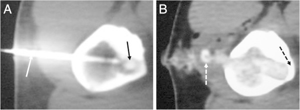
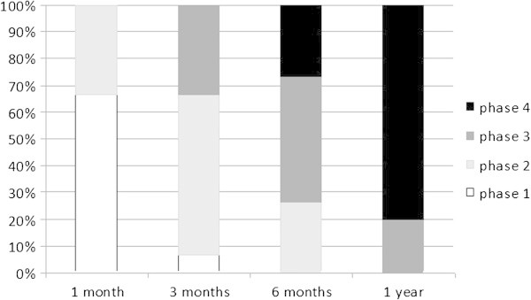
Computed tomography (CT)-guided percutaneous drilling is an alternative for osteoid osteoma treatment. This study aims to evaluate the remodeling of the drill orifice. The success rate and complications were also recorded and compared with other treatment methods. Fifteen patients with an average age of fourteen years (ranging from 4 to 25) submitted to CT-guided percutaneous drilling between 2003 and 2009 were retrospectively analyzed according to clinical and radiological criteria. Fourteen cases showed complete alleviation of pain one week after surgery. No relapse was detected even in the subject who continued complaining of pain. All patients were treated with a day-hospital regimen and were discharged with partial weight bearing. Total weight bearing was allowed after one month, and sports were allowed after consolidation, which occurred in all but one case after the third month. One patient, who did not follow our medical advice, returned to sports activities after two weeks and experienced a fracture as a result. Atrophy of the vastus lateralis muscle developed after the procedure in another patient. Our case series suggests that this method is reliable and safe. The level of complexity is comparable with other minimally invasive percutaneous procedures. The cost is low because there is no need to buy probes or other equipment. The negative points include weakening of the bone and the logistical problem of assembling the orthopedic surgeon, radiologist, and anesthesiologist in the tomography room.
Keywords: Bone neoplasms; Bone remodeling; Interventional radiography; Osteoid osteoma; Treatment outcome.
Figures
Similar articles
-
CT-Guided Percutaneous Drilling of Osteoid Osteoma: A Safe, Minimally Invasive and Cost-Effective Method.Indian J Orthop. 2020 Feb 11;54(2):194-199. doi: 10.1007/s43465-019-00029-x. eCollection 2020 Apr. Indian J Orthop. 2020. PMID: 32257037 Free PMC article.
-
Osteoid osteoma of the proximal femur: treatment by percutaneous bone resection and drilling (PBRD). A report of 44 cases.Orthop Traumatol Surg Res. 2014 Oct;100(6):641-5. doi: 10.1016/j.otsr.2014.05.017. Epub 2014 Sep 10. Orthop Traumatol Surg Res. 2014. PMID: 25217029
-
Percutaneous treatment of osteoid osteoma by CT-guided drilling resection in pediatric patients.Pediatr Radiol. 2006 Feb;36(2):115-8. doi: 10.1007/s00247-005-0032-y. Epub 2005 Nov 29. Pediatr Radiol. 2006. PMID: 16315060
-
Percutaneous computed tomography-guided resection of non-spinal osteoid osteomas in 54 patients and review of the literature.Arch Orthop Trauma Surg. 2013 Apr;133(4):449-55. doi: 10.1007/s00402-013-1686-9. Epub 2013 Jan 25. Arch Orthop Trauma Surg. 2013. PMID: 23354882 Review.
-
Percutaneous CT-guided curettage of osteoid osteoma with histological confirmation: a retrospective study and review of the literature.Int Orthop. 2006 Apr;30(2):139-42. doi: 10.1007/s00264-005-0051-1. Epub 2006 Feb 11. Int Orthop. 2006. PMID: 16474938 Free PMC article. Review.
Cited by
-
Midterm Clinical, Radiological, and Functional Results of Arthroscopic Excision of Osteoid Osteoma of the Hip Joint: a Case Series.Indian J Orthop. 2022 Nov 23;57(1):71-79. doi: 10.1007/s43465-022-00772-8. eCollection 2023 Jan. Indian J Orthop. 2022. PMID: 36660482 Free PMC article.
-
A comparison of percutaneous trephine excision and open surgery in the treatment of osteoid osteoma.Int Orthop. 2016 Jul;40(7):1481-7. doi: 10.1007/s00264-015-3044-8. Epub 2015 Nov 16. Int Orthop. 2016. PMID: 26572883
-
Remodeling of the Lumbar Facet Joint After Full Endoscopic Resection for Lumbar Osteoid Osteoma: Case Report and Literature Review.Int J Spine Surg. 2022 Apr;16(2):378-383. doi: 10.14444/8210. Epub 2022 Mar 10. Int J Spine Surg. 2022. PMID: 35273115 Free PMC article.
-
CT-Guided Percutaneous Drilling of Osteoid Osteoma: A Safe, Minimally Invasive and Cost-Effective Method.Indian J Orthop. 2020 Feb 11;54(2):194-199. doi: 10.1007/s43465-019-00029-x. eCollection 2020 Apr. Indian J Orthop. 2020. PMID: 32257037 Free PMC article.
-
Comparative Analysis of Percutaneous Excision and Radiofrequency Ablation for Osteoid Osteoma.Med Sci Monit. 2023 Jun 23;29:e940292. doi: 10.12659/MSM.940292. Med Sci Monit. 2023. PMID: 37349982 Free PMC article.
References
-
- Assoun J, Railhac JJ, Bonnevialle P, Poey C, de Salles GJ, Baunin C, et al. Osteoid osteoma: percutaneous resection with CT guidance. Radiology. 1993;188:541–547. - PubMed
LinkOut - more resources
Full Text Sources
Other Literature Sources
