Monocyte chemoattractant protein-1 is a mediator of the anabolic action of parathyroid hormone on bone
- PMID: 23519994
- PMCID: PMC3749291
- DOI: 10.1002/jbmr.1933
Monocyte chemoattractant protein-1 is a mediator of the anabolic action of parathyroid hormone on bone
Abstract
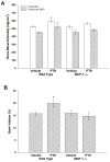
Parathyroid hormone (PTH) has a significant role as an anabolic hormone in bone when administered by intermittent injection. Previous microarray studies in our laboratory have shown that the most highly regulated gene, monocyte chemoattractant protein-1 (MCP-1), is rapidly and transiently induced when hPTH(1-34) is injected intermittently in rats. Through further in vivo studies, we found that rats treated with hPTH(1-34) showed a significant increase in serum MCP-1 levels 2 hours after PTH injection compared with basal levels. Using immunohistochemistry, increased MCP-1 expression in osteoblasts and osteocytes is evident after PTH treatment. PTH also increased the number of marrow macrophages. MCP-1 knockout mice injected daily with hPTH(1-34) showed less trabecular bone mineral density and bone volume compared with wild-type mice as measured by peripheral quantitative computed tomography (pQCT) and micro-computed tomography (µCT). Histomorphometric analysis revealed that the increase in osteoclast surface and osteoclast number observed with intermittent PTH treatment in the wild-type mice was completely eliminated in the MCP-1 null mice, as well as much lower numbers of macrophages. Consequently, the lack of osteoclast and macrophage activity in the MCP-1 null mice was paralleled by a reduction in bone formation. We conclude that osteoblast and osteocyte MCP-1 expression is an important mediator for the anabolic effects of PTH on bone.
Keywords: BONE ANABOLISM; BONE TURNOVER; CYTOKINES; MONOCYTE CHEMOATTRACTANT PROTEIN (MCP-1); PARATHYROID HORMONE (PTH).
Copyright © 2013 American Society for Bone and Mineral Research.
Conflict of interest statement
The authors state that they have no conflicts of interest pertaining to the work described.
Figures


Similar articles
-
Osteoblastic monocyte chemoattractant protein-1 (MCP-1) mediation of parathyroid hormone's anabolic actions in bone implicates TGF-β signaling.Bone. 2021 Feb;143:115762. doi: 10.1016/j.bone.2020.115762. Epub 2020 Nov 17. Bone. 2021. PMID: 33212319 Free PMC article.
-
Proteoglycan 4: a dynamic regulator of skeletogenesis and parathyroid hormone skeletal anabolism.J Bone Miner Res. 2012 Jan;27(1):11-25. doi: 10.1002/jbmr.508. J Bone Miner Res. 2012. PMID: 21932346 Free PMC article.
-
Black bear parathyroid hormone has greater anabolic effects on trabecular bone in dystrophin-deficient mice than in wild type mice.Bone. 2012 Sep;51(3):578-85. doi: 10.1016/j.bone.2012.05.003. Epub 2012 May 11. Bone. 2012. PMID: 22584007 Free PMC article.
-
Monocyte Chemoattractant Protein-1 (MCP-1/CCL2) Drives Activation of Bone Remodelling and Skeletal Metastasis.Curr Osteoporos Rep. 2019 Dec;17(6):538-547. doi: 10.1007/s11914-019-00545-7. Curr Osteoporos Rep. 2019. PMID: 31713180 Free PMC article. Review.
-
Studies on the mechanisms of the skeletal anabolic action of endogenous and exogenous parathyroid hormone.Arch Biochem Biophys. 2008 May 15;473(2):218-24. doi: 10.1016/j.abb.2008.03.003. Epub 2008 Mar 10. Arch Biochem Biophys. 2008. PMID: 18358824 Review.
Cited by
-
Tumor Microenvironment, Clinical Features, and Advances in Therapy for Bone Metastasis in Gastric Cancer.Cancers (Basel). 2022 Oct 6;14(19):4888. doi: 10.3390/cancers14194888. Cancers (Basel). 2022. PMID: 36230816 Free PMC article. Review.
-
Hydrogen-Rich Water Mitigates LPS-Induced Chronic Intestinal Inflammatory Response in Rats via Nrf-2 and NF-κB Signaling Pathways.Vet Sci. 2022 Nov 8;9(11):621. doi: 10.3390/vetsci9110621. Vet Sci. 2022. PMID: 36356098 Free PMC article.
-
CCL2/Monocyte Chemoattractant Protein 1 and Parathyroid Hormone Action on Bone.Front Endocrinol (Lausanne). 2017 Mar 29;8:49. doi: 10.3389/fendo.2017.00049. eCollection 2017. Front Endocrinol (Lausanne). 2017. PMID: 28424660 Free PMC article. Review.
-
Role of donor and host cells in muscle-derived stem cell-mediated bone repair: differentiation vs. paracrine effects.FASEB J. 2014 Aug;28(8):3792-809. doi: 10.1096/fj.13-247965. Epub 2014 May 19. FASEB J. 2014. PMID: 24843069 Free PMC article.
-
Interplay Between Skeletal and Hematopoietic Cells in the Bone Marrow Microenvironment in Homeostasis and Aging.Curr Osteoporos Rep. 2024 Aug;22(4):416-432. doi: 10.1007/s11914-024-00874-2. Epub 2024 May 23. Curr Osteoporos Rep. 2024. PMID: 38782850 Review.
References
-
- Tam CS, Heersche JN, Murray TM, Parsons JA. Parathyroid hormone stimulates the bone apposition rate independently of its resorptive action: differential effects of intermittent and continuous administration. Endocrinology. 1982;110(2):506–512. - PubMed
-
- Hock JM, Gera I. Effects of continuous and intermittent administration and inhibition of resorption on the anabolic response of bone to parathyroid hormone. J Bone Miner Res. 1992;7(1):65–72. - PubMed
-
- Dobnig H, Turner RT. The effects of programmed administration of human parathyroid hormone fragment (1-34) on bone histomorphometry and serum chemistry in rats. Endocrinology. 1997;138(11):4607–4612. - PubMed
-
- Shen V, Birchman R, Wu DD, Lindsay R. Skeletal effects of parathyroid hormone infusion in ovariectomized rats with or without estrogen repletion. J Bone Miner Res. 2000;15(4):740–760. - PubMed
-
- Sato M, Zheng GQ, Turner CH. Biosynthetic human parathyroid hormone (1-34) effects bone quality in aged ovariectomized rats. Endocrinology. 1997;138:4330–4337. - PubMed
Publication types
MeSH terms
Substances
Grants and funding
LinkOut - more resources
Full Text Sources
Other Literature Sources
Medical
Molecular Biology Databases
Research Materials
Miscellaneous

