. 2013 Apr 9;110(15):E1380-9.
doi: 10.1073/pnas.1218446110. Epub 2013 Mar 21.

Enterocyte loss of polarity and gut wound healing rely upon the F-actin-severing function of villin

Affiliations

Enterocyte loss of polarity and gut wound healing rely upon the F-actin-severing function of villin

Florent Ubelmann et al. Proc Natl Acad Sci U S A. .

Abstract

Efficient wound healing is required to maintain the integrity of the intestinal epithelial barrier because of its constant exposure to a large variety of environmental stresses. This process implies a partial cell depolarization and the acquisition of a motile phenotype that involves rearrangements of the actin cytoskeleton. Here we address how polarized enterocytes harboring actin-rich apical microvilli undergo extensive cell remodeling to drive injury repair. Using live imaging technologies, we demonstrate that enterocytes in vitro and in vivo rapidly depolarize their microvilli at the wound edge. Through its F-actin-severing activity, the microvillar actin-binding protein villin drives both apical microvilli disassembly in vitro and in vivo and promotes lamellipodial extension. Photoactivation experiments indicate that microvillar actin is mobilized at the lamellipodium, allowing optimal migration. Finally, efficient repair of colonic mechanical injuries requires villin severing of F-actin, emphasizing the importance of villin function in intestinal homeostasis. Thus, villin severs F-actin to ensure microvillus depolarization and enterocyte remodeling upon injury. This work highlights the importance of specialized apical pole disassembly for the repolarization of epithelial cells initiating migration.


Conflict of interest statement

The authors declare no conflict of interest.

Figures

Fig. 1.
Fig. 1.
Efficient colonic wound healing requires villin. (A) Serial endoscopic snapshots showing the site of biopsy-induced injury in mouse colon of the indicated genotypes at day 0 and 7 after wounding. Images are representative of at least four animals per genotype. Dashed lines delineate the wounded area. (B) Plot of the average changes in the wounded area over time for mice of the indicated genotype, normalized to the original lesion size at day 0. Values are for WT and villin−/− mice, respectively. Day 3: 54 ± 10 vs. 69 ± 7%; day 7: 17 ± 3 vs. 56 ± 9%. WT: n = 6; villin−/− : n = 4. Wilcoxon; *P < 0.05, NS, nonsignificant (P > 0.05). (C) Histological sections from WT and villin−/− mice, at day 4 postinjury stained for cleaved caspase 3 (Upper Left, red) or ki67 (Lower Left, red) and E-cadherin (Upper and Lower Right, green). DAPI stains nuclei (blue). Red asterisks highlight the injured area. (Scale bar: 100 μm.)
Fig. 2.
Fig. 2.
Transgenic villin proteins are functional, and villin severing activity is required for brush border disassembly upon carbachol treatment in vivo. (A) Histological frozen sections from jejunum (Left) or colon (Right) of tgvillinWT or tgvillinΔsev mice showing villin-mCherry fluorescence (red). DAPI labels nuclei (blue). (Scale bar: 100 μm.) (B and C) Histological frozen sections from untreated or carbachol-treated (+carb) small intestines stained for F-actin by phalloidin (green). DAPI labels nuclei (blue). White brackets indicate the width of the F-actin concentration at the apex of the enterocytes. (B) Sections from WT or villin−/− mice. (C) Sections from tgvillinWT or tgvillinΔsev mice. mCherry-villin fluorescence is shown (red). (Scale bar: 10 μm.) (D) Histogram depicting the width of the apical F-actin concentration of WT, villin−/−, tgvillinWT, or tgvillinΔsev mice untreated or treated with carbachol. Width values (in μm) are WT, 2.28 ± 0.08; WT+carb: 0.54 ± 0.07; villin−/−, 2.35 ± 0.09; villin−/−+carb, 2.20 ± 0.10; tgvillinWT, 2.10 ± 0.09; tgvillinWT+carb, 0.68 ± 0.05; tgvillinΔsev, 2.08 ± 0.08; tgvillinΔsev+carb, 1.92 ± 0.15. Numbers of measurements are WT: untreated; n = 15; WT+carb: n =15; villin−/− untreated: n = 8; villin−/−+carb: n =16; tgvillinWT: n = 11; tgvillinWT+carb: n =44; tgvillinΔsev, n = 15; tgvillinΔsev+carb: n =45. ***P < 0.001; NS, nonsignificant (P > 0.05 t test).
Fig. 3.
Fig. 3.
Efficient colonic wound healing requires villin through its severing activity. (A) Serial endoscopic snapshots showing the site of biopsy-induced injury in mouse colon of the indicated genotypes at day 0 and 7 after wounding. Images are representative of at least four animals per genotype. Dashed lines delineate the wounded area. (B) Plot of the average changes in the wounded area changes over time for mice of the indicated genotype, normalized to the original lesion size at day 0. Values are for tgvillinWT and tgvillinΔsev, respectively; day 3: 57 ± 6 vs. 74 ± 8%; day 7: 17 ± 8 vs. 62 ± 5%. Number of animals analyzed: WT: n = 4; tgvillinΔsev, n = 5. *P < 0.05. ns, nonsignificant (P > 0.05); Wilcoxon.
Fig. 4.
Fig. 4.
Microvilli disassemble upon cell migration via villin’s severing activity. (A) Migration of LLCPK-1 cells depleted for endogenous porcine villin (shRNA2) expressing EYFP-actin and mCherry-villinWT or villinΔsev visualized by spinning-disk time-lapse microscopy. EYFP and mCherry channels are shown. Images are maximal projections of the acquired z stacks. Serial images are shown at 12-min intervals. Red asterisks indicate the migrating cells analyzed. (Scale bar: 10 μm.) (B) Z reconstructions of the migrating cells highlighted in A at 0 and 48 min. Overlays of EYFP-actin (green) and mCherry-villin (red) are shown. White arrows point to the dorsal face of the migrating cell. (Scale bar: 10 μm.) (C) Plots depicting the intensity of villin and actin fluorescence over time at the dorsal face of the cells transfected for mCherry-villinWT or villinΔsev quantified from images acquired by spinning-disk time-lapse microscopy. Number of cells analyzed: villinWT, n =10; villinΔsev, n =19. (D) Histogram representing the penetrance of the phenotype illustrated by the presence or absence of apical microvilli positive for mCherry-villin on migrating cells transfected for mCherry-villinWT or villinΔsev at 30 min postinjury on fixed cells. Values are 62.5 ± 1.7 and 43.4 ± 1.7% for villinWT and villinΔsev, respectively. ***P < 0.001, t test. Number of cells analyzed: villinWT, n =121; villinΔsev, n = 95. (E) Relative number of lamellipodia formation per field over time in villinWT- or villinΔsev-expressing cells. Values are normalized to the number of lamellipodia at 20 min postinjury determined in villinWT-expressing cells. Values are averaged from three experiments.
Fig. 5.
Fig. 5.
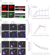
Microvillar actin is rapidly integrated at the lamellipodium following microvillus disassembly. (A) Biphotonic laser photoactivation of PA-GFP-actin at the apical face of a confluent monolayer of LLCPK1 cells depleted for endogenous villin and expressing villinWT-mCherry and PAGFP-actin. (Left) Maximal projections of the z stacks before and immediately after photoactivation. (Right) Z reconstruction of the z stacks before and immediately after photoactivation. (Scale bar: 10 μm.) (B) Serial micrographs of maximal z projections depicted as pseudocolored heat maps. Biphotonic laser photoactivation at the dorsal face of an early-migrating cell depleted for endogenous villin and expressing villinWT-mCherry (Upper) and villinΔsev (Lower). White arrows indicate the lamellipodium edge. (Scale bar: 10 μm.) (C). Plot of the proportion of apically photoactivated actin incorporated in the lamellipodia over time. Number of cells analyzed: villinWT, n =10; villinΔsev, n =16. *P < 0.01; ns, P > 0.05; t test. (D) Plot depicting the proportion of incorporation at the lamellipodia over time of actin photoactivated slightly away from the lamellipodial edge. Number of cells analyzed: villinWT, n = 4; villinΔsev, n = 4. (E) Plot depicting the relative intensity of photoactivated actin at the apical face of cells expressing villinWT or villinΔsev. Number of cells analyzed: villinWT, n =10; villinΔsev, n =16.
Fig. 6.
Fig. 6.
Villin is essential for brush border disassembly upon cell migration in vivo. (A) Histological paraffin sections of small intestine of WT or villin−/− mice luminally treated with DOC just after washout (Upper) or 30 min after washout (Lower) stained for E-cadherin (Ecadh, red) and sucrase-isomaltase (SI, green). DAPI labels nuclei (blue). Vertical dashed lines highlight the injured area; basal dashed lines delineate the lamina propria; arrows indicate the injury-adjacent enterocytes. (Scale bar: 10 μm.) (B) Histograms depicting the presence or the absence of apical staining of sucrase-isomaltase or ezrin quantified 30 min after washout on histological paraffin sections of small intestine of WT, villin−/−, tgvillinWT, or tgvillinΔsev mice treated with DOC. Values are WT, 23 ± 1%; villin−/−, 77 ± 4%; tgvillinWT, 21 ± 2%; and tgvillinΔsev, 79 ± 3% for apical sucrase-isomaltase and WT, 25 ± 2%; villin−/−; 84 ± 4%; tgvillinWT, 27 ± 3%; and tgvillinΔsev, 72 ± 3% for apical ezrin. For sucrase-isomaltase: WT, n = 84 cells analyzed from eight animals; villin−/−, n = 147 cells analyzed from 12 animals; tgvillinWT, n = 29 cells analyzed from three animals; tgvillinΔsev, n = 41 cells analyzed from three animals. For ezrin: WT, n = 45 cells analyzed from six animals; vil−/−, n = 26 cells analyzed from three animals; tgvillinWT, n = 22 cells analyzed from three animals; tgvillinΔsev, n = 35 cells analyzed from three animals.

Similar articles

Cited by

References

    1. Blikslager AT, Moeser AJ, Gookin JL, Jones SL, Odle J. Restoration of barrier function in injured intestinal mucosa. Physiol Rev. 2007;87(2):545–564. - PubMed
    1. Sturm A, Dignass AU. Epithelial restitution and wound healing in inflammatory bowel disease. World J Gastroenterol. 2008;14(3):348–353. - PMC - PubMed
    1. Iizuka M, Konno S. Wound healing of intestinal epithelial cells. World J Gastroenterol. 2011;17(17):2161–2171. - PMC - PubMed
    1. Lotz MM, Rabinovitz I, Mercurio AM. Intestinal restitution: Progression of actin cytoskeleton rearrangements and integrin function in a model of epithelial wound healing. Am J Pathol. 2000;156(3):985–996. - PMC - PubMed
    1. Fenteany G, Janmey PA, Stossel TP. Signaling pathways and cell mechanics involved in wound closure by epithelial cell sheets. Curr Biol. 2000;10(14):831–838. - PubMed

Publication types

MeSH terms

LinkOut - more resources