. 2013 Mar 22;339(6126):1445-8.
doi: 10.1126/science.1231077.

A localized Wnt signal orients asymmetric stem cell division in vitro

Affiliations

A localized Wnt signal orients asymmetric stem cell division in vitro

Shukry J Habib et al. Science. .

Erratum in

  • Science. 2013 May 24;340(6135):924

Abstract

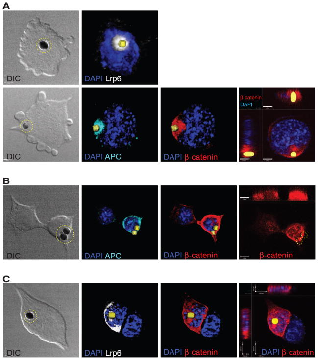
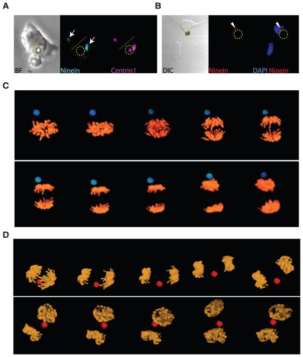
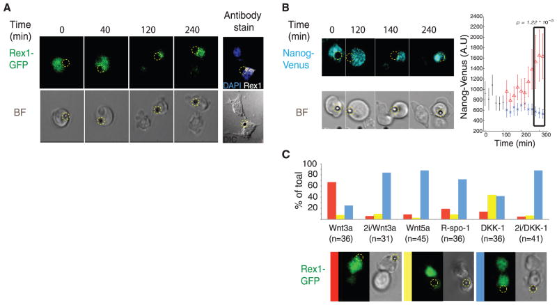
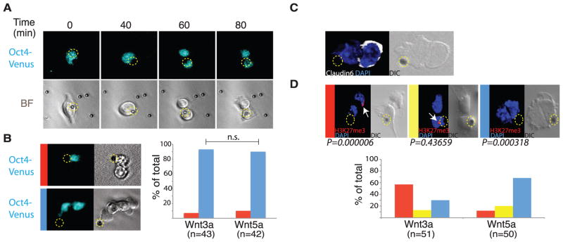
Developmental signals such as Wnts are often presented to cells in an oriented manner. To examine the consequences of local Wnt signaling, we immobilized Wnt proteins on beads and introduced them to embryonic stem cells in culture. At the single-cell level, the Wnt-bead induced asymmetric distribution of Wnt-β-catenin signaling components, oriented the plane of mitotic division, and directed asymmetric inheritance of centrosomes. Before cytokinesis was completed, the Wnt-proximal daughter cell expressed high levels of nuclear β-catenin and pluripotency genes, whereas the distal daughter cell acquired hallmarks of differentiation. We suggest that a spatially restricted Wnt signal induces an oriented cell division that generates distinct cell fates at predictable positions relative to the Wnt source.


Figures

Fig. 1
Fig. 1
Wnt3a beads induce asymmetric distribution of components of the Wnt/β-catenin pathway. Representative images of ES cells cocultured with Wnt3a beads (indicated by dashed yellow circle or reconstructed as a yellow square) and stained with antibodies to LRP6 (white), APC (cyan), and β-catenin (red). (A) Before division; (B) during division; (C) after division. The beads are 2.8 μm. The far right panels show optical sections through the nucleus. DAPI, 4′,6-diamidino-2-phenylindole; DIC, differential interference contrast.
Fig. 2
Fig. 2
Asymmetric inheritance of centrosomes and the orientation of the plane of mitotic division. Time-lapse imaging of dividing single ES cells cocultured with Wnt3a beads (indicated by a dashed yellow circle) and (A) expressing enhanced green fluorescent protein (EGFP)–Ninein (cyan; arrows) and DsRedex–Centrin1 (magenta). Dotted orange line indicates the boundary between two cells. (B) Immunostaining for endogenous Ninein (arrowheads). BF: bright-field image. (C and D) Representative images from three-dimensional time-lapse microscopy of segregating chromosomes in ES cells expressing H2B-Venus that were cocultured with Wnt3a beads (blue) or Wnt5a beads (red).
Fig. 3
Fig. 3
Expression of the pluripotency genes Rex1 and Nanog during ES cell division. (A) Selected frames from time-lapse imaging of a dividing Rex-1 GFP reporter ES cell in the presence of a Wnt3a bead (indicated by a dashed yellow circle). The far right panel shows antibody staining for endogenous Rex-1 protein. (B) Selected frames from time-lapse imaging of a dividing Nanog-Venus reporter ES cell in the presence of a Wnt3a bead. Signal intensities of all frames were individually determined, and the mean ± SD intensity values plotted. Red triangles represent signal intensities of the cell retaining contact with the bead after division. (C) Representative images of time-lapse microscopy of dividing Rex-1 GFP ES reporter cells cocultured in the presence of the indicated beads in 2i or 2i-free media. Cell divisions were classified based on the relative expression of GFP and plotted. Red bar: higher GFP amounts in the bead-proximal cell; yellow bar: higher amounts of GFP in the bead-distal cell; blue bar: similar amounts of GFP in either cell.
Fig. 4
Fig. 4
Distal cells express markers of epiblast stem cell fate. (A) Selected frames from time-lapse imaging of a dividing Oct4-Venus reporter ES cell cocultured with a Wnt3a bead (indicated by a dashed yellow circle). (B) Divisions of Oct4-Venus ES cells cocultured with Wnt3a or Wnt5a beads were classified based on the relative expression of Venus, and plotted. Red bar: higher Venus abundance in the bead-proximal cell; blue bar: similar abundance of Venus in both cells. (C) Antibody staining for Claudin6 in an ES cell cocultured with a Wnt3a bead. (D) Representative images of antibody staining for H3K27me3 (arrows) in LF2 female ES cells cocultured with Wnt3a or Wnt5a beads. Dividing cells were classified based on location of the H3K27me3 stain, and plotted. Red bar: H3K27me3 stain in the bead-distal cell; yellow bar: H3K27me3 stain in the bead-proximal cell; blue bar: no H3K27me3 stain in any cell.

Comment in

References

    1. Neumüller RA, Knoblich JA. Genes Dev. 2009;23:2675. - PMC - PubMed
    1. Werts AD, Goldstein B. Semin Cell Dev Biol. 2011;22:842. - PMC - PubMed
    1. Walston T, et al. Dev Cell. 2004;7:831. - PubMed
    1. Goldstein B, Takeshita H, Mizumoto K, Sawa H. Dev Cell. 2006;10:391. - PMC - PubMed
    1. Sugioka K, Mizumoto K, Sawa H. Cell. 2011;146:942. - PubMed

Publication types

MeSH terms