Multicenter Study
. 2013 Mar;24(4):550-8.
doi: 10.1681/ASN.2012010097. Epub 2013 Mar 21.

Renal hypodysplasia associates with a WNT4 variant that causes aberrant canonical WNT signaling

Affiliations
Multicenter Study

Renal hypodysplasia associates with a WNT4 variant that causes aberrant canonical WNT signaling

Asaf Vivante et al. J Am Soc Nephrol. 2013 Mar.

Abstract

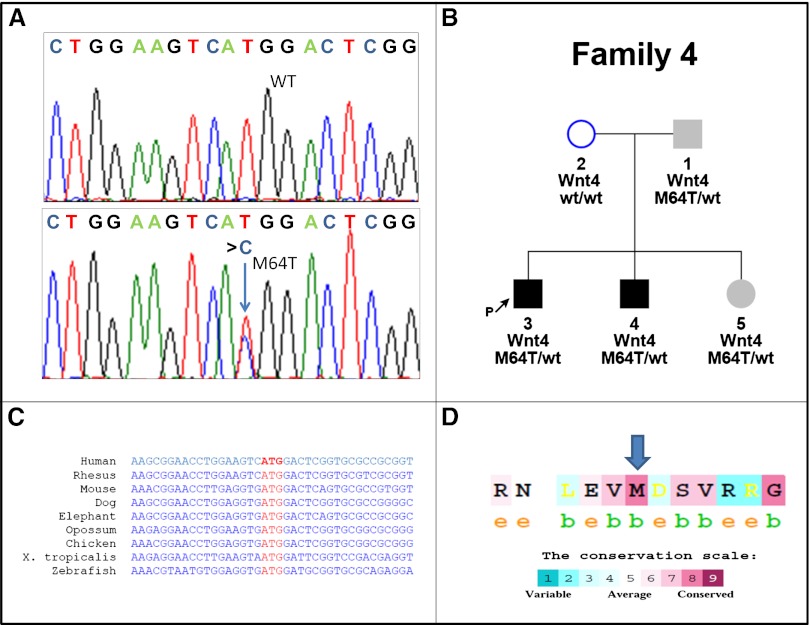
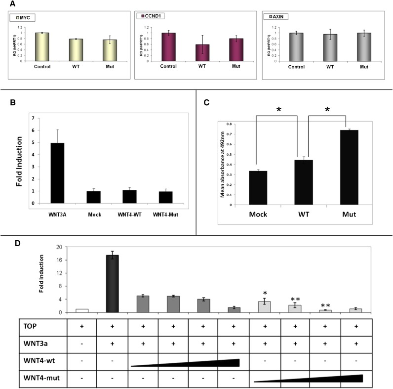
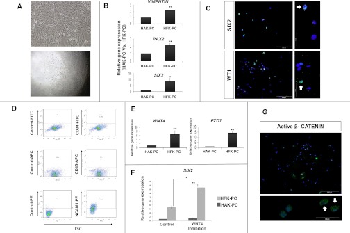
Abnormal differentiation of the renal stem/progenitor pool into kidney tissue can lead to renal hypodysplasia (RHD), but the underlying causes of RHD are not well understood. In this multicenter study, we identified 20 Israeli pedigrees with isolated familial, nonsyndromic RHD and screened for mutations in candidate genes involved in kidney development, including PAX2, HNF1B, EYA1, SIX1, SIX2, SALL1, GDNF, WNT4, and WT1. In addition to previously reported RHD-causing genes, we found that two affected brothers were heterozygous for a missense variant in the WNT4 gene. Functional analysis of this variant revealed both antagonistic and agonistic canonical WNT stimuli, dependent on cell type. In HEK293 cells, WNT4 inhibited WNT3A induced canonical activation, and the WNT4 variant significantly enhanced this inhibition of the canonical WNT pathway. In contrast, in primary cultures of human fetal kidney cells, which maintain WNT activation and more closely represent WNT signaling in renal progenitors during nephrogenesis, this mutation caused significant loss of function, resulting in diminished canonical WNT/β-catenin signaling. In conclusion, heterozygous WNT4 variants are likely to play a causative role in renal hypodysplasia.


Figures

Figure 1.
Figure 1.
Mutation analysis of the p.M64T, WNT4 variant shows high degree of conservation. (A) Sequence analysis reveals the WNT4 variant caused by heterozygous transition (c.t191c) (lower panel, blue arrow) resulting in amino acid substitution M64T. The wild-type (WT) sequence is given for comparison (upper panel). (B) WNT4 variant is found in patients 1, 3, 4, and 5. Whereas patients 3 and 4 have a clear isolated renal phenotype of severe left renal hypodysplasia, patient 1 has an isolated left renal cyst (sized 12.8×7 mm) in addition to normal BP and normal GFR. Patient 5, age 3 years, has normal pelvic and renal ultrasound results, normal BP, normal GFR, and low morning serum cortisol levels. Incomplete penetrance or variable expression can be considered in this family. Squares indicate male family members and circles female family members; black filled squares indicate that the patients are affected with RHD. Gray filled squares and circles indicate subtle clinical signs. (C) cDNA sequences of human WNT4 are compared with WNT4 orthologs in other species. (D) Conservation scale of WNT4 protein shows the amino acid methionine in position 64 (blue arrow) to be highly conserved (8 of 9), with “b” indicating a buried residue and “e” indicating an exposed residue. Note the high degree of conservation.
Figure 2.
Figure 2.
Functional analysis of the p.M64T,WNT4 variant in HEK293 shows enhanced inhibition of the canonical WNT pathway. (A) WNT4, wild-type and mutant, does not increase WNT canonical pathway target genes. Quantitative RT-PCR on HEK293 transfected cells with WNT4 wild-type (WT) and mutant (Mut) plasmids shows the mRNA expression levels of canonical WNT pathway target genes (MYC, CCND1, and AXIN). (B) WNT3A, but neither wild-type WNT4 nor mutant WNT4, activates the canonical WNT pathway. HEK293 cells are cotransfected on a 6-well plate, with TOPFlash reporter plasmid (4 µg) and either wild-type WNT4, mutant WNT4, or WNT3A expression plasmid (4 µg). pCMV-Renilla plasmid (0.4 µg) is used as the internal control. (C) Wild-type WNT4 and mutant variants increase cell proliferation compared with control. Mutant WNT4 leads to significantly increased proliferation compared with wild-type WNT4. *P<0.05. Results are presented as the mean absorbance at 492 nm using the MTS proliferation assay ± SEM of at least three replicates. (D) WNT4, wild-type and mutant, inhibit the canonical WNT pathway. WNT4 mutant compared with the wild-type leads to significantly enhanced inhibition of the canonical WNT pathway. Cells are cotransfected with TOPFlash reporter plasmid (500 ng), WNT3A expression plasmid (200 ng) to activate the canonical WNT pathway, and an increasing concentration of WNT4 wild-type or WNT4 mutant expression plasmid (200, 400, 800, and 1600 ng). pCMV-Renilla plasmid is used as an internal control (50 ng). Cell lysates are measured for luciferase activity 48 hours after transfection. Activities are expressed as fold activation of the relative luciferase activity. WNT3A and WNT4 does not activate the mutant TOPFlash reporter, FOPFlash, confirming assay specificity. Asterisks indicate a significant difference of the mutant WNT4 compared with dose-equivalent WNT4 wild-type transfection. *P<0.05; **P<0.01 (t test). Data are presented as the mean ± SEM from three separate experiments.
Figure 3.
Figure 3.
Human fetal kidney cells harbor renal epithelial stem/progenitor characteristics and exhibit basal canonical WNT activity. (A–D) HFK-PC cells express renal stem/progenitor markers. (A) Cellular appearance of cultured fetal kidney cells (upper panel) and clone formation by human fetal kidney cells, demonstrating their high clonogenic capacity. Culturing of 0.3 cells per well results in 5%–10% of single clone formation (lower panel). (B) Gene expression analysis of renal stem/progenitor genes. Quantitative RT-PCR (qat-PCR) analysis of Vimentin, PAX2, and SIX2, three representative genes expressed early during nephrogenesis. Normalization is performed against control GAPDH expression and real-time quantitative is calculated relative to the well differentiated HAK-PC cells. (C) Immunofluoresence staining for the expression of two representative early nephrogenesis transcriptional factors: WT1 and SIX2 (×20 and ×100). All nuclei are stained with DAPI (blue). The green florescence signal in the upper and lower panels corresponds to anti-SIX2 protein and anti-WT1 protein staining respectively. Both clearly show nuclear staining (representative stained nuclei are indicated with white arrows). (D) Flow cytometric analysis for CD34, CD45, and CD56/NCAM1 expression and corresponding isotype controls in HFK-PC cells. Results show negligible levels of CD34 (a well known marker of hematopoietic stem cells and vascular endothelial cells) and CD45 (leukocyte common antigen). Moreover, the HFK-PC contains 44% of CD56/NCAM1-positive cells. NCAM1 has been previously shown to be a stem/progenitor cell marker in the human fetal kidney. (E–G) HFK-PC cells harbor basal canonical WNT activity. (E) qRT-PCR analysis of WNT4 and frizzled 7 (FZD7), a representative receptor of the WNT signaling ligands. (F) Because WNT/β-catenin and SIX2 pathways have opposing actions (commitment and self-renewal of renal stem/progenitors, respectively), we sought to manipulate this balance so as to provide additional support that our HFK-PC cells contain a significant portion of cells with early renal stem/progenitor characteristics. Consequently, qRT-PCR analysis of SIX2, in HFK-PC after the addition of three different WNT pathway antagonists—dickkopf-related protein 1 (DKK1), secreted frizzled-related protein 1 (sFRP1), and Wnt inhibitory factor (WIF)—shows significant SIX2 increment compared with the control. Normalization is performed against control GAPDH expression and real-time quantitative is calculated relative to the well differentiated HAK-PC cells. Data are presented as the mean ± SEM from three separate experiments. (G) HFK-PC immunofluorescence staining for anti-active β-catenin (green) discloses its cytoplasmic and nuclear presence. All nuclei are stained with DAPI (blue). *P<0.05; **P<0.01 (t test). DAPI, 4',6-diamidino-2-phenylindole; NCAM-1, neural cell adhesion molecule 1.
Figure 4.
Figure 4.
Functional analysis of the p.M64T, WNT4 variant in HFK-PC shows significant loss of function and diminished canonical WNT/b-catenin signaling. (A) Wild-type WNT4, but neither WNT3A nor mutant WNT4, activates the canonical WNT pathway in HFK-PC. HFK-PC cells are cotransfected on a 6-well plate, with TOPFlash reporter plasmid (4 µg) and wild-type WNT4, mutant WNT4, or WNT3A expression plasmid (4 µg). pCMV-Renilla plasmid (0.4 µg) is used as internal control. *P<0.05 (t test). Data are presented as the mean ± SEM from three separate experiments. (B) Illustration of the conditioned media experiments. HEK293 cells are transfected separately with wild-type WNT4, mutant WNT4, or empty vector (control). Conditioned media containing WNT proteins were applied on HFK-PC 24 hours after transfection. Cells are harvested for total RNA and total protein 6–24 hours later. (C) Results of a representative Western blot analysis of the HFK-PC cells after they are treated with wild-type WNT4, mutant WNT4, and control conditioned media, with the use of anti-active β-catenin antibody. Cbl (95 kD) is used as a loading control. Active β-catenin protein levels are adjusted to Cbl and are quantified compared with the control (empty vector). Three separate experiments yield a similar result in which wild-type WNT4 induces a significant increase in active β-catenin as opposed to the p.M64T Wnt4 variant, which results in a nonsignificant change. (D) Quantification by real-time RT-PCR of the expression of mRNA axin2 and ccnd1 in HFK-PC after treatment with wild-type WNT4, mutant WNT4, and control conditioned media.

Similar articles

Cited by

References

    1. Hildebrandt F: Genetic kidney diseases. Lancet 375: 1287–1295, 2010 - PMC - PubMed
    1. Sanna-Cherchi S, Caridi G, Weng PL, Scolari F, Perfumo F, Gharavi AG, Ghiggeri GM: Genetic approaches to human renal agenesis/hypoplasia and dysplasia. Pediatr Nephrol 22: 1675–1684, 2007 - PMC - PubMed
    1. Weber S, Moriniere V, Knüppel T, Charbit M, Dusek J, Ghiggeri GM, Jankauskiené A, Mir S, Montini G, Peco-Antic A, Wühl E, Zurowska AM, Mehls O, Antignac C, Schaefer F, Salomon R: Prevalence of mutations in renal developmental genes in children with renal hypodysplasia: Results of the ESCAPE study. J Am Soc Nephrol 17: 2864–2870, 2006 - PubMed
    1. Eccles MR, Schimmenti LA: Renal-coloboma syndrome: A multi-system developmental disorder caused by PAX2 mutations. Clin Genet 56: 1–9, 1999 - PubMed
    1. Bingham C, Bulman MP, Ellard S, Allen LI, Lipkin GW, Hoff WG, Woolf AS, Rizzoni G, Novelli G, Nicholls AJ, Hattersley AT: Mutations in the hepatocyte nuclear factor-1beta gene are associated with familial hypoplastic glomerulocystic kidney disease. Am J Hum Genet 68: 219–224, 2001 - PMC - PubMed

Publication types

Supplementary concepts