. 2013 Apr 1;27(7):719-24.
doi: 10.1101/gad.207803.112. Epub 2013 Mar 21.

Robust cellular reprogramming occurs spontaneously during liver regeneration

Affiliations

Robust cellular reprogramming occurs spontaneously during liver regeneration

Kilangsungla Yanger et al. Genes Dev. .

Erratum in

  • Genes Dev. 2013 Jul 1;27(13):1537

Abstract

Cellular reprogramming-the ability to interconvert distinct cell types with defined factors-is transforming the field of regenerative medicine. However, this phenomenon has rarely been observed in vivo without exogenous factors. Here, we report that activation of Notch, a signaling pathway that mediates lineage segregation during liver development, is sufficient to reprogram hepatocytes into biliary epithelial cells (BECs). Moreover, using lineage tracing, we show that hepatocytes undergo widespread hepatocyte-to-BEC reprogramming following injuries that provoke a biliary response, a process requiring Notch. These results provide direct evidence that mammalian regeneration prompts extensive and dramatic changes in cellular identity under injury conditions.


Figures

Figure 1.
Figure 1.
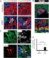
Notch signaling triggers hepatocyte-to-BEC reprogramming. (A) Schematic of simultaneous lineage labeling (YFP) and activation of Notch intracellular domain (NICD) expression by AAV administration. (B) Hes1 is expressed in most HNF4α+ hepatocytes 1 wk after Notch activation. (C) Notch activation in hepatocytes for 1 wk leads to an “intermediate” phenotype characterized by coexpression of BEC markers (OPN and Sox9) and HNF4α. Portal veins (PV) are denoted by white dotted lines. (D) YFP+ lineage-labeled hepatocytes express OPN and Sox9 upon Notch activation. (E) HNF4α+ hepatocytes express a BEC apical marker, Par6, after 2 wk of Notch activation. (F) Notch signaling reprograms hepatocytes into terminally differentiated BECs. Weak CK19 staining is observed in YFP+ cells 2 wk after infection, while robust CK19 expression in YFP+ cells appears 6 wk after infection. (Insets) Reprogrammed CK19+YFP+ cells are denoted with arrows, and “unreprogrammed” CK19+YFP- cells are denoted with arrowheads. These images are representative of at least five independent experiments. Bar, 25 μm.
Figure 2.
Figure 2.
Direct evidence for hepatocyte-to-ADC reprogramming in vivo. (A) Mice treated with DDC for 1 wk exhibit costaining with a hepatocyte marker (HNF4α) and one of three biliary markers (OPN, Sox9, or A6). Double-positive cells have a hepatocyte morphology (arrowheads), and many are binucleated (arrows). The images are representative of multiple experiments. (B) Costaining of the biliary markers OPN, A6, Sox9, and CK19 with the YFP lineage label was observed following DDC treatment (6 wk), BDL (2 wk), or PHx (2 wk); costained cells are denoted with arrowheads. Note that many of the costained cells are smaller than neighboring hepatocytes. (C) Biliary markers are induced in a stepwise fashion following injury. The percentage of marker+ cells (i.e., cells that stained positive for OPN, Sox9, A6, or CK19) that coexpress the YFP lineage label is shown. Both CK19hi and CK19lo cells were included in the analysis. Double-positive cells were not detected in vehicle-treated livers. Each bar represents the mean (±SE) of at least three mice; a minimum of 950 marker+ cells were examined for each data point. Bars, 25 μm.
Figure 3.
Figure 3.
Hepatocytes undergo morphological changes during reprogramming. (A) YFP+ hepatocytes change their morphology after DDC injury, organizing into duct-like structures with neo-lumens marked by the apical polarity markers PKCζ and Par6 (arrowheads). (B) Reprogrammed cells have cilia, marked by acetylated tubulin (6 wk, Ac-tub; pink asterisk). (C) Reprogramming leads to binucleated ADCs. Sections of liver from AAV8-TBG-Cre-labeled animals given DDC were costained with DAPI (gray/blue), YFP (green), and CK19 (red) and examined by confocal microscopy. Binucleated ADCs were present in the YFP+ population. (D) Quantification of binucleated BECs in labeled mice given normal chow (AAV vehicle) or DDC (AAV DDC); the number of cells counted (N = 3 mice in each group) is shown. Bars, 10 μm.
Figure 4.
Figure 4.
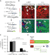
Hepatocyte reprogramming requires Notch signaling. (A) Schematic of simultaneous deletion of RBPJκ and lineage tracing of hepatocytes. (B) Mice with hepatocyte-specific deletion of RBP-Jκ have a significant reduction in the number of “intermediate” cells. Concomitant hepatocyte labeling and RBP-Jκ deletion results in a reduction in the percentage of hepatocyte-derived Sox9+ or OPN+ cells after DDC treatment (3 wk). (C) Quantification of B, depicting the percentage of Sox9+ or OPN+ cells that carry the YFP lineage marker (N = 4 mice each group). (D) Model for hepatocyte-to-ADC reprogramming, a Notch-dependent cascade of transcriptional changes and alterations in cellular morphology. Bars, 25 μm.

Comment in

References

    1. Desmet VJ 1985. Intrahepatic bile ducts under the lens. J Hepatol 1: 545–559 - PubMed
    1. Factor V, Radaeva S, Thorgeirsson S 1994. Origin and fate of oval cells in dipin-induced hepatocarcinogenesis in the mouse. Am J Pathol 145: 409–422 - PMC - PubMed
    1. Fan B, Malato Y, Calvisi DF, Naqvi S, Razumilava N, Ribback S, Gores GJ, Dombrowski F, Evert M, Chen X, et al. 2012. Cholangiocarcinomas can originate from hepatocytes in mice. J Clin Invest 122: 2911–2915 - PMC - PubMed
    1. Farber E 1956. Similarities in the sequence of early histological changes induced in the liver of the rat by ethionine, 2-acetylamino-fluorene, and 3′-methyl-4-dimethylaminoazobenzene. Cancer Res 16: 142–148 - PubMed
    1. Fukuda K, Sugihara A, Nakasho K, Tsujimura T, Yamada N, Okaya A, Sakagami M, Terada N 2004. The origin of biliary ductular cells that appear in the spleen after transplantation of hepatocytes. Cell Transplant 13: 27–33 - PubMed

Publication types

Substances