

Enamel pearls in permanent dentition: case report and micro-CT evaluation
- PMID: 23520396
- PMCID: PMC3667525
- DOI: 10.1259/dmfr.20120332
Enamel pearls in permanent dentition: case report and micro-CT evaluation
Abstract
Objectives: To investigate the frequency, position, number and morphology of enamel pearls (EPs) using micro-CT (µCT) and to report a case of an EP mimicking an endodontic-periodontic lesion.
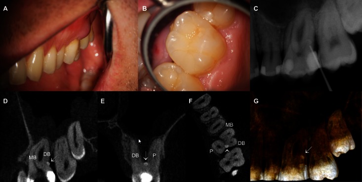
Methods: Cone beam CT (CBCT) was performed in a patient to evaluate a radio-opaque nodule observed on the left maxillary first molar during the radiographic examination. Additionally, 23 EPs were evaluated regarding frequency, position, number and morphology by means of µCT. The results were statistically compared using the Student's t-test for independent samples.
Results: 1 pearl was presented in 13 specimens, while 5 specimens presented 2 pearls. The most frequent location of the EPs was the furcation between the disto-buccal and the palatal roots of the maxillary molars. Overall, the mean major diameter, volume and surface area were 1.98 ± 0.85 mm, 1.76 ± 1.36 mm³ and 11.40 ± 7.59 mm², respectively, with no statistical difference between maxillary second and third molars (p > 0.05). In the case report, CBCT revealed an EP between the disto-buccal and the palatal roots of the maxillary first left molar associated with advanced localized periodontitis. The tooth was referred for extraction.
Conclusions: EPs, located generally in the furcation area, were observed in 0.74% of the sample. The majority was an enamel-dentin pearl type and no difference was found in maxillary second and third molars regarding diameter, volume and surface area of the pearls. In this report, the EP mimicked an endodontic-periodontic lesion and was a secondary aetiological factor in the periodontal breakdown.
Figures


References
-
- Cavanha AO. Enamel pearls. Oral Surg Oral Med Oral Pathol 1965; 19: 373–382 - PubMed
-
- Chrcanovic BR, Abreu MH, Custodio AL. Prevalence of enamel pearls in teeth from a human teeth bank. J Oral Sci 2010; 52: 257–260 - PubMed
-
- Goldstein AR. Enamel pearls as contributing factor in periodontal breakdown. J Am Dent Assoc 1979; 99: 210–211 - PubMed
-
- Matthews DC, Tabesh M. Detection of localized tooth-related factors that predispose to periodontal infections. Periodontol 2000 2004; 34: 136–150 - PubMed
-
- Moskow BS, Canut PM. Studies on root enamel (2). Enamel pearls. A review of their morphology, localization, nomenclature, occurrence, classification, histogenesis and incidence. J Clin Periodontol 1990; 17: 275–281 - PubMed
Publication types
MeSH terms
LinkOut - more resources
Full Text Sources
Other Literature Sources
