. 2013;8(3):e58712.
doi: 10.1371/journal.pone.0058712. Epub 2013 Mar 8.

AQP4-dependent water transport plays a functional role in exercise-induced skeletal muscle adaptations

Affiliations

AQP4-dependent water transport plays a functional role in exercise-induced skeletal muscle adaptations

Davide Basco et al. PLoS One. 2013.

Erratum in

  • PLoS One. 2013;8(6). doi:10.1371/annotation/86fc2632-913c-490d-8b9b-e925b38baec5

Abstract

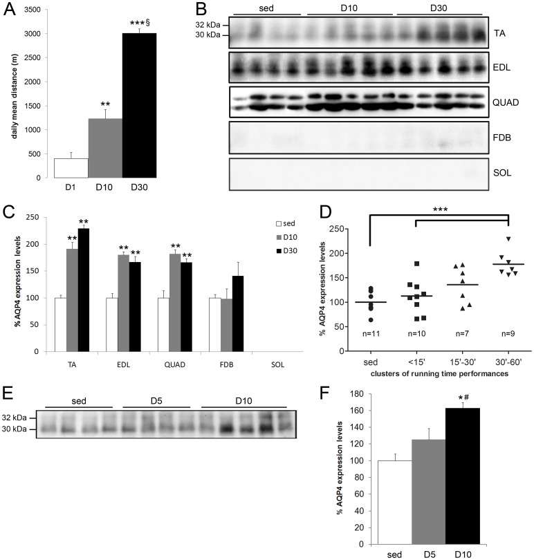
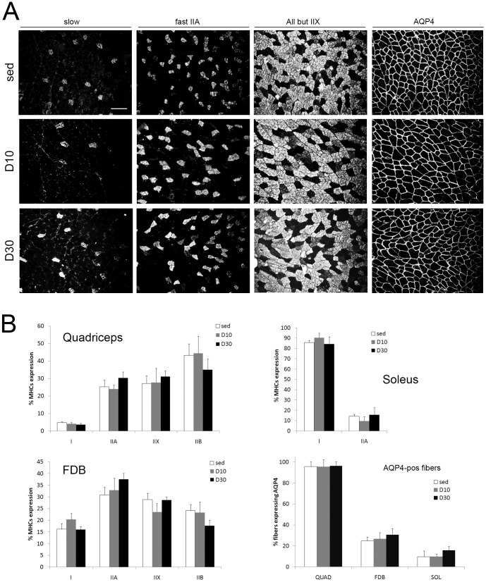
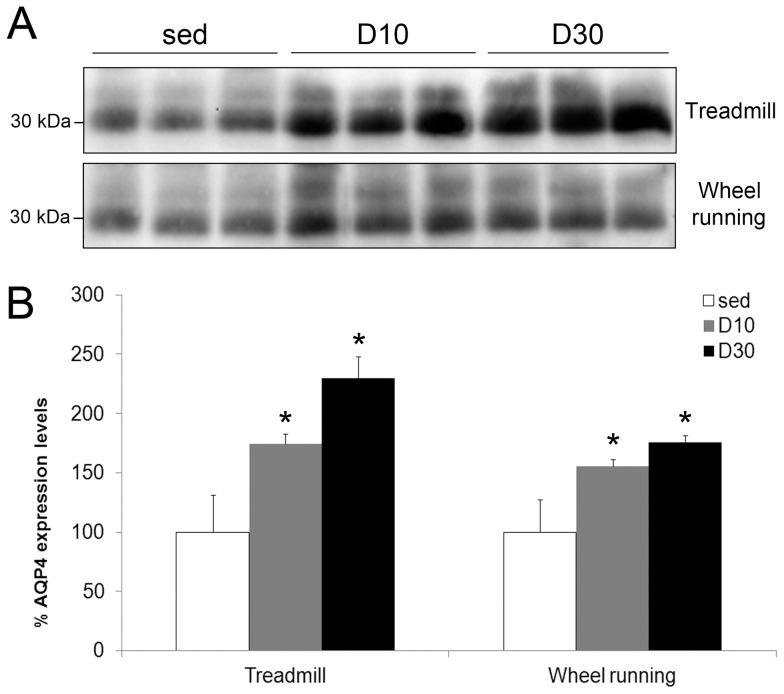
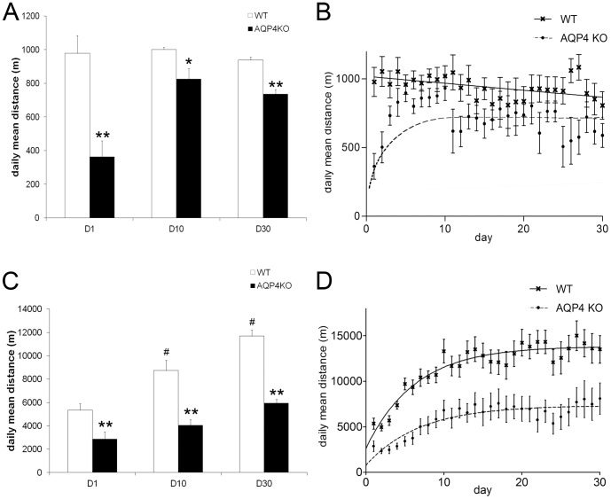
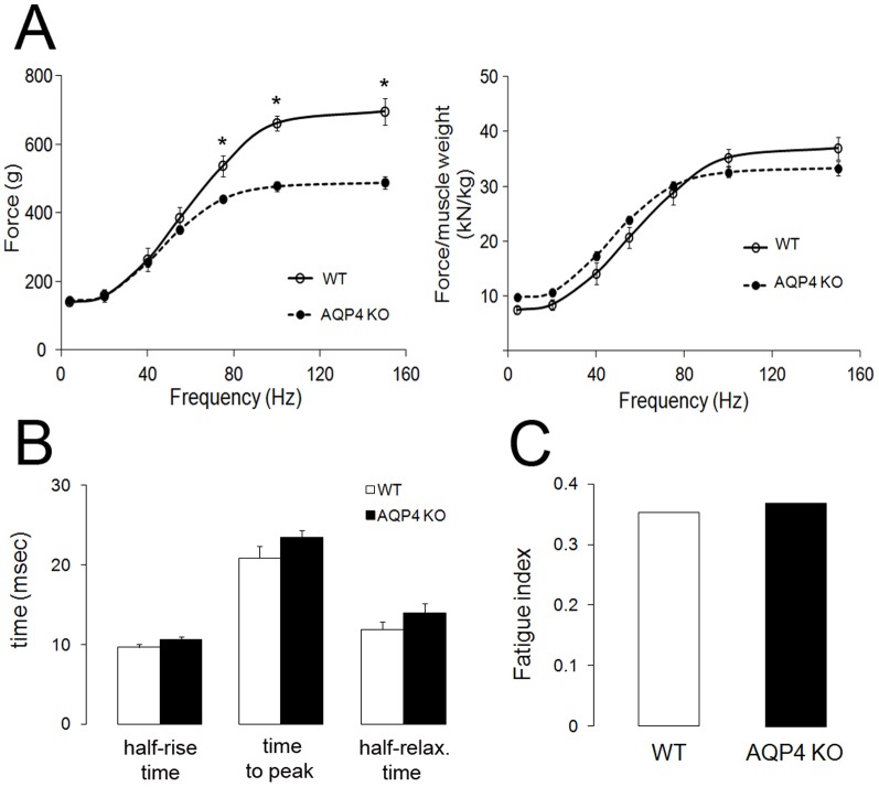
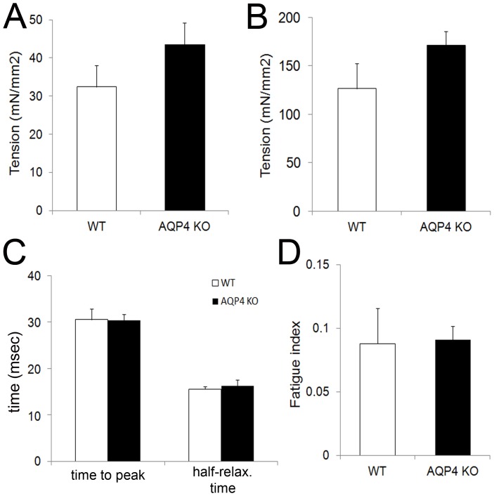
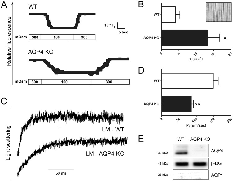
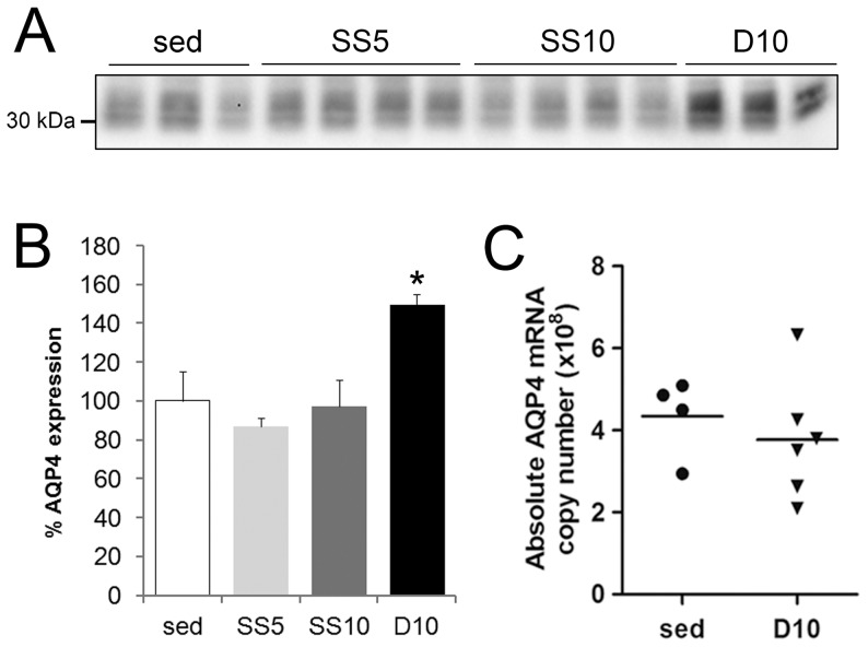
In this study we assess the functional role of Aquaporin-4 (AQP4) in the skeletal muscle by analyzing whether physical activity modulates AQP4 expression and whether the absence of AQP4 has an effect on osmotic behavior, muscle contractile properties, and physical activity. To this purpose, rats and mice were trained on the treadmill for 10 (D10) and 30 (D30) days and tested with exercise to exhaustion, and muscles were used for immunoblotting, RT-PCR, and fiber-type distribution analysis. Taking advantage of the AQP4 KO murine model, functional analysis of AQP4 was performed on dissected muscle fibers and sarcolemma vesicles. Moreover, WT and AQP4 KO mice were subjected to both voluntary and forced activity. Rat fast-twitch muscles showed a twofold increase in AQP4 protein in D10 and D30 rats compared to sedentary rats. Such increase positively correlated with the animal performance, since highest level of AQP4 protein was found in high runner rats. Interestingly, no shift in muscle fiber composition nor an increase in AQP4-positive fibers was found. Furthermore, no changes in AQP4 mRNA after exercise were detected, suggesting that post-translational events are likely to be responsible for AQP4 modulation. Experiments performed on AQP4 KO mice revealed a strong impairment in osmotic responses as well as in forced and voluntary activities compared to WT mice, even though force development amplitude and contractile properties were unvaried. Our findings definitively demonstrate the physiological role of AQP4 in supporting muscle contractile activity and metabolic changes that occur in fast-twitch skeletal muscle during prolonged exercise.


Conflict of interest statement

Competing Interests: The authors have declared that no competing interests exist.

Figures

Figure 1
Figure 1. AQP4 protein expression after endurance exercise in rats.
A) Daily mean distance at day 1 (D1), day 10 (D10) and day 30 (D30). Note that rats significantly increased performance during the training period; n = 6-8 per group. ***p<0.0001 vs D1. **p<0.001 vs D1. §p<0.001 vs D10. B) Immunoblotting analysis of AQP4 protein expression on TA, EDL, QUAD, FDB and SOL muscles. Note the presence of the two AQP4 isoforms of 30 and 32 kDa. Except for FDB, AQP4 was found to be significantly over-expressed in fast muscles, as reported by densitometric analysis (C); n =  6-8 per muscle; **p<0.001 vs sed.. D) Protein levels of AQP4 in QUAD muscles of four groups of treadmill runners based on their mean time run per day of both D10 and D30 animals. Significant changes in AQP4 protein levels were obtained after 30 min/day of endurance exercise; n = 7–11 per cluster. *p<0.01 vs sed and <15’ cluster. E–F) Effect of short-term exercise on AQP4 expression in QUAD muscles. Note that 5 days of endurance exercise (D5) did not determine a significant increase in AQP4 expression, in contrast with observations in D10 rats; n = 6–8 per group. *p<0.01 vs sed. #p<0.05 vs D5. In all the immunoblotting experiments, protein levels were corrected for whole protein loading determined by staining membrane with Ponceau S.
Figure 3
Figure 3. AQP4 mRNA and protein levels in rat QUAD muscles after stress conditioning and exercise.
A) Representative immunoblotting analysis for sed (n = 8), SS5 (n = 8), SS10 (n = 6) and D10 (n = 8) rats (*p<0.05; see histogram in B). C) Absolute AQP4 mRNA copy number measured by Real-Time PCR in sed and D10 rats.
Figure 4
Figure 4. MHCs distribution in rat quadriceps muscles after treadmill activity.
A) Representative immunofluorescence photomicrographs of MHC isoforms and AQP4 in quadriceps muscle from sed, D10 and D30 rats groups. Sections were immunostained for slow MHC, MHC IIA, and all MHC isoforms except MHC IIX. Scale bar, 50 µm. B) Percentage of fibers expressing MHC isoforms and AQP4 in rat muscles. Note that the distribution of MHCs and AQP4 did not change in either fast or slow-twitch muscles after D10 and D30 compared to sed rats (n = 5 muscles/group).
Figure 5
Figure 5. Skeletal muscle AQP4 protein expression during mice exercise.
A) Representative immunoblotting analysis of AQP4 protein levels in quadriceps muscles of sed, D10 and D30 mice. B) Densitometric analysis (n =  4–5 per group) was performed normalizing to whole protein amount by staining membrane with Ponceau S solution. Note a significant AQP4 accumulation at D10 and D30 of voluntary and forced exercise (*p<0.05 vs sed).
Figure 6
Figure 6. Treadmill and wheel running exercise in WT and AQP4 KO mice.
A) Daily mean distance at D1, D10 and D30 of treadmill activity. Note that AQP4 KO mice ran significantly less than WT mice at D1 and throughout the training period; n = 6 per group. *p<0.05 and **p<0.01 vs the relative WT group. B) Progression of daily mean distance in WT and AQP4 KO mice; n = 24 per group. Note a two-phase behavior of AQP4 KO mice in treadmill exercise. C) Daily mean distance at D1, D10 and D30 of voluntary activity. AQP4 KO mice ran significantly less than WT mice during all the training period; n = 6 per group. **p<0.01 vs the relatives WT group. #p<0.01 vs D1 WT group. D) Progression of daily mean distance in WT and AQP4 KO mice; n = 24 per group.
Figure 7
Figure 7. Force generation by WT and AQP4 KO gastrocnemius muscles “in vivo”.
A)Force-frequency curves (n = 8–10): absolute force (left) and force normalized to muscle mass (right). B) Twitch times (n = 8–10). C) Low-frequency fatigue (n = 8–10). In each case, no difference was found between WT and AQP4 KO muscles. *p<0.05.
Figure 8
Figure 8. Force generation by WT and AQP4 KO EDL muscles “ex vivo”.
A)Twitch tension (n = 12). B) Maximal tetanic tension(n = 12). C) Twitch times (n = 12). D) Fatigue index (n = 12). In each case, no difference was found between WT and AQP4 KO muscles.
None
Osmotic properties of skeletal muscle from WT and AQP4 KO mice.
A) Representative TIR fluorescence time course in response to a 200-mOsm inwardly directed NaCl gradient at 10°C. B) Mean values ± SEM of the time constant, τ (n = 4). Inset: micrograph of a single layer muscle fibers immobilized on cover glass (scale bar: 50 µm). C) Time course of scattered light intensity in response to a 225 mOsm sucrose gradient at 10°C. Representative curves are shown for LM vesicles from WT and AQP4 KO skeletal muscles (in one set of experiments typical of three). D) Pf data are mean ± SEM; **p <0.01 compared with WT vesicles. E) Immunoblot analysis of LM fractions from WT and AQP4 KO skeletal muscles using AQP4, β-dystroglycan, and AQP1 antibodies. Protein levels were corrected for whole protein loading determined by staining membrane with Ponceau S.

References

    1. Usher-Smith JA, Huang CL, Fraser JA (2009) Control of cell volume in skeletal muscle. Biol Rev Camb Philos Soc 84: 143–159. - PubMed
    1. Frigeri A, Gropper MA, Umenishi F, Kawashima M, Brown D, et al. (1995) Localization of MIWC and GLIP water channel homologs in neuromuscular, epithelial and glandular tissues. J Cell Sci 108 ( Pt 9): 2993–3002. - PubMed
    1. Jung JS, Bhat RV, Preston GM, Guggino WB, Baraban JM, et al. (1994) Molecular characterization of an aquaporin cDNA from brain: candidate osmoreceptor and regulator of water balance. Proc Natl Acad Sci U S A 91: 13052–13056. - PMC - PubMed
    1. Verbavatz JM, Ma T, Gobin R, Verkman AS (1997) Absence of orthogonal arrays in kidney, brain and muscle from transgenic knockout mice lacking water channel aquaporin-4. J Cell Sci 110 ( Pt 22): 2855–2860. - PubMed
    1. Silberstein C, Bouley R, Huang Y, Fang P, Pastor-Soler N, et al. (2004) Membrane organization and function of M1 and M23 isoforms of aquaporin-4 in epithelial cells. Am J Physiol Renal Physiol 287: F501–511. - PubMed

Publication types

LinkOut - more resources