. 2013;8(3):e59003.
doi: 10.1371/journal.pone.0059003. Epub 2013 Mar 8.

Depletion of cellular iron by curcumin leads to alteration in histone acetylation and degradation of Sml1p in Saccharomyces cerevisiae

Affiliations

Depletion of cellular iron by curcumin leads to alteration in histone acetylation and degradation of Sml1p in Saccharomyces cerevisiae

Gajendra Kumar Azad et al. PLoS One. 2013.

Erratum in

  • PLoS One. 2013;8(10). doi:10.1371/annotation/ac974676-18a6-4aee-b38e-57a1142a3dc1

Abstract

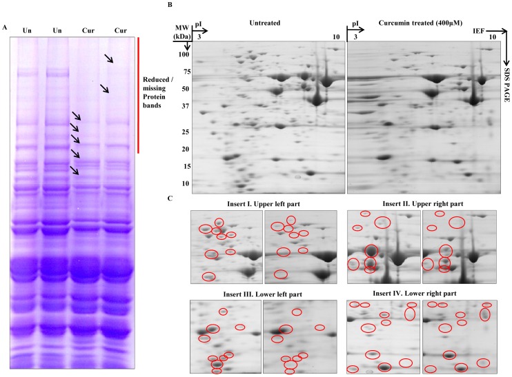
Curcumin, a naturally occurring polyphenolic compound, is known to possess diverse pharmacological properties. There is a scarcity of literature documenting the exact mechanism by which curcumin modulates its biological effects. In the present study, we have used yeast as a model organism to dissect the mechanism underlying the action of curcumin. We found that the yeast mutants of histone proteins and chromatin modifying enzymes were sensitive to curcumin and further supplementation of iron resulted in reversal of the changes induced by curcumin. Additionally, treatment of curcumin caused the iron starvation induced expression of FET3, FRE1 genes. We also demonstrated that curcumin induces degradation of Sml1p, a ribonucleotide reductase inhibitor involved in regulating dNTPs production. The degradation of Sml1p was mediated through proteasome and vacuole dependent protein degradation pathways. Furthermore, curcumin exerts biological effect by altering global proteome profile without affecting chromatin architecture. These findings suggest that the medicinal properties of curcumin are largely contributed by its cumulative effect of iron starvation and epigenetic modifications.


Conflict of interest statement

Competing Interests: The authors have declared that no competing interests exist.

Figures

Figure 1
Figure 1. Curcumin inhibits growth of yeast cells and its effect is antagonized by iron supplementation.
A) Growth Assay; 1588-4C (Wild-type) was grown up to log phase. 3 µl of each undiluted and 10-fold serially diluted culture was spotted onto control SCA plates, SCA plates containing 200, 400, 600 μM curcumin, and SCA plates impregnated with iron (100 μM) in combination with 600 μM curcumin. All plates were incubated at 30°C for 72 hr and photographed. B) Viability Assay; 1588-4C (Wild-type) was cultured up to mid-log phase and treated with 10, 25, 50, 100 and 200 μM curcumin for 3 hr and cells were stained with 0.3% methylene blue for checking viability. Untreated and heat killed cells were taken as negative and positive controls respectively; viability was observed under light microscope (400X) and photographed. C) Clonogenic assay; equal number of cells from mid-log phase of untreated and methylene blue stained curcumin (400 μM) treated cultures were spread on standard SCA plates in triplicate. All plates were incubated at 30°C and the colony forming ability was analyzed after 36 hr. No. of colonies were counted and shown in the form of bar diagram. D) Viability Assay; 1588-4C (Wild-type) was cultured up to mid-log phase, treated with 200 μM curcumin or iron (100 μM) in combination with 200 μM curcumin. A small number of cells were collected and stained with 0.3% methylene blue at different time points (10, 30, 60, 90 and 120 min) of curcumin treatment for viability examination. Untreated and heat killed cells were taken as negative and positive controls respectively; viability was observed under light microscope (400X) and photographed.
Figure 2
Figure 2. Mutations in histone proteins and chromatin modifiers causes increased sensitivity to curcumin.
A & B) Growth Assay; wild-type and different mutant yeast strains were grown up to log phase. 3 µl of each undiluted and 10-fold serially diluted culture was spotted onto control SCA (DMSO) plates or SCA plates containing 400 and 600 μM curcumin. All plates were incubated at 30°C for 72 hr and photographed.
Figure 3
Figure 3. Supplementation of iron suppresses the effects of curcumin.
Growth Assay; wild-type and different mutant yeast strains were grown up to log phase. 3 µl of each undiluted and 10-fold serially diluted culture was spotted onto control SCA (DMSO), SCA plates containing 600 μM curcumin and plates containing iron (100 μM) in combination with 600 μM curcumin. All plates were incubated at 30°C for 72 hr and photographed.
Figure 4
Figure 4. Curcumin reduces H3K56Ac by inhibiting Rtt109.
A) Growth Assay; wild-type, Rtt109Δ and H3K56Q mutant yeast strains were grown up to log phase. 3 µl of each undiluted and 10-fold serially diluted culture was spotted onto control SCA (DMSO) plates or SCA plates containing 200, 400 and 600 μM of curcumin. All plates were incubated at 30°C for 72 hr and photographed. B) Wild-type (1588-4C) cells were cultured up to log phase and treated with either DMSO or curcumin (400 μM) for 3 hr in triplicate. Whole cell extracts were prepared by TCA extraction method and samples were subjected to western blot anlaysis using indicated antibodies. Tbp, Gapdh and Rap1 protein levels were used as loading controls.
Figure 5
Figure 5. Curcumin induced alterations in epigenetic modifications are restored upon iron supplementation.
Wild-type (1588-4C) cells were cultured up to log phase and treated with either DMSO or curcumin (400 μM). Samples were collected after 1, 2 and 3 hr of curcumin treatment. Cultures were supplemented with iron (100 μM) after 3 hr of curcumin treatment. Again samples were collected after 1, 2, 3, 4 hr of iron supplementation. Whole cell extracts were prepared by TCA extraction method and samples were subjected to western blot anlaysis using indicated antibodies. Tbp, Gapdh, H3 and Rap1 levels were used as loading controls. Same extracts were loaded on different gels and transferred separately for western blotting.
Figure 6
Figure 6. Curcumin induces iron starvation and Sml1p degradation.
A & B) RT-PCR Analysis; Logarithmically grown wild-type (1588-4C) cells in standard SC liquid media were treated with either DMSO or curcumin (400 μM) for 3 hr. Total RNA were extracted and reverse transcribed to cDNA. Semi-quantitative PCR analysis was performed to assess the levels of FRE1, FET3, ACO1, RNR1/2/3/4, HUG1 and ACT1 transcripts. C) Wild-type (1588-4C) cells were cultured up to log phase and treated with either DMSO or curcumin (400 μM) for 3 hr in triplicate. Whole cell extracts were prepared by TCA extraction method and samples were subjected to western blot anlaysis using antibodies against Sml1, Rad53, Tbp and Rap1. D) Wild-type (1588-4C) cells were grown up to log phase, cultures of cells were divided equally and pre-incubated with either 1 mM PMSF or 100 mM MG132 for 90 min and then treated with curcumin (400 μM) for 3 hr. In addition, some cells were treated with iron (100 μM) and rapamycin (50 ng/ml) in presence of curcumin (400 μM) for 3 hr. Sml1 protein levels were analyzed by western blotting and cellular levels of Rap1, Tbp and Gapdh were used as loading controls.
Figure 7
Figure 7. Curcumin alters global proteomics.
Whole cell lysate was prepared by disrupting mid-log phase untreated and curcumin (400 μM) treated cultures using chilled glass beads. Protein extracts were prepared by precipitation and clean-up as mentioned in ‘Materials and methods’. A) Equal quantity of protein extracts were resolved by SDS-PAGE. B) 200 µg of total cellular proteins from untreated and curcumin-treated cultures was resolved by two-dimensional gel electrophoresis; first by isoelectric focusing (IEF) on a 7 cm long ImmobilineTM pH 3–10 DryStrip, followed by SDS-PAGE. The gel was stained with Coomassie Brilliant Blue R-250. C) Enlarged images of the upper left (Inset I), upper right (Inset II), lower left (Inset III), and lower right (Inset IV) regions of the two-dimensional gels containing proteins from untreated and curcumin-treated cells are shown. The differentially expressed spots are indicated by red circles.
Figure 8
Figure 8. Proposed model for action of curcumin in budding yeast
. Curcumin causes iron starvation by chelating it which leads to transcriptional alteration of iron-regulated genes. Histone hypo-acetylation by curcumin results in delayed growth phenotype. Curcumin induces Sml1p degradation through vacuole and proteasome-mediated protein degradation pathways. We propose that after Sml1p degradation the released Rnr1 associates with Rnr2 and Rnr4 to form an active RNR enzyme allowing the production of dNTPs.

References

    1. Bednar J, Horowitz RA, Grigoryev SA, Carruthers LM, Hansen JC, et al. (1998) Nucleosomes, linker DNA, and linker histone form a unique structural motif that directs the higher-order folding and compaction of chromatin. Proc Natl Acad Sci U S A 95: 14173–14178. - PMC - PubMed
    1. Luger K, Mader AW, Richmond RK, Sargent DF, Richmond TJ (1997) Crystal structure of the nucleosome core particle at 2.8 A resolution. Nature 389: 251–260. - PubMed
    1. Clapier CR, Cairns BR (2009) The biology of chromatin remodeling complexes. Annu Rev Biochem 78: 273–304. - PubMed
    1. Huertas D, Sendra R, Munoz P (2009) Chromatin dynamics coupled to DNA repair. Epigenetics 4: 31–42. - PubMed
    1. Ehrenhofer-Murray AE (2004) Chromatin dynamics at DNA replication, transcription and repair. Eur J Biochem 271: 2335–2349. - PubMed

Publication types

MeSH terms