. 2013 May;222(5):487-94.
doi: 10.1111/joa.12042. Epub 2013 Mar 24.

The human lung during the embryonic period: vasculogenesis and primitive erythroblasts circulation

Affiliations

The human lung during the embryonic period: vasculogenesis and primitive erythroblasts circulation

J Pereda et al. J Anat. 2013 May.

Abstract

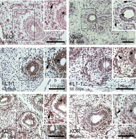
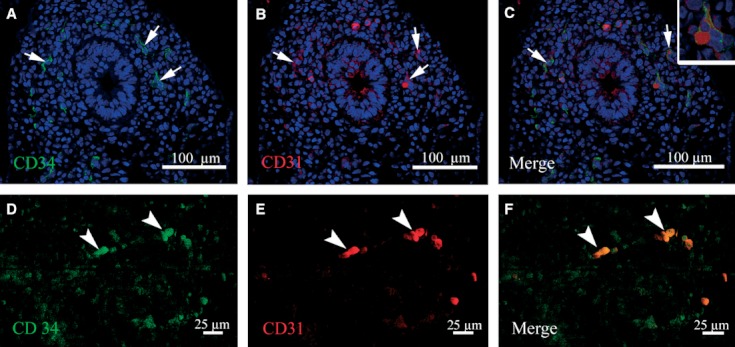
Vascularization and blood cell circulation are crucial steps during lung development. However, how blood vessels are generated and when lung circulation is initiated is still a matter of debate. A morpho-functional analysis of pulmonary vasculature was done using human lung samples between 31 and 56 days post-fertilization (pf). The immunolocalization and expression of CD31, CD34, FLT-1, KDR and the vascular growth factor (VEGF) were investigated. The results showed that at day 31 pf, a capillary plexus is already installed, and a few primitive erythroblasts were seen for the first time within the lumen of some blood vessels. Around day 45 pf, an increase in the amount of primitive erythroblasts was detected in the parenchyma surrounding the distal segment of the bronchial tree. The expression of FLT-1, KDR, CD31 and CD34 was observed in endothelial cells of the capillary plexus and the VEGF was detected in the endodermal epithelium. Our results support the hypothesis that the initial formation of the capillary plexus around the tip of the growing airway bud occurs by vasculogenesis, probably regulated by VEGF and KDR. We also showed a very early onset of blood circulation, starting from day 34 pf, concomitant with the generation of new lung buds. In addition, the increasing number of primitive erythroblasts from week 6 onward, associated with a change in the shape of the blood vessels, suggests a remodeling process and that the generation of new distal vessels at the tip of the lung bud occurs mainly by a process of angiogenesis.


Figures

Fig. 1
Fig. 1
Histological and SEM images illustrating the lobar organization of the embryonic lung. (A) A transverse section of the lung stained with H/E at day 34 pf. Two bronchi (arrows) on both sides of the esophagus (e) are seen. Note that the esophagus and the main bronchi are entirely surrounded by splanchnopleuric mesenchyme (m). Abundant blood cells are seen in the auricle (a). Pleural cavity (pc). Magnification, 20×. (B,C) SEM images of the lung at day 50 pf. Three pulmonary lobes can be seen in the right lung (B), and only two (numbered) in the left lung (C).
Fig. 2
Fig. 2
Histological sections of human lung stained with H/E. The insets correspond to higher magnification of the area enclosed in the rectangles. (A,B,C) The capillary plexus (arrows) around a lung terminal bud (tb) of embryos aged 34, 40 and 42 days pf. Primitive erythroblasts are seen within the lumen of some blood vessels (inset). (D) Primitive erythroblasts arranged in a single line within the capillary network of the lung (inset) at day 45 pf. From this embryonic age on, an increase of the amount of blood cells in the distal vascular plexus as well of their diameter was observed. Note that some definitive erythrocytes can be seen within the capillaries (inset) (E).
Fig. 3
Fig. 3
SEM images of the human lung at day 50 pf. (A) Two lung buds at the periphery of the lung (arrows). (B) Higher magnification of the tip of the blind projection of the lung bud enclosed in (A). The tip of the lung bud appears covered by a basal membrane and no mesenchymal cells were observed at this location. A higher magnification of the tip image is shown in (C). Some mesenchymal cells that look like migratory cells are attached to the basal lamina. (D) A transverse section through a terminal bud, showing a very regular-shaped endodermal epithelium surrounded by a strong basal lamina (arrows). Note that the mesenchymal tissue forms a sheath around the bud.
Fig. 4
Fig. 4
Immunostaining of VEGF, FLT-1 and KDR in embryonic lung sections. The insets correspond to a higher magnification of the area enclosed in the rectangles. In lungs from 42 (A) to 56 days pf (B), VEGF was expressed in the airway epithelial cells (*) and in some blood vessels (arrow). (C) FLT-1+ group endothelial cells (arrows) around the terminal buds, forming a vascular plexus at 42 days pf. (D) An increased number of FLT-1+ endothelial cells (arrow) are seen in the distal mesenchyma surrounding the developing airway at 56 days pf. Insets (C,D) show FLT-1+ endodermal cells (*). (E) KDR+ group endothelial cells (arrow) around the terminal buds, forming a vascular plexus at 49 days pf. (F) An increased number of KDR+ endothelial cells (arrow) is seen in the distal mesenchyma surrounding the developing airway at 56 days pf. Insets (E,F) shows KDR+ endodermal cells (*).
Fig. 5
Fig. 5
Confocal images of double immunofluorescence for CD34 and CD31 in human embryonic lungs at 35 days pf. (A) CD34+ cells (green) in the vascular plexus around the terminal bud. (B) Immunoreactivity for CD31 (red) in some endothelial cells. (C) The merged image of CD34 and CD31 in endothelial cells of the capillary plexus. A higher magnification of the endothelial cell is shown in the inset. (D,E) Single primitive CD34+ (green) and CD31+ (red) erythroblasts (arrow head). (F) Merged image of (D) and (E) showing CD34 and CD31 colocalization (arrowhead). Nuclei staining with TO-PRO-3-iodide.

References

    1. Ahlbrecht K, Schmitz J, Seay U, et al. Spatiotemporal expression of flk-1 in pulmonary epithelial cells during lung development. Am J Respir Cell Mol Biol. 2008;39:163–170. - PubMed
    1. Breier G, Clauss M, Risau W. Coordinate expression of vascular endothelial growth factor receptor-1 (flt-1) and its ligand suggests a paracrine regulation of murine vascular development. Dev Dyn. 1995;204:228–239. - PubMed
    1. Cardoso WV, Lu J. Regulation of early lung morphogenesis: questions, facts and controversies. Development. 2006;133:1611–1624. - PubMed
    1. Del Moral PM, Sala FG, Tefft D, et al. VEGF-A signaling through Flk-1 is a critical facilitator of early embryonic lung epithelial to endothelial crosstalk and branching morphogenesis. Dev Biol. 2006;290:177–188. - PubMed
    1. Demello DE, Reid LM. Embryonic and early fetal development of human lung vasculature and its functional implications. Pediatr Dev Pathol. 2000;3:439–449. - PubMed

Publication types

MeSH terms

Substances

LinkOut - more resources