

Neuronal morphology goes digital: a research hub for cellular and system neuroscience
- PMID: 23522039
- PMCID: PMC3653619
- DOI: 10.1016/j.neuron.2013.03.008
Neuronal morphology goes digital: a research hub for cellular and system neuroscience
Erratum in
- Neuron. 2013 Apr 10;78(1):206
Abstract
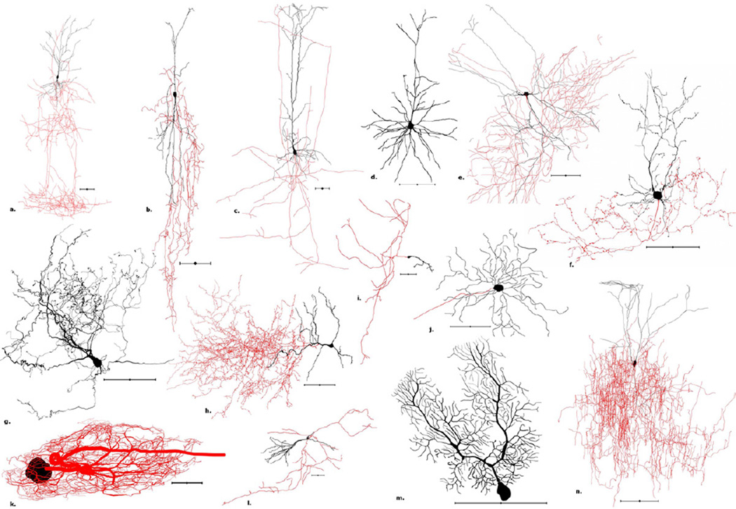
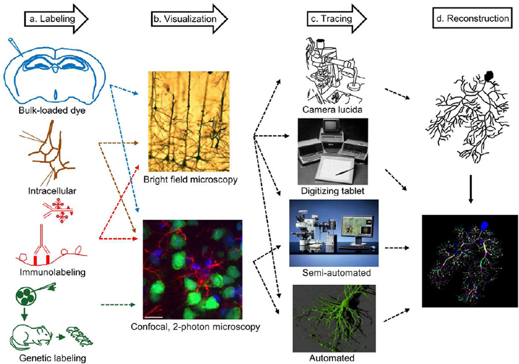
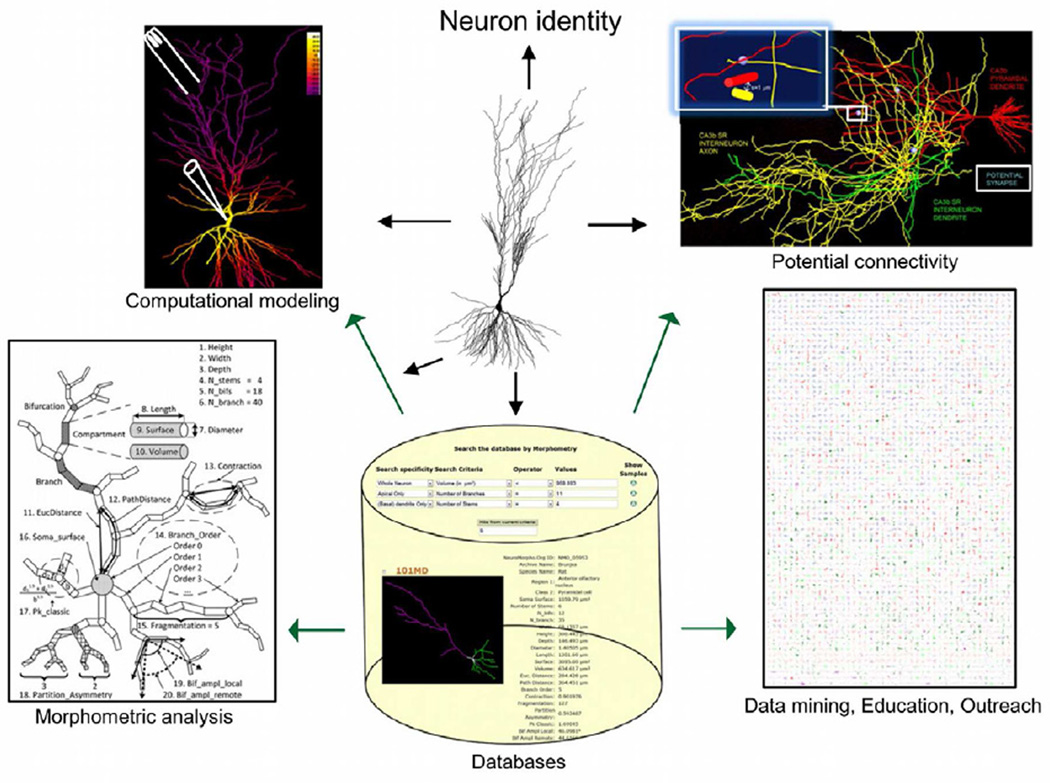
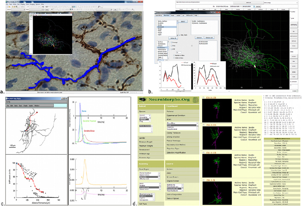
The importance of neuronal morphology in brain function has been recognized for over a century. The broad applicability of "digital reconstructions" of neuron morphology across neuroscience subdisciplines has stimulated the rapid development of numerous synergistic tools for data acquisition, anatomical analysis, three-dimensional rendering, electrophysiological simulation, growth models, and data sharing. Here we discuss the processes of histological labeling, microscopic imaging, and semiautomated tracing. Moreover, we provide an annotated compilation of currently available resources in this rich research "ecosystem" as a central reference for experimental and computational neuroscience.
Copyright © 2013 Elsevier Inc. All rights reserved.
Figures
Similar articles
-
Mobilizing the base of neuroscience data: the case of neuronal morphologies.Nat Rev Neurosci. 2006 Apr;7(4):318-24. doi: 10.1038/nrn1885. Nat Rev Neurosci. 2006. PMID: 16552417 Review.
-
High-content analysis in neuroscience.Nat Rev Neurosci. 2008 Oct;9(10):779-88. doi: 10.1038/nrn2492. Epub 2008 Sep 11. Nat Rev Neurosci. 2008. PMID: 18784656 Review.
-
Neuron tracing in perspective.Cytometry A. 2010 Jul;77(7):693-704. doi: 10.1002/cyto.a.20895. Cytometry A. 2010. PMID: 20583273 Review.
-
Adding new dimension to neuroscience.J Neurosci Res. 2018 Jul;96(7):1123-1124. doi: 10.1002/jnr.24238. Epub 2018 Mar 23. J Neurosci Res. 2018. PMID: 29570824 Free PMC article. No abstract available.
-
Special issue on computational neuroscience.J Neurosci Methods. 2012 Sep 15;210(1):1-2. doi: 10.1016/j.jneumeth.2012.08.008. Epub 2012 Aug 10. J Neurosci Methods. 2012. PMID: 22903194 No abstract available.
Cited by
-
Subdiffraction Imaging of Cleared and Expanded Large-Scale Tissues.Chem Biomed Imaging. 2024 Jun 18;2(8):542-559. doi: 10.1021/cbmi.4c00013. eCollection 2024 Aug 26. Chem Biomed Imaging. 2024. PMID: 39473992 Free PMC article. Review.
-
Topological characterization of neuronal arbor morphology via sequence representation: II--global alignment.BMC Bioinformatics. 2015 Jul 4;16(1):209. doi: 10.1186/s12859-015-0605-1. BMC Bioinformatics. 2015. PMID: 26141505 Free PMC article.
-
In search of a periodic table of the neurons: Axonal-dendritic circuitry as the organizing principle: Patterns of axons and dendrites within distinct anatomical parcels provide the blueprint for circuit-based neuronal classification.Bioessays. 2016 Oct;38(10):969-76. doi: 10.1002/bies.201600067. Epub 2016 Aug 12. Bioessays. 2016. PMID: 27516119 Free PMC article. Review.
-
CellRemorph: A Toolkit for Transforming, Selecting, and Slicing 3D Cell Structures on the Road to Morphologically Detailed Astrocyte Simulations.Neuroinformatics. 2023 Jul;21(3):483-500. doi: 10.1007/s12021-023-09627-5. Epub 2023 May 3. Neuroinformatics. 2023. PMID: 37133688 Free PMC article.
-
SmartTracing: self-learning-based Neuron reconstruction.Brain Inform. 2015 Sep;2(3):135-144. doi: 10.1007/s40708-015-0018-y. Epub 2015 Aug 19. Brain Inform. 2015. PMID: 27747506 Free PMC article.
References
-
- Abbott A, Schiermeier Q. Research prize boost for Europe. Nature. 2013;493:585–586. - PubMed
-
- Alle H, Roth A, Geiger JR. Energy-efficient action potentials in hippocampal mossy fibers. Science. 2009;325:1405–1408. - PubMed
-
- Ascoli GA. Computational Neuroanatomy: Principles and methods. New York: Humana Press; 2002.
-
- Ascoli GA. Mobilizing the base of neuroscience data: the case of neuronal morphologies. Nat. Rev. Neurosci. 2006;7:318–324. - PubMed
Publication types
MeSH terms
Grants and funding
LinkOut - more resources
Full Text Sources
Other Literature Sources
Molecular Biology Databases
