. 2013 Mar 20;77(6):1039-46.
doi: 10.1016/j.neuron.2013.01.013.

Regulation of postsynaptic retrograde signaling by presynaptic exosome release

Affiliations

Regulation of postsynaptic retrograde signaling by presynaptic exosome release

Ceren Korkut et al. Neuron. .

Abstract

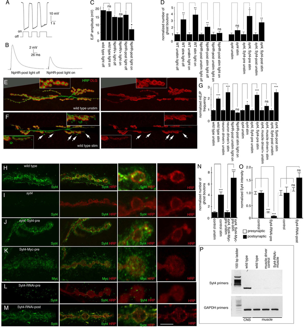
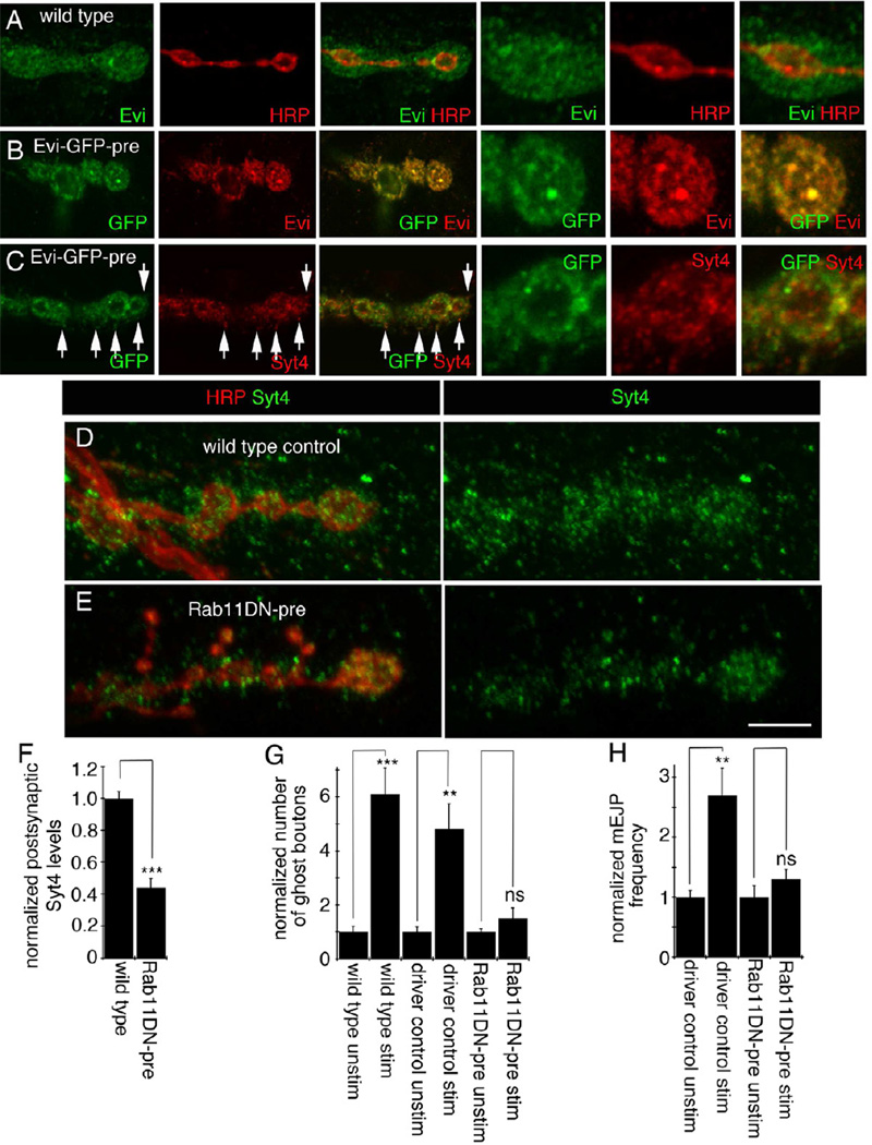
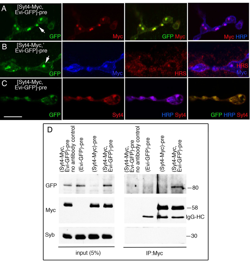
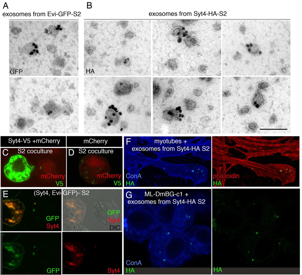
Retrograde signals from postsynaptic targets are critical during development and plasticity of synaptic connections. These signals serve to adjust the activity of presynaptic cells according to postsynaptic cell outputs and to maintain synaptic function within a dynamic range. Despite their importance, the mechanisms that trigger the release of retrograde signals and the role of presynaptic cells in this signaling event are unknown. Here we show that a retrograde signal mediated by Synaptotagmin 4 (Syt4) is transmitted to the postsynaptic cell through anterograde delivery of Syt4 via exosomes. Thus, by transferring an essential component of retrograde signaling through exosomes, presynaptic cells enable retrograde signaling.


Figures

Figure 1
Figure 1. Retrograde control of synaptic growth and function by trans-synaptic Syt4 transfer
(A) Hyperpolarization of muscles (upper trace) upon 560 nm illumination (bottom trace), in a 3rd-instar larva expressing NpHR in muscle. (B) Nerve-evoked EJPs in control and upon activating NpHR in muscles. (C) Nerve-evoked EJP amplitude in wild-type, NpHR/+, and postsynaptic NpHR-expressing larvae. N (left to right)=5,5,5,5,7,7. Also see Figure SF1A. (D) Number of ghost boutons normalized to unstimulated controls induced after spaced stimulation of controls, animals expressing NpHR in muscles, and syt4 mutants. N (left to right)=14,15,11,12,27,20,16,15,28,25,15,15,15,13. (E,F) NMJs from 3rd-instar larval muscles 6/7 (A3) labeled with anti-HRP and anti-DLG in wild-type (E) unstimulated and (F) after spaced stimulation, showing ghost bouton (arrows) induction after stimulation. Insets = high magnification of NMJ branches. (G) mEJP frequency normalized to unstimulated controls. N (left to right)= 27,30,8,9,24,26,21,23,21,21,8,8,6,7. (H–M) 3rd-instar larval NMJ branches at muscles 6/7 (A3) shown at low (left two columns) or high (right two columns) magnification labeled with anti-HRP and (H-J, L,M) anti-Syt4 or (K) anti-Myc. (H) wild-type control; (I) syt4 null mutant; (J) syt4 null mutant expressing a wild-type Syt4 transgene in neurons; (K) a larva expressing Syt4-Myc in neurons; also see Figure SF1B, C. (L) a larva expressing Syt4-RNAi in neurons; (M) a larva expressing Syt4-RNAi in muscles. (N) Number of ghost boutons normalized to unstimulated controls in unstimulated and stimulated wild-type controls, as well as in unstimulated and stimulated syt4 mutants expressing the Syt4-Myc transgene in neurons. N (left to right)=15,14,21,15. (O) Syt4 immunoreactivity levels normalized to control levels. N (left to right)=21,12,11,15. (P) RT-PCR from larval CNS and muscles showing Syt4 mRNA in neurons but not in muscles, with GAPDH mRNA as control. Calibration bar is 20µm for E,F and 10µm for insets; 6 µm for H–M left two columns; 2.5 µm for H–M right two columns. ***=p<0.001; **=p<0.01; *=p<0.05. Bars in plots represent mean±SEM.
Figure 2
Figure 2. Syt4 and Evi partially colocalize at the NMJ and interfering with Rab11 function in neurons inhibits Syt4 transfer from pre- to postsynaptic compartments as well as retrograde signaling
(A–E) 3rd-instar larval NMJs at muscles 6 or 7 (A3) in (A) wild-type, (B,C) larvae expressing Evi-GFP in neurons, (D) neuronal driver control, and (E) larvae expressing Rab11DN in neurons, labeled with (A) anti-Evi and anti-HRP; (B) anti-GFP and anti-Evi, (C) anti-GFP and anti-Syt4. Arrows=colocalization of transgenic Evi and endogenous Syt4; (D,E) anti-Syt4 and anti-HRP. Also see Figure SF2A,B. Calibration Bar is 9 µm for A–C (left panels), 5µm for A–C (right panels) and D, E. (F) Normalized postsynaptic Syt4 levels. N (left to right)=25,25. (G) Number of ghost boutons normalized to unstimulated preparations in controls and animals expressing Rab11DN in neurons. N (left to right)=20,21,15,10,16,12. (H) mEJP frequency normalized to unstimulated preparations in controls and larvae expressing Rab11DN in neurons. N (left to right)= 6,9,6,8.
Figure 3
Figure 3. Trapping Syt4 in presynaptic boutons reveals absence of endogenous Syt4 in postsynaptic muscles
(A–C) 3rd-instar larval NMJ branches at muscles 6 or 7 (A3) in larvae expressing both Evi-GFP and Syt4-Myc in neurons labeled with antibodies to (A) GFP, Myc and HRP; (B) GFP, Myc and HRS; (C) GFP, Syt4 (labeling both endogenous and transgenic Syt4), and HRP. Calibration bar is 6.5 µm. (D) Co-immunoprecipitation of Evi-GFP by Myc antibodies from body wall muscle and CNS extracts obtained from larvae expressing both Evi-GFP and Syt4-Myc in neurons. Numbers at the right= molecular weight in kDa. IgG-HC= IgG heavy chain. Also see Figure SF3A–C.
Figure 4
Figure 4. Syt4 is present in purified S2 cell exosomes and purified exosomes from Syt4-HA S2 cells are taken up by S2 cells, primary myoblast cell cultures and a neuronal cell line
(A,B) Electron micrographs of purified, permeabilized, and negatively stained exosome fraction from the culture medium of (A) Evi-GFP-S2 cells labeled with anti-GFP, and (B) Syt4-HA-S2 cells labeled with anti-HA. Also see Figure SF4. (C–E) S2 cells labeled with (C,D) anti-V5 and mCherry in co-cultures of Syt4-V5-S2 and mCherry-S2 cells. In (C) both a Syt4-V5 transfected and a mCherry transfected cell are observed. Note that V5 positive puncta are visualized within the mCherry cell, suggesting that Syt4-V5 is transferred transcellularly. In (D) a mCherry cell from the co-culture in (C) is shown, demonstrating the presence of transferred Syt4-V5 puncta. (E) Shows the transfer of Evi-GFP and/or Syt4 containing puncta to an untransfected cell, from S2 cells co-expressing Evi-GFP and untagged Syt4. (F,G) Confocal image of (F) myotubes from gastrula embryos and (G) cells from a Drosophila neuronal cell line, incubated with purified exosome fraction from Syt4-HA-S2 cells, labeled with (F, G) fluorescently conjugated concanavalin A (ConA) to stain membranes, and anti-HA, as well as (F) fluorescent phalloidin to label myofibrils. Calibration bar is 0.17µm for A,B; 12µm for C–E, 15µm for F and 8µm for G.

Similar articles

Cited by

References

    1. Adolfsen B, Saraswati S, Yoshihara M, Littleton JT. Synaptotagmins are trafficked to distinct subcellular domains including the postsynaptic compartment. J Cell Biol. 2004;166:249–260. - PMC - PubMed
    1. Ataman B, Ashley J, Gorczyca M, Ramachandran P, Fouquet W, Sigrist SJ, Budnik V. Rapid activity-dependent modifications in synaptic structure and function require bidirectional wnt signaling. Neuron. 2008;57:705–718. - PMC - PubMed
    1. Babity JM, Armstrong JN, Plumier JC, Currie RW, Robertson HA. A novel seizure-induced synaptotagmin gene identified by differential display. Proc Natl Acad Sci U S A. 1997;94:2638–2641. - PMC - PubMed
    1. Bai J, Sepp KJ, Perrimon N. Culture of Drosophila primary cells dissociated from gastrula embryos and their use in RNAi screening. Nat Protoc. 2009;4:1502–1512. - PubMed
    1. Barber CF, Jorquera RA, Melom JE, Littleton JT. Postsynaptic regulation of synaptic plasticity by synaptotagmin 4 requires both C2 domains. J Cell Biol. 2009;187:295–310. - PMC - PubMed

Publication types

MeSH terms

LinkOut - more resources