. 2013 Apr 2;21(4):672-9.
doi: 10.1016/j.str.2013.02.014. Epub 2013 Mar 21.

Structure of the catalytic region of DNA ligase IV in complex with an Artemis fragment sheds light on double-strand break repair

Affiliations

Structure of the catalytic region of DNA ligase IV in complex with an Artemis fragment sheds light on double-strand break repair

Takashi Ochi et al. Structure. .

Abstract

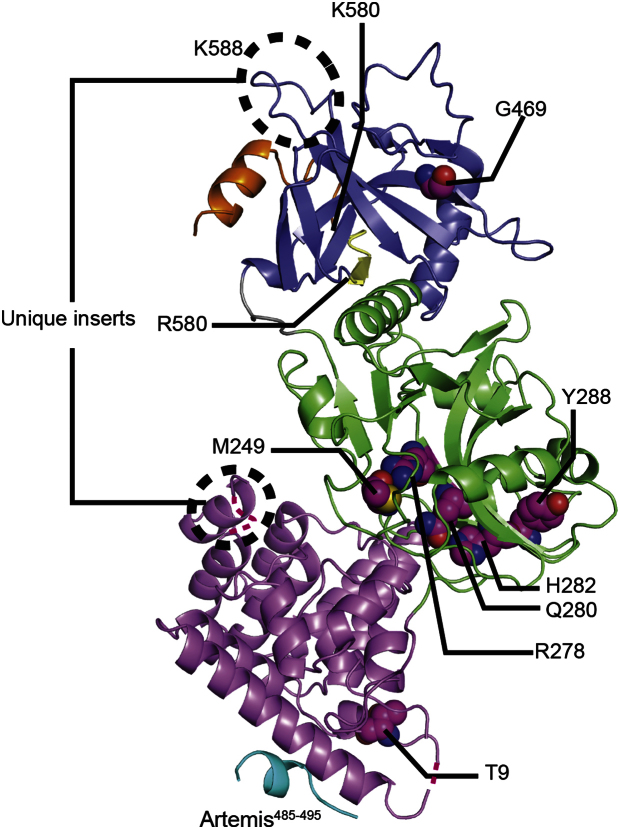
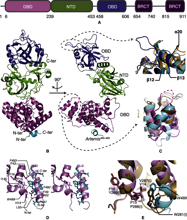
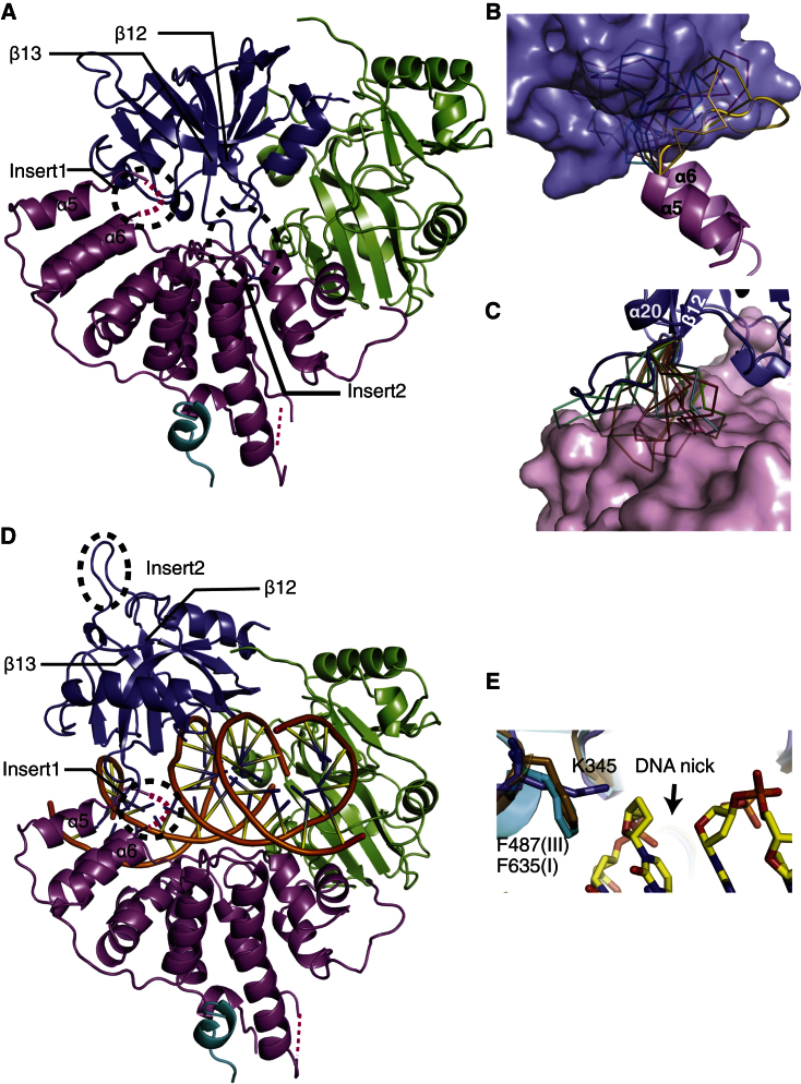
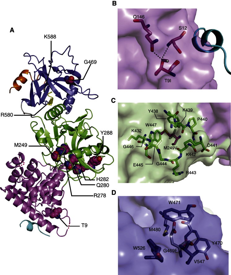
Nonhomologous end joining (NHEJ) is central to the repair of double-stranded DNA breaks throughout the cell cycle and plays roles in the development of the immune system. Although three-dimensional structures of most components of NHEJ have been defined, those of the catalytic region of DNA ligase IV (LigIV), a specialized DNA ligase known to work in NHEJ, and of Artemis have remained unresolved. Here, we report the crystal structure at 2.4 Å resolution of the catalytic region of LigIV (residues 1-609) in complex with an Artemis peptide. We describe interactions of the DNA-binding domain of LigIV with the continuous epitope of Artemis, which, together, form a three-helix bundle. A kink in the first helix of LigIV introduced by a conserved VPF motif gives rise to a hydrophobic pocket, which accommodates a conserved tryptophan from Artemis. We provide structural insights into features of LigIV among human DNA ligases.


Figures

None
Graphical abstract
Figure 1
Figure 1
Purification of LigIV Constructs (A) Profile of the UV absorbance at 280 nm during heparin affinity chromatography. The absorbance during heparin is shown in blue. Arrows indicate the ranges of fractions used for SDS-PAGE. (B) SDS-PAGE gel of fractions eluted from a heparin column. The molecular weight markers are in column “M” and the molecular weights (kDa) of the gel are shown on the left of the gel. Protein bands are indicated on the right of the gel: LigIV1–609 (residues 1–609 of LigIV), BRCT (residue 645–911 of LigIV) and XRCC4 (residue 1–213 of XRCC4, all cysteines of which are mutated to alanines). (C) Profile of the UV absorbance at 280 nm during size exclusion chromatography. The absorbance during size-exclusion chromatography is shown in red. (D) SDS-PAGE gel of fractions eluted from a Superdex 200 column. The molecular weight markers are identical to those shown in (B). Protein bands are indicated on the right of the gel. (E) SDS-PAGE gels of purified LigIV1–609. A total of 2 μg of both LigIV1–609 (left) and LigIV1–620 (right) are shown with their molecular weights (kDa). See also Figure S1.
Figure 2
Figure 2
Structure of the LigIV1–609/Artemis485–495 Complex (A) Schematic representation of the domains of LigIV. The same color scheme for the domains is used in all figures throughout this paper. (B) Overall structure of LigIV1–609. Missing parts are represented by dotted lines. (C) Unique inserts of LigIV1–609 and comparisons with LigI and LigIII. Top: an insert in OBD (blue) is shown. LigI and LigIII are shown in gold and cyan, respectively. Bottom: the difference in orientation of the α5 is shown. The loop between α5 and α6, shown as a pink dotted line, is disordered in the crystal structure of LigIV1–609. (D) Stereo image of LigIV1–609 with the Artemis485–495. The first 53 residues of LigIV1–609 (pink) are shown with Artemis485–495 (cyan). A disordered loop between α2 and α3 is represented by a gray dotted line. A hydrogen bond between D18 and W489 is shown by a black dotted line. Residues of LigIV1–609 are labeled in regular characters, whereas those of Artemis485–495 are in italic. (E) Conserved interaction motif VP(F/Y) in LigIV1–609 and LigI. The color scheme of LigIV1–609/Artemis485–495 is the same as in (D). LigI is shown in gold. Residues with “(I)” are from LigI. See also Figure S2.
Figure 3
Figure 3
Structural Models of Closed and DNA-Bound Conformations of LigIV1–609 (A) Model of the closed conformation of LigIV1–609. Insert1 (residues 111–121) and Insert2 (residues 490–494) are circled with black dotted lines. (B) Ten RapperTK models of Insert1. The modeled loop that has the least contact with OBD (surface presentation) is shown in cartoon representation; the others are displayed in ribbon representation. (C) Ten RapperTK models of Insert2. The modeled loop that has the least contact with DBD (surface presentation) is shown in cartoon representation; others are displayed in ribbon representations. (D) Model of the DNA-bound conformation of LigIV1–609. DNA is from the structure of human LigIII (PDB code 3L2P). (E) DNA-bound model of LigIV1–609 (blue) compared with the structures of LigI (gold) and LigIII (cyan). Residues shown are key residues interacting with the base sugar of the 3′ end of the DNA nick.
Figure 4
Figure 4
LIG4 Syndrome Mutations in the Structure of LigIV1–609 (A) Map of residues related to LIG4 syndrome. Residues, mutations of which cause LIG4 syndrome, are shown in a stick representation and magenta. The residues after R580 and R588 are colored in yellow and orange. (B) Mutation T9I. I9 (gray) is superimposed onto T9 (magenta). S12 and Q146 are residues having hydrogen bonds with T9. The Artemis peptide is shown in a cartoon representation. (C) Mutation M249V. V249 (gray) is superimposed onto M249 (magenta). (D) Mutation G469E. E469 (gray) is superimposed on G469.

References

    1. Adams P.D., Afonine P.V., Bunkóczi G., Chen V.B., Davis I.W., Echols N., Headd J.J., Hung L.W., Kapral G.J., Grosse-Kunstleve R.W. PHENIX: a comprehensive Python-based system for macromolecular structure solution. Acta Crystallogr. D Biol. Crystallogr. 2010;66:213–221. - PMC - PubMed
    1. Battye T.G., Kontogiannis L., Johnson O., Powell H.R., Leslie A.G. iMOSFLM: a new graphical interface for diffraction-image processing with MOSFLM. Acta Crystallogr. D Biol. Crystallogr. 2011;67:271–281. - PMC - PubMed
    1. Bryans M., Valenzano M.C., Stamato T.D. Absence of DNA ligase IV protein in XR-1 cells: evidence for stabilization by XRCC4. Mutat. Res. 1999;433:53–58. - PubMed
    1. Chistiakov D.A., Voronova N.V., Chistiakov A.P. Ligase IV syndrome. Eur. J. Med. Genet. 2009;52:373–378. - PubMed
    1. Cotner-Gohara E., Kim I.-K.K., Hammel M., Tainer J.A., Tomkinson A.E., Ellenberger T. Human DNA ligase III recognizes DNA ends by dynamic switching between two DNA-bound states. Biochemistry. 2010;49:6165–6176. - PMC - PubMed

Publication types