. 2013 Jul;9(7):7151-7.
doi: 10.1016/j.actbio.2013.03.019. Epub 2013 Mar 21.

In vivo response to dynamic hyaluronic acid hydrogels

Affiliations

In vivo response to dynamic hyaluronic acid hydrogels

Jennifer L Young et al. Acta Biomater. 2013 Jul.

Abstract

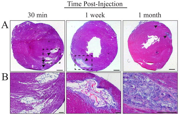
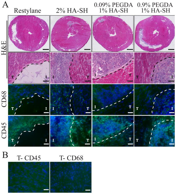
Tissue-specific elasticity arises in part from developmental changes in extracellular matrix over time, e.g. ~10-fold myocardial stiffening in the chicken embryo. When this time-dependent stiffening has been mimicked in vitro with thiolated hyaluronic acid (HA-SH) hydrogels, improved cardiomyocyte maturation has been observed. However, host interactions, matrix polymerization, and the stiffening kinetics remain uncertain in vivo, and each plays a critical role in therapeutic applications using HA-SH. Hematological and histological analysis of subcutaneously injected HA-SH hydrogels showed minimal systemic immune response and host cell infiltration. Most importantly, subcutaneously injected HA-SH hydrogels exhibited time-dependent porosity and stiffness changes at a rate similar to hydrogels polymerized in vitro. When injected intramyocardially host cells begin to actively degrade HA-SH hydrogels within 1week post-injection, continuing this process while producing matrix to nearly replace the hydrogel within 1month post-injection. While non-thiolated HA did not degrade after injection into the myocardium, it also did not elicit an immune response, unlike HA-SH, where visible granulomas and macrophage infiltration were present 1month post-injection, likely due to reactive thiol groups. Altogether these data suggest that the HA-SH hydrogel responds appropriately in a less vascularized niche and stiffens as had been demonstrated in vitro, but in more vascularized tissues, in vivo applicability appears limited.


Figures

Figure 1
Figure 1
Hematological cell counts of subcutaneously HA-injected (black) and non-injected rats (gray) for whole blood (top), monocytes (middle) and neutrophils (bottom) are shown. Values are post-injection at each indicated time point, were always taken prior to sample removal, and were normalized to pre-injection values. None of the subcutaneous HA-injected data was statistically different from non-injected rats, i.e. whole blood had p>0.06, monocytes had p > 0.14, and neutrophils had p > 0.16 as determined by student’s t-test. n = 4 for each group.
Figure 2
Figure 2
(top) Hematoxylin and eosin (H&E) stained sections of subcutaneously injected samples at 1 (left) and 14 (right) days post-injection. (middle) Immunohistochemical staining for CD45 (green) and Hoescht (blue) shows the presence of a limited number of inflammatory cells within and around the hydrogel (arrowheads). (bottom) Accompanying brightfield images for the middle panel of images. Scale bars are 500 μm for the top and 100 μm for the middle and bottom images. The dashed white line denotes the host tissue (T) and injection (I) regions.
Figure 3
Figure 3
Hematoxylin and eosin stained cross-sections of in vitro (left) and subcutaneously injected in vivo (right) samples are shown as a function of time. Scale bars are 500 μm.
Figure 4
Figure 4
Elastic modulus of subcutaneously injected HA (gray) compared to in vitro (black) on a log-log plot. Data points are fitted with a power law: y=xA, where A=0.36 in vivo and A=0.32 in vitro. Student’s t-tests analysis indicates that these data are not statistically different (p>0.97). In vitro and in vivo samples had n = 3 and 6, respectively.
Figure 5
Figure 5
(A) HA-SH hydrogel in myocardium 30 min (left), 1 week (middle) and 1 month (right) post-injection (indicated by arrowheads in top panel). (B) Images of the area indicated by the black dashed box have been enlarged. 1 month post-injection shows material degradation and cell infiltration (3). Scale bars in panels A and B are 500 and 250 μm, respectively.
Figure 6
Figure 6
(A) Histological sections of hearts injected with Restylane® (left), 2% HA-SH hydrogels (left center), 0.09% PEGDA/1% HA-SH hydrogel (right center), and 0.9% PEGDA/1% HA-SH (original formulation, right) are shown 3 weeks post-intramyocardial injection. For H&E stained sections, dashed box in the top row (scale bar is 2000 μm) indicates the enlarged area depicted in the second row (scale bar is 100 μm). Immunohistochemical staining 3 weeks post-intramyocardial injection for CD68 (green, third row) and CD45 (green, fourth row) show macrophages and lymphocytes, respectively, as well as other invasive cells (Hoescht-labeled nuclei in blue) present within and around the hydrogels as indicated. The dashed line separates the injection (I) from surrounding host tissue (T). (B) Immunohistochemical staining for CD45 (left) and CD68 (right) in non-injected regions of the myocardium. Image intensity is scaled according to the injected images in panel A. Scale bars are 50 μm for all immunofluorescent images.

References

    1. Tse JR, Engler AJ. Stiffness gradients mimicking in vivo tissue variation regulate mesenchymal stem cell fate. PLoS One. 2011;6:e15978. - PMC - PubMed
    1. Peyton SR, Putnam AJ. Extracellular matrix rigidity governs smooth muscle cell motility in a biphasic fashion. J Cell Physiol. 2005;204:198–209. - PubMed
    1. Zaari N, Rajagopalan P, Kim SK, Engler AJ, Wong JY. Photopolymerization in microfluidic gradient generators: Microscale control of substrate compliance to manipulate cell response. Advanced Materials. 2004;16:2133–7.
    1. Rowlands AS, George PA, Cooper-White JJ. Directing osteogenic and myogenic differentiation of MSCs: interplay of stiffness and adhesive ligand presentation. Am J Physiol Cell Physiol. 2008;295:C1037–44. - PubMed
    1. Huebsch N, Arany PR, Mao AS, Shvartsman D, Ali OA, Bencherif SA, et al. Harnessing traction-mediated manipulation of the cell/matrix interface to control stem-cell fate. Nat Mater. 2010;9:518–26. - PMC - PubMed

Publication types

MeSH terms

LinkOut - more resources