. 2013 Apr;20(4):426-32.
doi: 10.1038/nsmb.2529. Epub 2013 Mar 24.

Evidence for monomeric actin function in INO80 chromatin remodeling

Affiliations

Evidence for monomeric actin function in INO80 chromatin remodeling

Prabodh Kapoor et al. Nat Struct Mol Biol. 2013 Apr.

Abstract

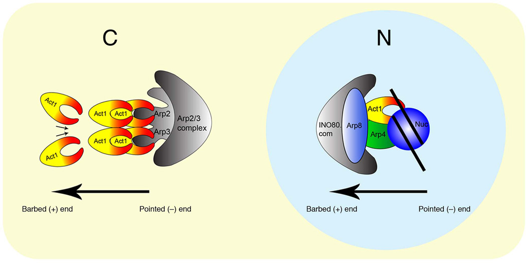
Actin has well-established functions in the cytoplasm, but its roles in the nucleus remain poorly defined. Here, by studying the nuclear actin-containing yeast INO80 chromatin remodeling complex, we provide genetic and biochemical evidence for a role of monomeric actin in INO80 chromatin remodeling. We demonstrate that, in contrast to cytoplasmic actin, nuclear actin is present as a monomer in the INO80 complex, and its barbed end is not accessible for polymerization. We identify an actin mutation in subdomain 2 affecting in vivo nuclear functions and reducing the chromatin remodeling activity of the INO80 complex in vitro. Notably, the highly conserved subdomain 2 at the pointed end of actin contributes to the interaction of INO80 with chromatin. Our results establish an evolutionarily conserved function of nuclear actin in its monomeric form and suggest that nuclear actin can utilize a fundamentally distinct mechanism from that of cytoplasmic actin.


Figures

Figure 1
Figure 1. Analysis of the actin subunit in INO80 complex
(a) Top shows a schematic representation of the Ino80 ATPase with the split ATPase-helicase domain (shaded) and the N-terminal region in black. Bottom shows the amino acid sequence alignment of the TELY motifs within the N-terminal region of the Ino80 ATPases from yeast (Ino80), Drosophila Ino80 and human Ino80. The conserved amino acids TELY is underlined. Region of HSA domain is marked above the N-terminal region (amino acid 496-588 from the N-terminus of Ino80). (b,c) SDS-PAGE and silver staining showing, (b) wild type and ∆TELY INO80 complexes, and (c) wild type INO80 and N-terminal (N.com) complexes. Relevant subunits are shown on the right. (d,e) Phenotypic analysis of (d) ino80 mutants lacking the N-terminal region (∆N) or the TELY motif (∆TELY) on YEPD (30°C), HU (100 mM, 30 °C) and YEPD (37 °C) plates, and (e) actin mutants on YEPD, HU (100 mM) plates at 30 °C and on a YEPD plate at 37 °C. (f) Schematic representation of the structure of actin. The four subdomains of actin are indicated by S1, S2, S3 and S4. The positions of actin mutations described in this study are indicated by boxes labeled with allele numbers: 2 for act1-2 (A58T), 1 for act1-1 (P32A), 101 for act1-101 (D363A E364A) and 136 for act1-136 (D2A). Pointed (−) end region and barbed (+) end region are marked respectively. Adapted from Kabsch et al., (1990) with permission. (g) Northern blot analysis of PHO5 and ACT1 expression in wild type (WT) and mutant strains at 30 °C after 4 hours of induction, using RPL3 as loading control.
Figure 2
Figure 2. Actin contributes to INO80 chromatin remodeling
(a) SDS-PAGE and silver staining showing wild type (WT) and mutant (act1-2) INO80 complexes. (b) Native PAGE showing 359 bp INO1 DNA (5 nM) in the presence of increasing equimolar concentrations of WT and act1-2 INO80 complexes from 2 nM to 20 nM at 30 °C. (c,d) Graph showing (c) % DNA bound to WT and act1-2 INO80 complexes as shown in (b), data presented is the mean of five independent experiments ± S.D., (d) Kd values for mono-nucleosome binding of WT and act1-2 INO80 complexes using nucleosomes with linker DNA (207 Nuc), assessed using gel shift assay, data presented is the mean of five independent experiments ± S.D. (e) Native PAGE showing nucleosome mobilization by wild type and mutant INO80 complexes (1× equals 5 nM) at 30 °C using the INO1 mono-nucleosome substrate (5.8 nM). Chromatin remodeling by the INO80 complex is indicated by the reduction of N3 band intensity and the increase in N1, N2 band intensity, which is represented by an increase in (N1+N2)/N3 ratios (bottom), ± S.D. from three independent experiments. (f) Graphical representation for mono-nucleosome mobilization as shown in (e), data presented is the mean of three independent experiments ± S.D. (g) SDS-PAGE and deep purple staining of the INO80 complex (1× equals 10 ng actin). Numbers on the left indicate the stoichiometry of labeled subunits with the actin subunit normalized as 1. Note that the Rvb1 and Rvb2 helicases co-migrate as a single band - Rvb1/2. (h) Northern blot analysis of PHO5 expression in wild type (WT) and mutant strains at 30 °C after 3 hours of induction, and at 37 °C after 1.5 hours of induction. RPL3 is a loading control.
Figure 3
Figure 3. Actin in INO80 complex exists in a unique microenvironment
(a) Schematic representation of the structure of actin as described in Fig. 1f. The position of the C4 epitope (Asp24 and Asp25) is now indicated in a box labeled with C4. DNase I binding and profilin binding regions are indicated by lines which represents the pointed (−) end region and barbed (+) end region. (b) Dot blot for actin and INO80 complex probed with antibodies as indicated on the right. (c) SDS-PAGE and silver staining of actin-profilin and INO80 complexes purified from cells grown at 30 °C. (d) Dot blot for INO80, and actin-profilin complexes probed with anti-C4 antibodies and fluorescently labeled DNase I as indicated. 1× refers to actin equivalent of the spotted complexes. (e) Western blot analysis of whole cell extracts using the monoclonal anti-C4 actin antibodies. Coomassie staining indicates equal loading of whole cell extracts. (f) Dot blot for INO80 complexes probed with anti-C4 antibodies and fluorescently labeled DNase I as indicated. (g,h) Top panel shows dot blot analysis for INO80 complexes purified from wild type and act1-2 mutant cells probed with anti-C4 antibodies and fluorescently labeled DNase I as indicated. Bottom panel shows equal loading of the complexes in the top panel, as judged by silver staining.
Figure 4
Figure 4. Actin subdomain 2 is crucial for INO80 interaction with chromatin
(a,b) Western blot analysis of, (a) purified INO80-chromatin complex in presence and absence of MNase after complete subtilisin digestion, probed with polyclonal anti-β-actin antibodies, (b) subtilisin accessibility to actin subdomain 2 in purified INO80 complex without its substrate chromatin, in presence or absence of 147 Nuc or 207 Nuc. (c) SDS-PAGE and silver staining for INO80 complex (lane I), and INO80: DNase I complex (lane II). (d) Graph showing relative DNA or nucleosome (359 bp INO1) dependent ATPase activity of INO80: DNase I compared to INO80 complex using equimolar amount of both the complexes and data presented is the mean of five independent experiments ± S.D.
Figure 5
Figure 5. A model for nuclear actin in INO80 complex
A model showing unique positioning of actin in the INO80 complex in the nucleus compared to the positioning of actin with Arp2/3 complex in the cytoplasm. Whereas the barbed end of actin with Arp2/3 complex is free to polymerize, the barbed end of actin with INO80 complex in the nucleus is masked by other subunits and the pointed end is engaged with chromatin.

Comment in

References

    1. Bettinger BT, Gilbert DM, Amberg DC. Actin up in the nucleus. Nat Rev Mol Cell Biol. 2004;5:410–415. - PubMed
    1. Blessing CA, Ugrinova GT, Goodson HV. Actin and ARPs: action in the nucleus. Trends Cell Biol. 2004;14:435–442. - PubMed
    1. Olave IA, Reck-Peterson SL, Crabtree GR. Nuclear actin and actin-related proteins in chromatin remodeling. Annu Rev Biochem. 2002;71:755–781. - PubMed
    1. Shumaker DK, Kuczmarski ER, Goldman RD. The nucleoskeleton: lamins and actin are major players in essential nuclear functions. Curr Opin Cell Biol. 2003;15:358–366. - PubMed
    1. Clark TG, Merriam RW. Diffusible and bound actin nuclei of Xenopus laevis oocytes. Cell. 1977;12:883–891. - PubMed

Methods-only references

    1. Shen X. Preparation and analysis of the INO80 complex. Methods Enzymol. 2004;377:401–412. - PubMed
    1. Winkler DD, Muthurajan UM, Hieb AR, Luger K. Histone chaperone FACT coordinates nucleosome interaction through multiple synergistic binding events. J Biol Chem. 2011;286:41883–41892. - PMC - PubMed
    1. Clarke AS, Lowell JE, Jacobson SJ, Pillus L. Esa1p is an essential histone acetyltransferase required for cell cycle progression. Mol Cell Biol. 1999;19:2515–2526. - PMC - PubMed

Publication types