. 2013 Apr;34(4):522-30.
doi: 10.1038/aps.2012.208. Epub 2013 Mar 25.

Astragaloside II triggers T cell activation through regulation of CD45 protein tyrosine phosphatase activity

Affiliations

Astragaloside II triggers T cell activation through regulation of CD45 protein tyrosine phosphatase activity

Chun-ping Wan et al. Acta Pharmacol Sin. 2013 Apr.

Abstract

Aim: To investigate the immunomodulating activity of astragalosides, the active compounds from a traditional tonic herb Astragalus membranaceus Bge, and to explore the molecular mechanisms underlying the actions, focusing on CD45 protein tyrosine phosphatase (CD45 PTPase), which plays a critical role in T lymphocyte activation.

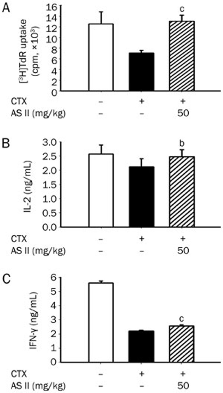
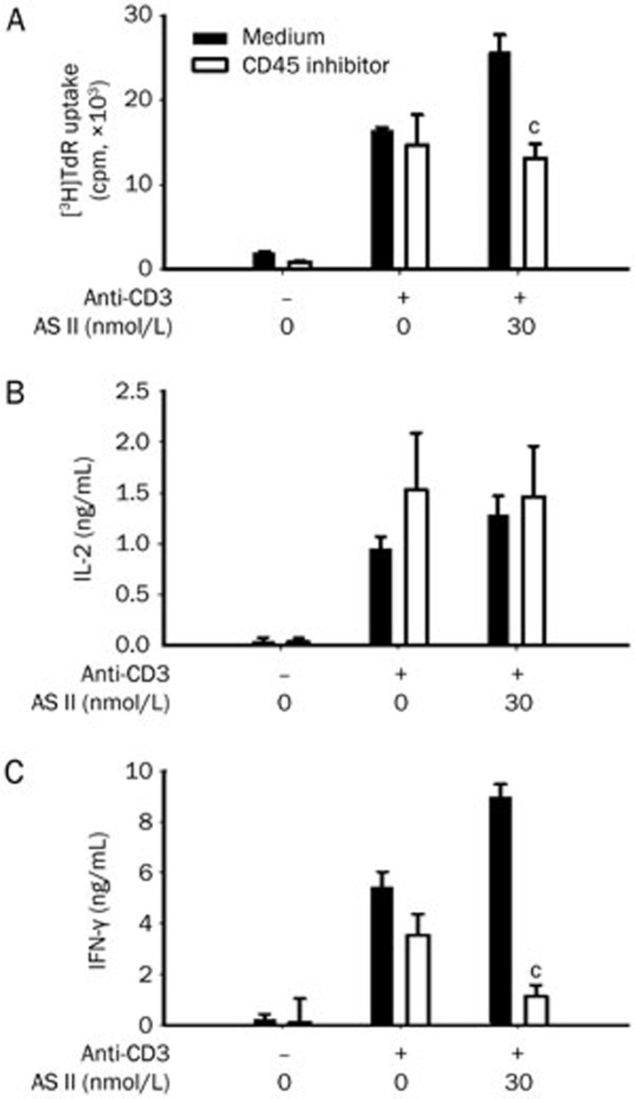
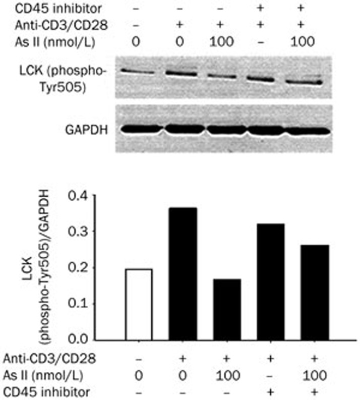
Methods: Primary splenocytes and T cells were prepared from mice. CD45 PTPase activity was assessed using a colorimetric assay. Cell proliferation was measured using a [(3)H]-thymidine incorporation assay. Cytokine proteins and mRNAs were examined with ELISA and RT-PCR, respectively. Activation markers, including CD25 and CD69, were analyzed using flow cytometry. Activation of LCK (Tyr505) was detected using Western blot analysis. Mice were injected with the immunosuppressant cyclophosphamide (CTX, 80 mg/kg), and administered astragaloside II (50 mg/kg).

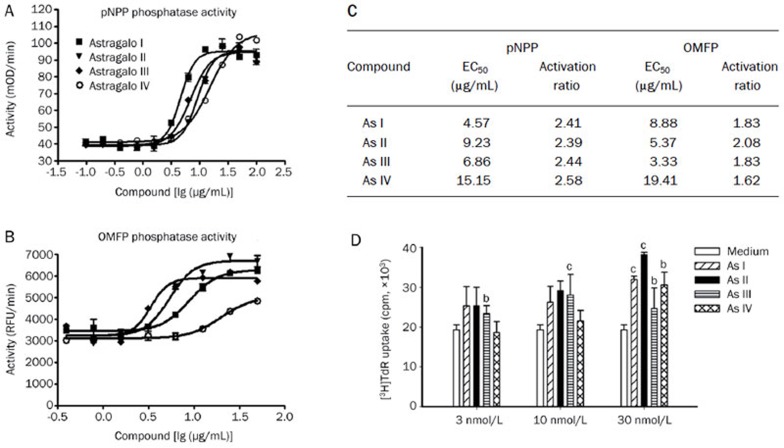
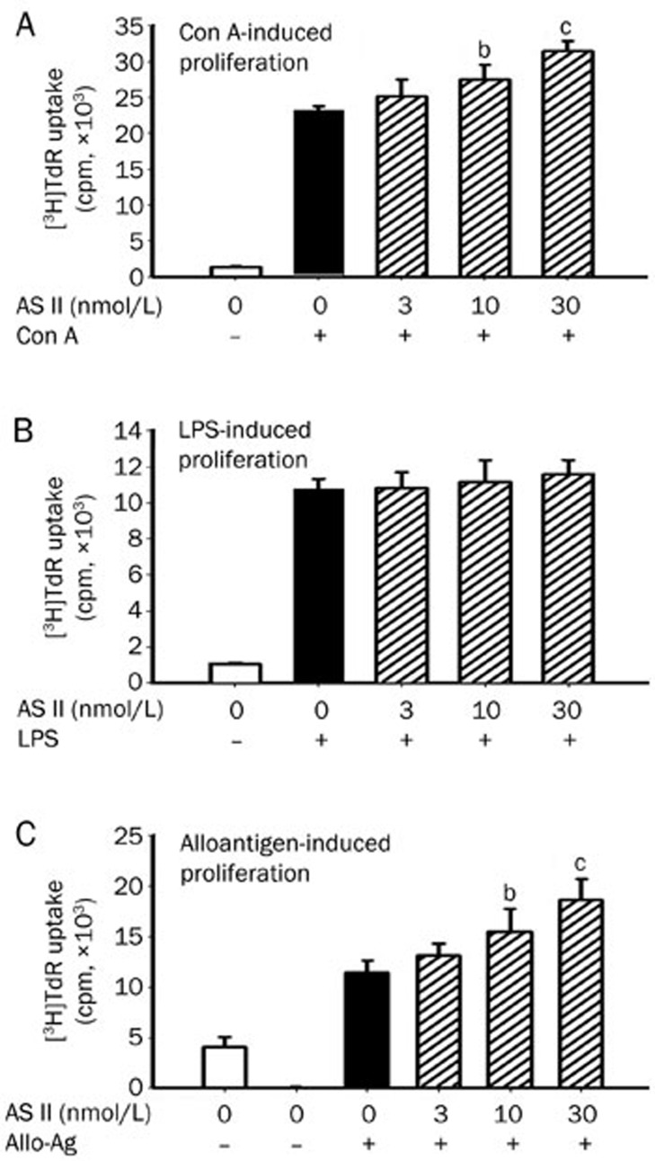
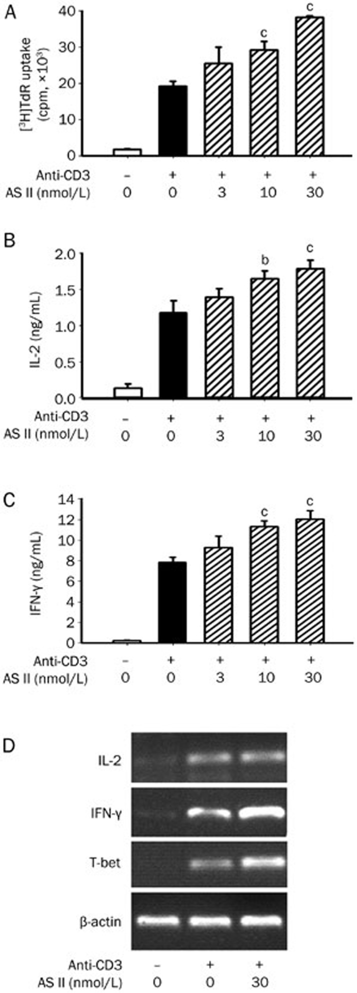
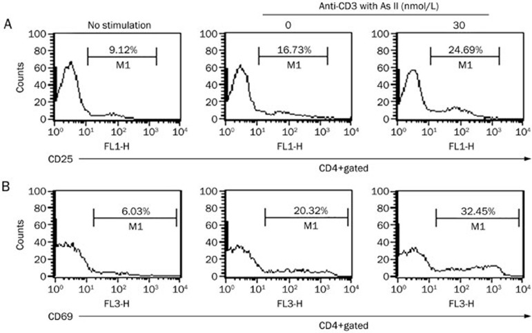
Results: Astragaloside I, II, III, and IV concentration-dependently increased the CD45-mediated of pNPP/OMFP hydrolysis with the EC50 values ranged from 3.33 to 10.42 μg/mL. Astragaloside II (10 and 30 nmol/L) significantly enhanced the proliferation of primary splenocytes induced by ConA, alloantigen or anti-CD3. Astragaloside II (30 nmol/L) significantly increased IL-2 and IFN-γ secretion, upregulated the mRNA levels of IFN-γ and T-bet in primary splenocytes, and promoted CD25 and CD69 expression on primary CD4(+) T cells upon TCR stimulation. Furthermore, astragaloside II (100 nmol/L) promoted CD45-mediated dephosphorylation of LCK (Tyr505) in primary T cells, which could be blocked by a specific CD45 PTPase inhibitor. In CTX-induced immunosuppressed mice, oral administration of astragaloside II restored the proliferation of splenic T cells and the production of IFN-γ and IL-2. However, astragaloside II had no apparent effects on B cell proliferation.

Conclusion: Astragaloside II enhances T cell activation by regulating the activity of CD45 PTPase, which may explain why Astragalus membranaceus Bge is used as a tonic herb in treating immunosuppressive diseases.


Figures

Figure 1
Figure 1
AstragalosideI, II, III, and IV significantly enhanced CD45 phosphatase activity towards the substrates pNPP (A) and OMFP (B). CD45 activity was detected by monitoring the dephosphorylation of pNPP and OMFP, and the EC50 value (C) was also analyzed. The lymphocyte proliferation assay was used to investigate anti-CD3-stimulated splenocyte proliferation (D). The results are expressed as the mean±SEM of three independent experiments. bP<0.05, cP<0.01 vs the medium group. The EC50 is the concentration of compound required to increase enzyme activity by 50%. The activation ratio was calculated as Top Value/Bottom Value.
Figure 2
Figure 2
Astragaloside II enhanced the splenocyte proliferation induced by ConA, LPS, and alloantigen treatment. BALB/c mouse splenocytes (4×10⁵ cells/well) were stimulated with ConA (1 μg/mL) (A), LPS (5 μg/mL) (B), or irradiated C57BL/6 splenocytes (1:1) (C) for 48 h or 96 h, in the absence or presence of astragaloside II. Cells were plated in triplicate in a 96-well plate. The results are presented as the mean±SEM. n=3. bP<0.05, cP<0.01 vs the control group.
Figure 3
Figure 3
Administration of astragaloside II promoted recovery of splenic T cell function in immunosuppressed mice. BALB/c mice received and intraperitoneal injection (ip) of CTX (80 mg/kg) on d 1 and d 4; astragaloside II (50 mg/kg) was given orally for 8 d (5 mice per group). Splenocytes (4×10⁵ cells/well) from each group were incubated with ConA (1 μg/mL) in triplicate for 24 h. (A) A proliferation assay. An ELISA was used to detect IL-2 (B) and IFN-γ (C) in supernatants. Results presented are the mean±SEM. n=3. bP<0.05, cP<0.01 vs CTX-treated mice orally administered saline vehicle.
Figure 4
Figure 4
Astragaloside II significantly promoted anti-CD3-stimulated splenocyte activation. (A) For the proliferation assay, BALB/c splenocytes (4×10⁵ cells/well) were cultured in a plate that was coated with anti-CD3 (5 μg/mL) in the absence or presence of astragaloside II for 48 h. For cytokine measurements, the culture supernatants were harvested at 36 h to measure the IL-2 (B) and IFN-γ (C) levels by ELISA. IL-2, IFN-γ, and T-bet expression in splenocytes at the mRNA level were examined by RT-PCR after 16 h of stimulation (D). The results are expressed as the mean±SEM of three independent experiments. bP<0.05, cP<0.01 vs control group.
Figure 5
Figure 5
Astragaloside II enhanced the expression of activation markers on CD4+ T cells. BALB/c splenocytes (4×10⁶ cell/well) were cultured in 24-well flat-bottom plates that were coated with anti-CD3 (5 μg/mL) in the absence or presence of astragaloside II (30 nmol/L) for 36 h. Subsequently, CD25 (A) and CD69 (B) expression on CD4+ T cells were analyzed by flow cytometry. The result presented here is representative of three individual experiments.
Figure 6
Figure 6
Astragaloside II triggered T cell activation by regulating the activity of CD45 PTPase. Splenocytes (4×10⁵ cells/well) were stimulated with anti-CD3 (5 μg/mL) in the absence or presence of the CD45 PTPase inhibitor (1.5 μmol/L). The proliferative response (A) and cytokine level of IL-2 (B) and IFN-γ (C) were detected after 48 h or 36 h. The results are expressed as the mean±SEM of three independent experiments. cP<0.01 vs medium control.
Figure 7
Figure 7
Astragaloside II facilitated CD45-mediated LCK dephosphorylation. Purified primary T cells were pretreated with astragaloside II (100 nmol/L) in the absence or presence of CD45 PTPase inhibitor (1.5 μmol/L) for 2 h, followed by anti-CD3 stimulation (5 μg/mL) and anti-CD28 (2 μg/mL) for 30 min. The cells were lysed and assayed for LCK (phospho-Tyr505) phosphorylation and GAPDH by western blotting assay. The result presented here is representative of three individual experiments.

References

    1. Bedir E, Calis I, Aquino R, Piacente S, Pizza C. Cycloartane triterpene glycosides from the roots of Astragalus brachypterus and Astragalus microcephalus. J Nat Prod. 1998;61:1469–72. - PubMed
    1. Yesilada E, Bedir E, Caliş I, Takaishi Y, Ohmoto Y. Effects of triterpene saponins from Astragalus species on in vitro cytokine release. J Ethnopharmacol. 2005;96:71–7. - PubMed
    1. Wang YP, Li XY, Song CQ, Hu ZB. Effect of astragaloside IV on T, B lymphocyte proliferation and peritoneal macrophage function in mice. Acta Pharmacol Sin. 2002;23:263–6. - PubMed
    1. Nalbantsoy A, Nesil T, Erden S, Calış I, Bedir E. Adjuvant effects of Astragalus saponins macrophyllosaponin B and astragaloside VII. J Ethnopharmacol. 2011;134:897–903. - PubMed
    1. Nalbantsoy A, Nesil T, Yılmaz-Dilsiz O, Aksu G, Khan S, Bedir E. Evaluation of the immunomodulatory properties in mice and in vitro anti-inflammatory activity of cycloartane type saponins from Astragalus species. J Ethnopharmacol. 2012;139:574–81. - PubMed

Publication types

MeSH terms