. 2014 Jan 2;33(1):116-28.
doi: 10.1038/onc.2013.96. Epub 2013 Mar 25.

Generation of cancer stem-like cells through the formation of polyploid giant cancer cells

Affiliations

Generation of cancer stem-like cells through the formation of polyploid giant cancer cells

S Zhang et al. Oncogene. .

Erratum in

  • Oncogene. 2014 Jan 2;33(1):134

Abstract

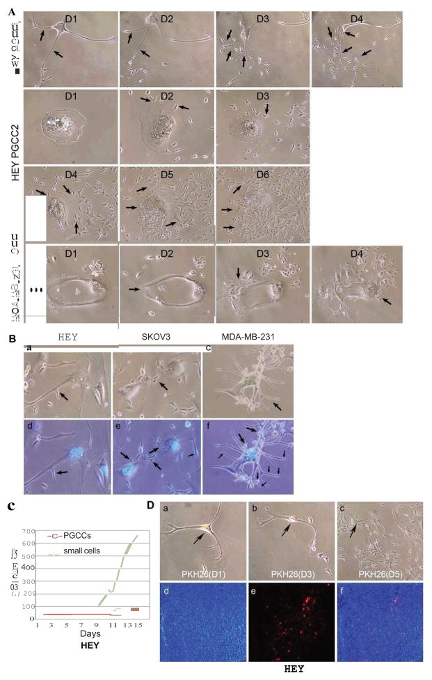
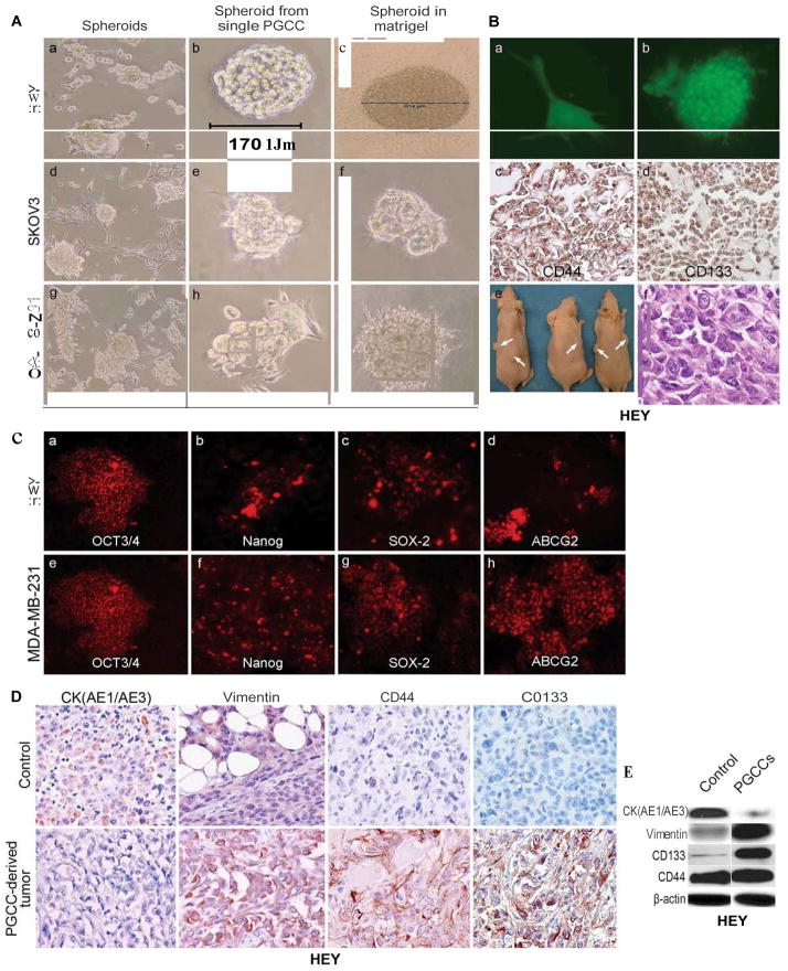
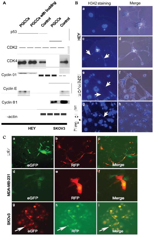
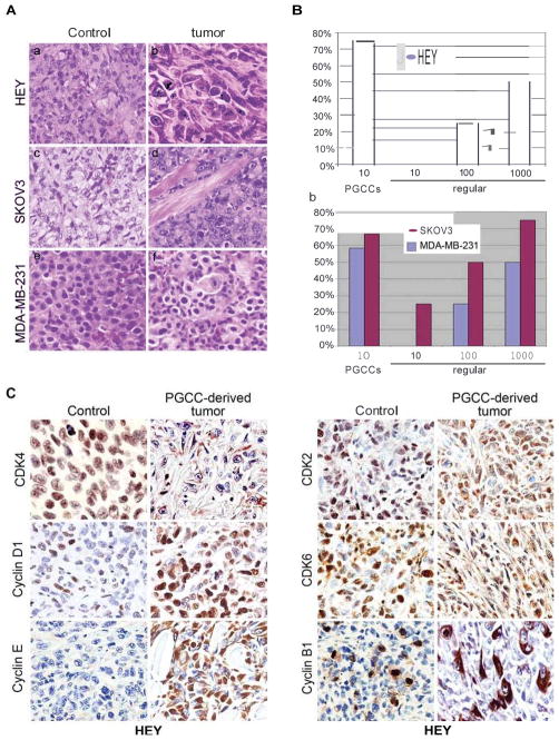
Polyploid giant cancer cells (PGCCs) have been observed by pathologists for over a century. PGCCs contribute to solid tumor heterogeneity, but their functions are largely undefined. Little attention has been given to these cells, largely because PGCCs have been generally thought to originate from repeated failure of mitosis/cytokinesis and have no capacity for long-term survival or proliferation. Here we report our successful purification and culture of PGCCs from human ovarian cancer cell lines and primary ovarian cancer. These cells are highly resistant to oxygen deprivation and could form through endoreduplication or cell fusion, generating regular-sized cancer cells quickly through budding or bursting similar to simple organisms like fungi. They express normal and cancer stem cell markers, they divide asymmetrically and they cycle slowly. They can differentiate into adipose, cartilage and bone. A single PGCC formed cancer spheroids in vitro and generated tumors in immunodeficient mice. These PGCC-derived tumors gained a mesenchymal phenotype with increased expression of cancer stem cell markers CD44 and CD133 and become more resistant to treatment with cisplatin. Taken together, our results reveal that PGCCs represent a resistant form of human cancer using an ancient, evolutionarily conserved mechanism in response to hypoxia stress; they can contribute to the generation of cancer stem-like cells, and also play a fundamental role in regulating tumor heterogeneity, tumor growth and chemoresistance in human cancer.


Conflict of interest statement

Conflict of interest

The authors declare no conflict of interest.

Figures

Figure 1
Figure 1
A. Morphologic characteristics of regular cells and PGCCs from HEY, SKOv3 and MDA-MB-231 before and after CoCl2 treatment. PGCCs were mixed with regular-sized cells before CoCl2 treatment (Black arrows) (10×). B. Flow cytometry analysis before and after CoCl2 treatment. C. X chromosome fluorescence in situ hybridization of PGCCs and diploid control HEY cells (a). D. β-galactosidase staining. No staining was observed in HEY (b), SKOv3 (c) and MDA-MB-231 (d) PGCCs compared with positive staining from senescent fibroblasts (a) (10×). E. Western blot analysis of CD133, ElF-2α and HIF-1α expression in HEY and SKOv3 PGCCs, and control cells.
Figure 2
Figure 2
A. Patterns of cell division of HEY and MDA-MB-231 PGCCs. HEY and MDA-MB-231 PGCC generated small-sized daughter cells via budding and bursting (black arrows) over a 4-day (HEY PGCC1 and MDA-MB-231) and 6-day period (HEY PGCC2) (10×). B. Nuclear morphology of HEY, SKOv3 and MDA-MB-231 PGCCs with budding daughter cells (black arrows) stained with Hoechst 33342. (a and d) Giant Multinucleated cells of HEY PGCCs and budding daughter cells (10×). (b and e) Budding daughter cells from SKOv3 PGCCs (10×). (c and f) Giant nucleated cells of MDA-MB-231 PGCCs and budding daughter cells (10×). C. Cell counts from HEY PGCCs. The total number of small-sized cells and PGCCs were counted for 15 days in a T25 flask after treatment with 450 μM CoCl2 for 72 hours. D. Slow-cycling nature of HEY PGCCs. (a–c) Single HEY PGCCs stained with PKH26 for 5 days (10×). (d–f) PKH26 fluorescence was detected in some PGCCs but not daughters in xenografts from HEY PGCCs (10×).
Figure 3
Figure 3
A. Formation of spheroids from PGCCs. (a, d, and g) Spheroids formed from HEY, SKOv3 and MDA-MB-231 PGCCs in stem cell media after the removal of CoCl2 (10×). Please refer Fig. 1A for control pictures of PGCCs in the presence of CoCl2. (b, e and h). Growth of spheroids from a single HEY, SKOv3 and MDA-MB-231 PGCCs in stem cell medium (10×). (c, f and i) Growth of spheroids from a single HEY, SKOv3 or MDA-MB-231 PGCC in Matrigel (10×). B. Tumor formation from single spheroid derived from a single PGCC. (a) Growth of a single eGFP-labeled HEY PGCC in Matrigel (10×). (b) Growth of spheroids from the single PGCC of (a) (10×). (c and d) Representative pictures of immunohistochemical staining against CD44 (c) and CD133 (d) on sections from the spheroids of (b) (10×). (e) A single spheroid (6/6) can form a tumor in mice. (f) H&E staining showed the morphologic characteristics of tumor from (e) (20×). C. Expression of stem cell markers in cancer spheroids. (a–h) HEY and MDA-MB-231 spheroids stained with antibodies against OCT3/4 (a and e), Nanog (b and f), SOX-2(c and g), and ABCG2 (d and h) (10×). D. Expression of cytokeratin (AE1/AE3), vimentin, CD44 and CD133 in tumors derived from control HEY cells and a single spheroid (20×). E. Western blot analysis of vimentin, CK (AE1/AE3), CD133, and CD44 in control HEY and PGCCs.
Figure 4
Figure 4
A. Western blot analysis of protein expression related with cell cycle. The total proteins in purified PGCCs, PGCCs with 70% budding cells, control HEY and SKOv3 PGCCs, control SKOv3 cancer cells were extracted and subject for western blot analysis. B. Visualization of DNA transport via branches in PGCCs after staining with Hoechst 33342. (a and b) Lack of DNA was indicated by an absence of H342staining in the HEY PGCC branches at 8 hours after trypsin digestion (10×). (c and d) DNA was reappearance in the branches of PGCCs at 4th days after trypsin digestion (white arrow points, 10×). (e and f) DNA transportation in the branches from MDA-MB-231 PGCCs (white arrow heads) (H342staining, 10×). (g and h) Giant nucleated cells of PGCCs and DNA transportation in the branches from human ovarian cancer primary culture (white arrow heads) (H342staining, 10×). C. Generation of PGCCs via cell fusion as indicated by yellow fluorescence (10×). (a–c) Formation of HEY PGCCs fused by regular HEY cells labeled with eGFP and regular HEY cells labeled RFP (10×). (d–f). Formation of MDA-MB-231 PGCCs fused by regular MDA-MB-231 cells labeled with eGFP and regular MDA-MB-231 cells labeled RFP (10×). (g–i) Formation of SKOv3 PGCCs fused by regular SKOv3 cells labeled with eGFP and regular SKOv3 cells labeled RFP (10×).
Figure 5
Figure 5
Morphology of tumors derived from 1×10⁶ regular HEY (a) and 10 HEY PGCCs (b), 1×10⁶ regular SKOv3 (c) and a single SKOv3 PGCC (d), 1×10⁶ regular MDA-MB-231(e) and a single MDA-MB-231 PGCC (f). B. Comparison of tumor formation ability (a) Comparison of tumor formation ability between 10, 100, or 1000 regular HEY and 10 HEY PGCCs; (b) Comparison of tumor formation ability between 10,100, or 1000 regular SKOv3 and MDA-MB-231 and a single SKOv3 and MDA-MB-231 PGCC. C. Expression of cell cycle-related proteins in xenograft. Immunohistochemical stainings performed on tumor tissues derived from PGCCs and regular HEY cells with antibodies against proteins involved the cell cycle regulation (20×).
Figure 6
Figure 6
A. Adipose differentiation of HEY PGCCs in vitro and in vivo. Staining of regular HEY cell and PGCCs with oil red O (a and b) or FABP4 (c and d) following incubation with adipogenesis medium. (e) Histopathology of PGCC-derived xenograft tumors (H&E, 20×) and immunohistochemical staining against FABP4 (f). (g and h) immunohistochemical staining against human-specific vimentin (g:10× and h:20×). B. Cartilage differentiation of HEY PGCCs in vitro. (a) Regular HEY cells cultured in chondrogenesis medium (10×). (b) Formation of chondrogenic pellets from PGCCs cultured in chondrogenesis medium (10×). Chondrogenic pellets stained with (c) Alcian blue and (d) Alcian blue/PAS (10×). C. Cartilage and bone differentiation in vivo. (a) Cartilage-like tumor near the rib cage in nude mice. (b)Bony differentiation on sections stained with H&E (10×), (c) Safranin red and fast green (10×), and (d) anti-eGFP (10×). (e) Formation of loose tumor bodies in the abdomen following injection of chondrogenic pellets. (f) Histopathology of the loose tumor bodies (10×). (g) Cartilage differentiation (black arrow heads, 10×). (h) Bony differentiation revealed by anti-osteopontin staining (10×). D. Osteogenesis, chondrogenesis and adipose differentiation of MDA-MB-231 PGCCs. (a) Chondrogenic pellets of MDA-MB-231 PGCCs cultured with chondrogenesis medium (10×). Chondrogenic pellets stained with (b) Alcian blue and (c) Alcian blue/PAS (10×). (d) Formation of loose tumor bodies following abdominal injection of pellets. (e) Computed tomography image showing osteogenesis differentiation (white arrow heads). (f) Calcification of tumor derived from MDA-MB-231 PGCCs (black arrow heads) (10×). (g) Osteoid differentiation revealed by anti-osteopontin staining in loose bodies (10×). (h) Adipose differentiation revealed by human specific anti-vimentin staining in loose bodies (20×).
Figure 7
Figure 7
A. PGCCs in human ovarian cancers (black arrows). (a) Normal fallopian tube (20×). (b) Cystadenoma (20×). (c) PGCCs in human high-grade serous ovarian cancinoma (20×). (d) PGCCs in metastatic ovarian cancer (20×). B. Primary culture of human ovarian cancer and PGCC-derived xenograft. (a) Histology of human ovarian cancer used for primary culture. PGCCs indicated by black arrows (20×). (b) PGCCs (large arrow heads) mixed with regular cancer cells (small arrow heads) in the primary culture of (a) (10×). (c) Primary culture of mice xenografted tumor from 50 PGCCs of (b) injection showed PGCCs (large arrows) and regular cancer cells (small arrows) (10×). (d) The recovered PGCCs have numerous small branches (budding daughter cells) after CoCl2 treatment (10×). (e) Hoechst 33342 staining showed the multinucleated PGCCs and budding daughter cells (black arrows point) (10×). (f) Formation of spheroids from (d) in stem cell medium (10×). C. Cyclin B1 immunohistochemical staining in human ovarian cancers. (a) Normal human ovarian cysts negative for cyclin B1 expression (20×). (b) Nuclear expression in low-grade ovarian serous carcinoma (20×). (c) Cyclin B1 was expressed in the cytoplasm of giant nucleated cells from high-grade serous carcinoma (20×) and (d) metastatic ovarian cancer (20×).

References

    1. Heppner GH. Tumor heterogeneity. Cancer Res. 1984;44:2259–2265. - PubMed
    1. Marusyk A, Almendro V, Polyak K. Intra-tumour heterogeneity: a looking glass for cancer? Nat Rev Cancer. 2012;12:323–334. - PubMed
    1. Kumar V, Abbas AL, Fausto N, Aster JC. Robbins and Cotran Pathologic Basis of of Disease. 8. 2010. pp. 262–270.
    1. Malpica A, Deavers MT, Lu K, Bodurka DC, Atkinson EN, Gershenson DM, et al. Grading ovarian serous carcinoma using a two-tier system. Am J Surg Pathol. 2004;28:496–504. - PubMed
    1. Geigl JB, Obenauf AC, Schwarzbraun T, Speicher MR. Defining ‘chromosomal instability’. Trends Genet. 2008;24:64–69. - PubMed

Publication types

MeSH terms