. 2013 Apr 17;32(8):1103-14.
doi: 10.1038/emboj.2013.62. Epub 2013 Mar 22.

OTUB1 modulates c-IAP1 stability to regulate signalling pathways

Affiliations

OTUB1 modulates c-IAP1 stability to regulate signalling pathways

Tatiana Goncharov et al. EMBO J. .

Abstract

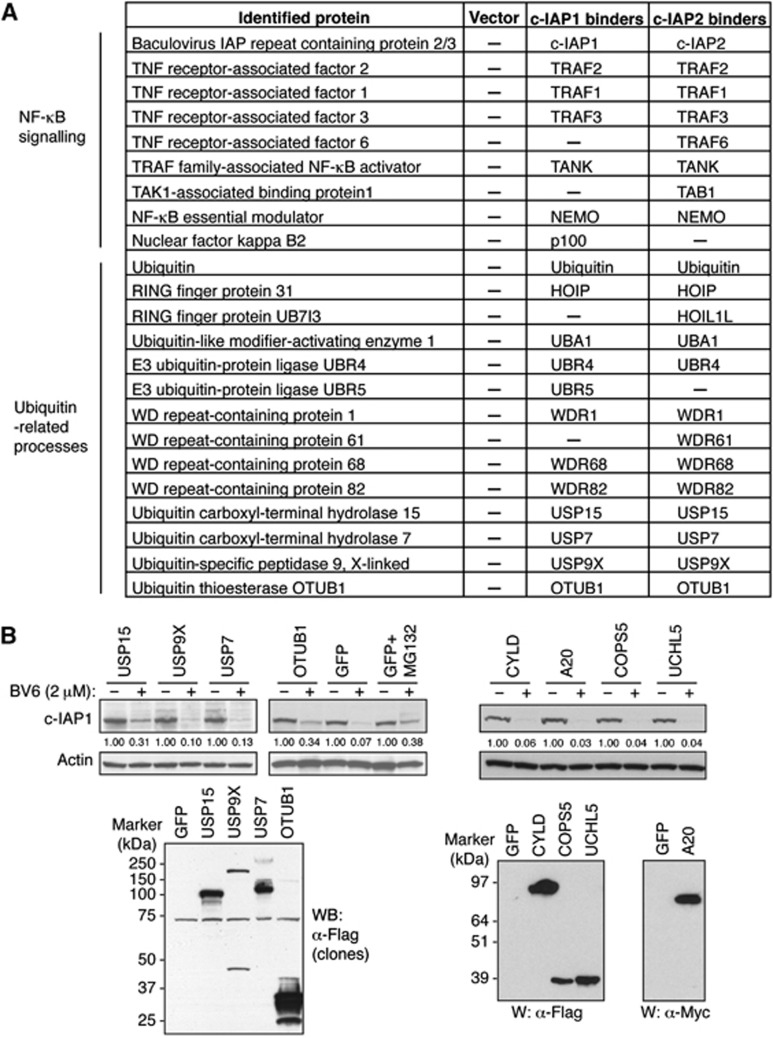
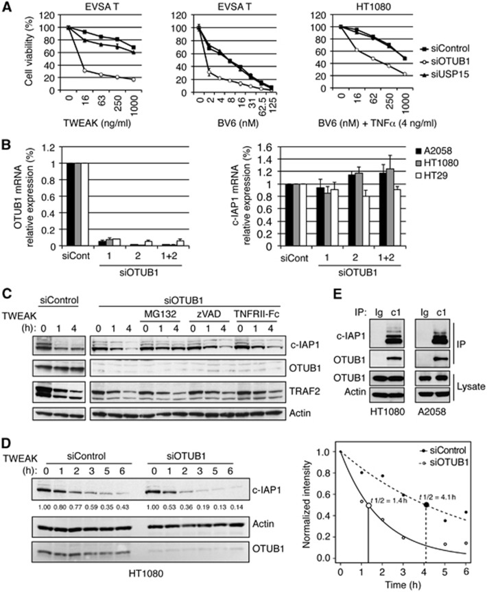
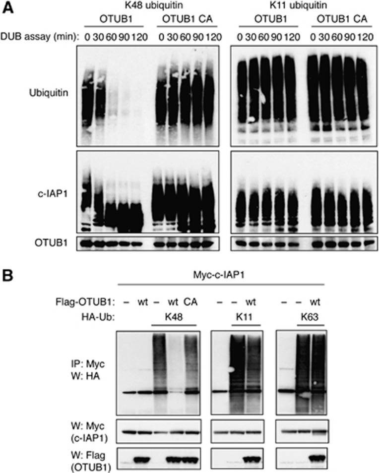
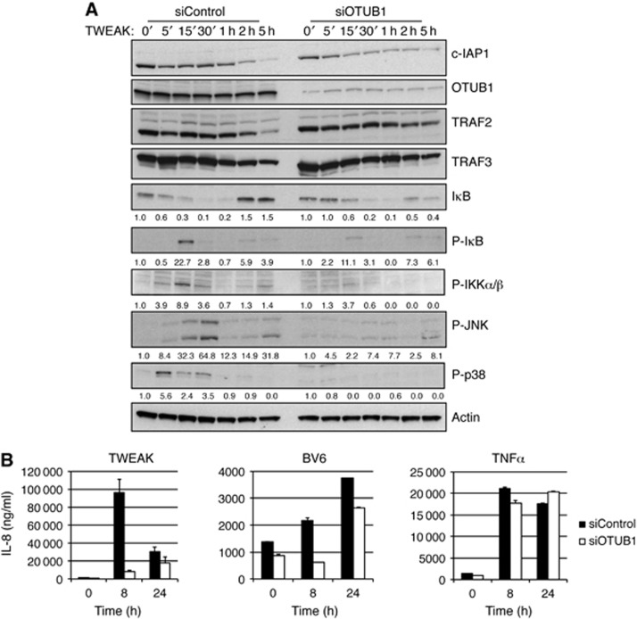
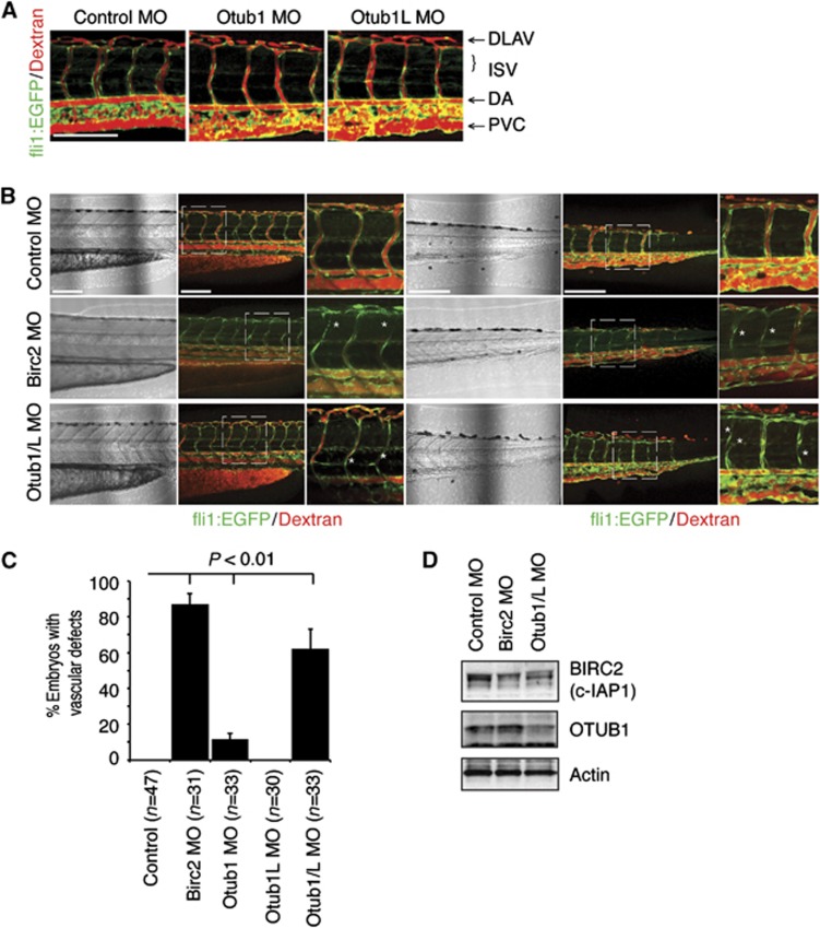
The cellular inhibitor of apoptosis (c-IAP) proteins are E3 ubiquitin ligases that are critical regulators of tumour necrosis factor (TNF) receptor (TNFR)-mediated signalling. Through their E3 ligase activity c-IAP proteins promote ubiquitination of receptor-interaction protein 1 (RIP1), NF-κB-inducing kinase (NIK) and themselves, and regulate the assembly of TNFR signalling complexes. Consequently, in the absence of c-IAP proteins, TNFR-mediated activation of NF-κB and MAPK pathways and the induction of gene expression are severely reduced. Here, we describe the identification of OTUB1 as a c-IAP-associated deubiquitinating enzyme that regulates c-IAP1 stability. OTUB1 disassembles K48-linked polyubiquitin chains from c-IAP1 in vitro and in vivo within the TWEAK receptor-signalling complex. Downregulation of OTUB1 promotes TWEAK- and IAP antagonist-stimulated caspase activation and cell death, and enhances c-IAP1 degradation. Furthermore, knockdown of OTUB1 reduces TWEAK-induced activation of canonical NF-κB and MAPK signalling pathways and modulates TWEAK-induced gene expression. Finally, suppression of OTUB1 expression in zebrafish destabilizes c-IAP (Birc2) protein levels and disrupts fish vasculature. These results suggest that OTUB1 regulates NF-κB and MAPK signalling pathways and TNF-dependent cell death by modulating c-IAP1 stability.


Conflict of interest statement

All of the authors are employees of Genentech, Inc.

Figures

Figure 1
Figure 1
Identification of c-IAP-associated regulators of protein stability. (A) Proteins captured in pull-downs from KMS18 cells stably transfected with Flag-tagged c-IAP1 or c-IAP2 were identified by mass spectrometry and are grouped according to their reported roles in cellular processes. (B) DUB screen. A collection of DUB constructs including the DUBs identified in (A) was transfected into 293T cells. Forty-eight hours later cells were treated with BV6 (2 μM) for 10 min, and lysates were analysed by western blotting with c-IAP1, Actin, Flag (DUBs), and Myc (A20) antibodies. The intensities of c-IAP1 bands were quantified using densitometry and indicated under western blots.
Figure 2
Figure 2
OTUB1 modulates c-IAP1 stability and cellular viability. (A) EVSA T and HT1080 cells were transfected with control, OTUB1-specific, or USP15-specific siRNA oligos and 48 h later treated with TWEAK or BV6 (EVSA T), or BV6 and TNFα (HT1080). Cell death was assessed using CellTiter-Glo luminescent cell viability assay 24 h following the start of treatment. (B) Indicated cell lines were transfected with control or OTUB1-specific siRNA oligos and 48 h later mRNA levels of OTUB1 and c-IAP1 were determined by quantitative RT–PCR. All values were normalized to an RPL19 RNA internal control. Columns represent mean from triplicate experiments and bars represent standard deviation. (C) HT1080 cells were transfected with control or OTUB1-specific siRNA oligos and 48 h later with TWEAK (100 ng/ml) for 1 or 4 h in the presence of MG132 (20 μM), zVAD (20 μM), or TNFRII-Fc (20 μg/ml). Protein levels were determined by western blotting with c-IAP1, OTUB1, TRAF2, or Actin-specific antibodies. (D) HT1080 cells were transfected with control or OTUB1-specific siRNA oligos and 32 h later pre-incubated with transcription inhibitor Actinomycin D (1 μg/ml) for 16 h and then treated with TWEAK (100 ng/ml) for 1–6 h. The effect of OTUB1 knockdown on c-IAP1 half-life was examined by western blotting (left panel) and subsequent densitometry of band intensities was indicated under c-IAP1 western blot and plotted as a function of time using RGraph (t1/2 differences are significant with Z=5.815 and P<0.0001) (right panel). (E) Endogenous association of c-IAP1 and OTUB1. A2058 and HT1080 cells were treated with MG132 (20 μM) for 1 h, lysed and immunoprecipitated with c-IAP1-specific (c1) or isotype control (Ig) antibodies. Cellular lysates and immunoprecipitates were examined by western blotting using antibodies specific for c-IAP1, OTUB1, or Actin.
Figure 3
Figure 3
Knockdown of OTUB1 promotes TNF-dependent apoptosis. (A) Indicated cell lines were transfected with control or OTUB1-specific siRNA oligos and 48 h later treated with TWEAK. Cell death was assessed using CellTiter-Glo luminescent cell viability assay 24 h following the start of treatment. (B) A2058 cells were transfected with control or OTUB1-specific siRNA oligos and 48 h later treated with TWEAK (100 ng/ml) in combination with TNFRII-Fc, zVAD, or DMSO. Cell death was assessed as in (A). (C) A2058 cells were transfected with control or OTUB1-specific siRNA oligos and 48 h later treated with TWEAK (100 ng/ml) for indicated time periods. Cellular lysates were examined by western blotting with antibodies against OTUB1, caspase-8, caspase-3, and Actin.
Figure 4
Figure 4
OTUB1 disassembles K48-linked polyubiquitin chains from c-IAP1. (A) OTUB1 removes K48-linked chains from c-IAP1 in vitro. Recombinant c-IAP1 was incubated in reconstituted ubiquitination reaction for 20 min at 30°C with K48-only or K11-only ubiquitin followed by the addition of apyrase, for 1 h to stop the ubiquitination reaction. Recombinant OTUB1 wild-type or C91A catalytic mutant was then added and deubiquitination reactions were carried out for indicated time periods and subjected to western blotting with ubiquitin, c-IAP1, or OTUB1-specific antibodies. (B) 293T cells were transiently transfected with Myc-c-IAP1, Flag-OTUB1 (wild-type or C91A mutant), K48-, K11-, or K63-only ubiquitin constructs and vector plasmid for 40 h. Cells were lysed and lysates were immunoprecipitated with anti-Myc affinity resin. Inputs from cellular lysates and immunoprecipitates were examined by western blotting with Myc, Flag, and HA antibodies.
Figure 5
Figure 5
OTUB1 regulates endogenous K48-linked polyubiquitination of c-IAP1. (A) c-IAP1 undergoes polyubiquitination with multiple linkages in response to IAP antagonist BV6. HT1080 cells were pre-treated with MG132 for 30 min and then treated with BV6 (2 μM) for 5 min. Cells were lysed in 6 M urea buffer, lysates were diluted twice and immunoprecipitated using linkage-specific anti-ubiquitin antibodies or control antibody (anti-Herceptin). Immunoprecipitated material was detected using anti-c-IAP1 antibody. (B) TWEAK stimulates K48-linked c-IAP1 polyubiquitination. HT1080 cells were pre-treated with MG132 for 30 min followed by the TNFα (50 ng/ml) or TWEAK (100 ng/ml) treatment for 7 min. Cells were lysed and immunoprecipitated as in (A) and immunoprecipitated proteins were detected using anti-RIP1 and c-IAP1 antibodies. (C) A2058 cells were transfected with control or OTUB1-specific siRNA oligos and 48 h later treated with MG132 (20 μM) for 1 h and subsequently with α-Flag antibody cross-linked TWEAK for indicated time periods. Cellular lysates were immunoprecipitated with TWEAK as described in Materials and methods, precipitates disrupted in 6 M urea and re-immunoprecipitated with K48 ubiquitin chain-specific antibodies. Lysates and precipitates were examined by western blotting with indicated antibodies. (D) A2058 cells were treated with MG132 (20 μM) for 1 h and with TWEAK for indicated time periods. Cellular lysates were immunoprecipitated with TWEAK as in (B) and examined by western blotting with indicated antibodies.
Figure 6
Figure 6
Downregulation of OTUB1 diminishes TWEAK signalling. (A) A2058 cells were transfected with control or OTUB1-specific siRNA oligos and 48 h later treated with TWEAK (100 ng/ml) for indicated time periods. Cellular lysates were examined by western blotting with indicated antibodies. (B) HT1080 cells were transfected with control or OTUB1-specific siRNA oligos and 48 h later treated with TWEAK (100 ng/ml), BV6 (2.5 μM), or TNFα (20 ng/ml) for indicated time periods. Supernatant from cells was examined for production of IL-8 cytokine by Luminex Technology.
Figure 7
Figure 7
OTUB1 regulates zebrafish vascular stability. (A) Tg(fli1:EGFP) zebrafish were injected with Control (2 ng), OTUB1 (0.8 ng), or OTUB1L (2 ng) morpholinos. At 72 hpf angiography was performed and vascular development and blood flow were analysed by confocal microscopy. ISV, intersegmental vessel; DLAV, dorsal longitudinal vessel; DA, dorsal aorta; PVC, posterior cardinal vein. Scale bar: 150 μm. (B) Tg(fli1:EGFP) zebrafish were injected with Control (4 ng), Otub1/L (2.8 ng), or Birc2 (4 ng) morpholinos. At 72 hpf angiography was performed and vascular development and blood flow were analysed by confocal microscopy in two zebrafish trunk regions. *Indicates sites of lumen narrowing and retraction of ISV vessels. Scale bar: 150 μm. (C) Quantification of ISV blood flow. Percentage of embryos with two or more ISV vessels that have no observable blood flow (Dextran-rhodamine). Error bars represent the standard deviation. Statistical significance of experimental groups versus control, P<0.01, t-test. (D) Western blot analysis of Birc2 (with α-c-IAP1/2 pan antibody from R&D Systems) and Otub1 (with α-OTUB1 antibody from Bethyl) protein expression at 48 hpf in Tg(fli1:EGFP) zebrafish were injected with Control (4 ng), Otub1/L (2.8 ng), or Birc2 (4 ng) morpholinos.

References

    1. Annunziata CM, Davis RE, Demchenko Y, Bellamy W, Gabrea A, Zhan F, Lenz G, Hanamura I, Wright G, Xiao W, Dave S, Hurt EM, Tan B, Zhao H, Stephens O, Santra M, Williams DR, Dang L, Barlogie B, Shaughnessy JD Jr. et al. (2007) Frequent engagement of the classical and alternative NF-kappaB pathways by diverse genetic abnormalities in multiple myeloma. Cancer Cell 12: 115–130 - PMC - PubMed
    1. Bertrand MJ, Milutinovic S, Dickson KM, Ho WC, Boudreault A, Durkin J, Gillard JW, Jaquith JB, Morris SJ, Barker PA (2008) cIAP1 and cIAP2 facilitate cancer cell survival by functioning as E3 ligases that promote RIP1 ubiquitination. Mol Cell 30: 689–700 - PubMed
    1. Blankenship JW, Varfolomeev E, Goncharov T, Fedorova AV, Kirkpatrick DS, Izrael-Tomasevic A, Phu L, Arnott D, Aghajan M, Zobel K, Bazan JF, Fairbrother WJ, Deshayes K, Vucic D (2009) Ubiquitin binding modulates IAP antagonist-stimulated proteasomal degradation of c-IAP1 and c-IAP2. Biochem J 417: 149–160 - PubMed
    1. Deshaies RJ, Joazeiro CA (2009) RING domain E3 ubiquitin ligases. Annu Rev Biochem 78: 399–434 - PubMed
    1. Dynek JN, Goncharov T, Dueber EC, Fedorova AV, Izrael-Tomasevic A, Phu L, Helgason E, Fairbrother WJ, Deshayes K, Kirkpatrick DS, Vucic D (2010) c-IAP1 and UbcH5 promote K11-linked polyubiquitination of RIP1 in TNF signalling. EMBO J 29: 4198–4209 - PMC - PubMed