. 2013 May 29;32(11):1514-28.
doi: 10.1038/emboj.2013.65. Epub 2013 Mar 22.

SENP3-mediated deSUMOylation of dynamin-related protein 1 promotes cell death following ischaemia

Affiliations

SENP3-mediated deSUMOylation of dynamin-related protein 1 promotes cell death following ischaemia

Chun Guo et al. EMBO J. .

Abstract

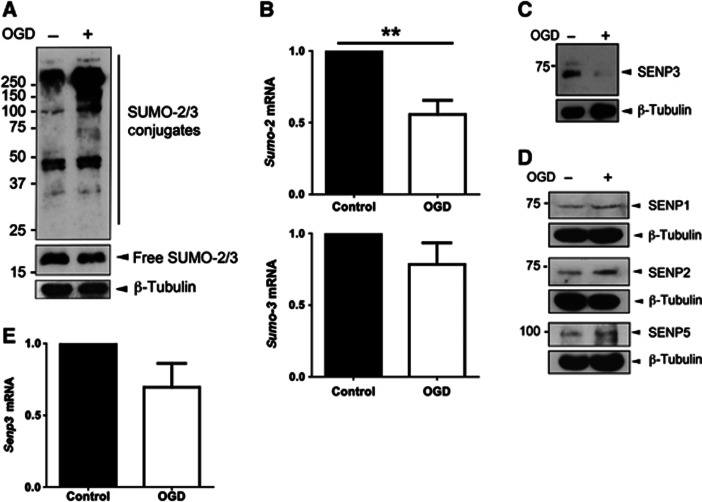
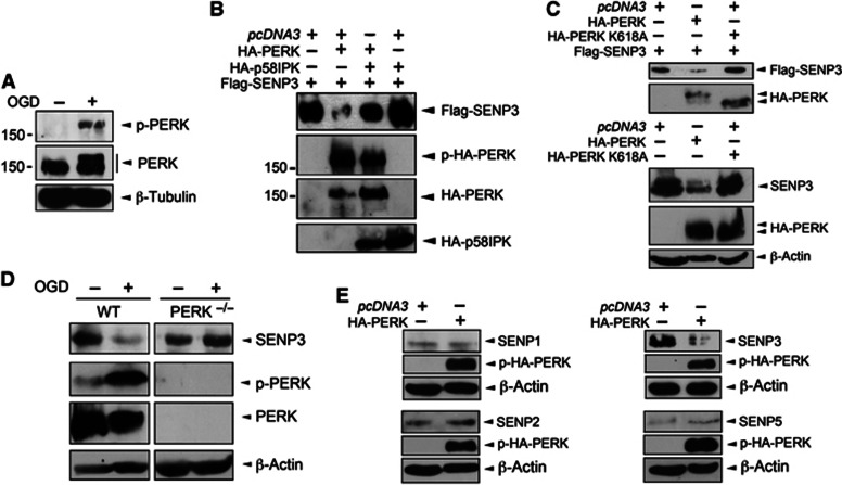
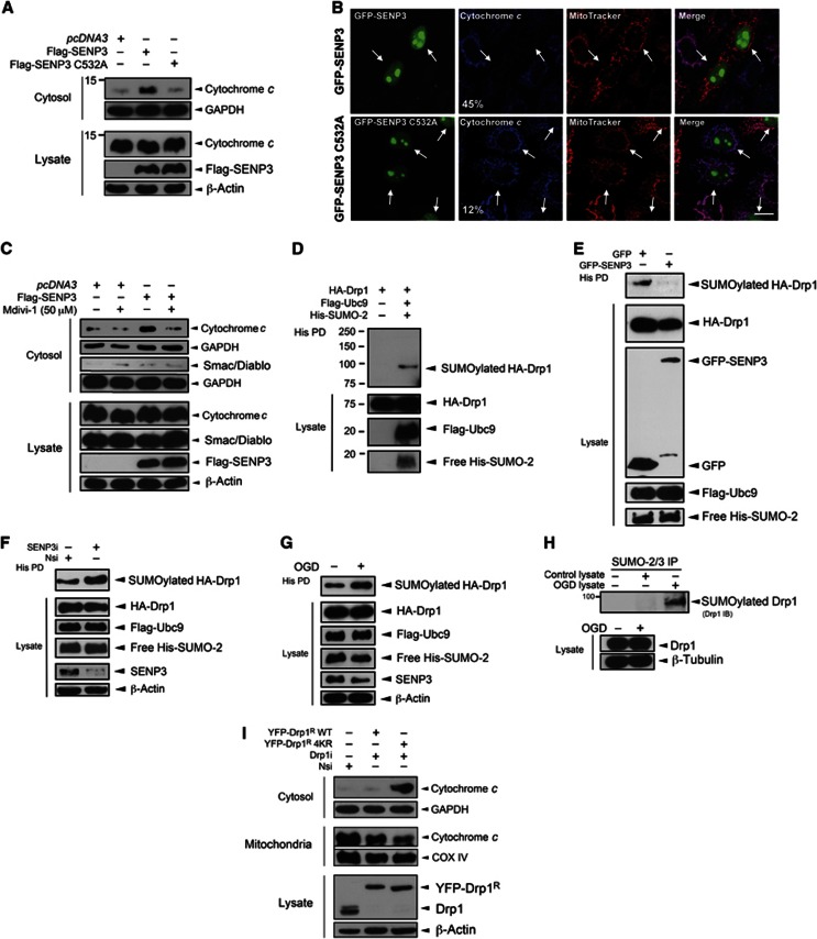
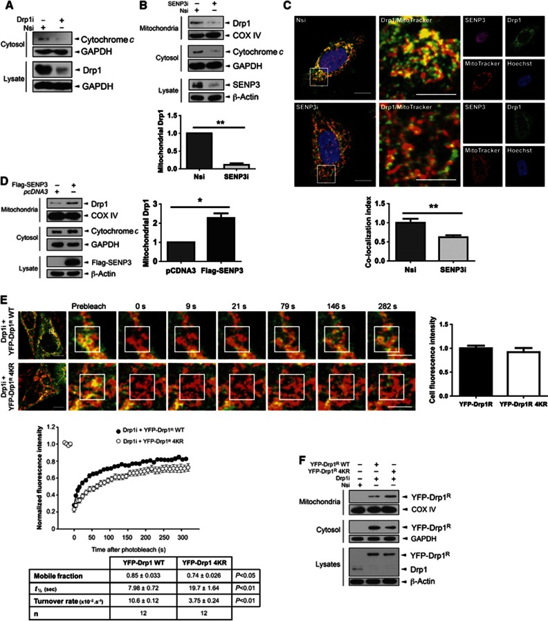
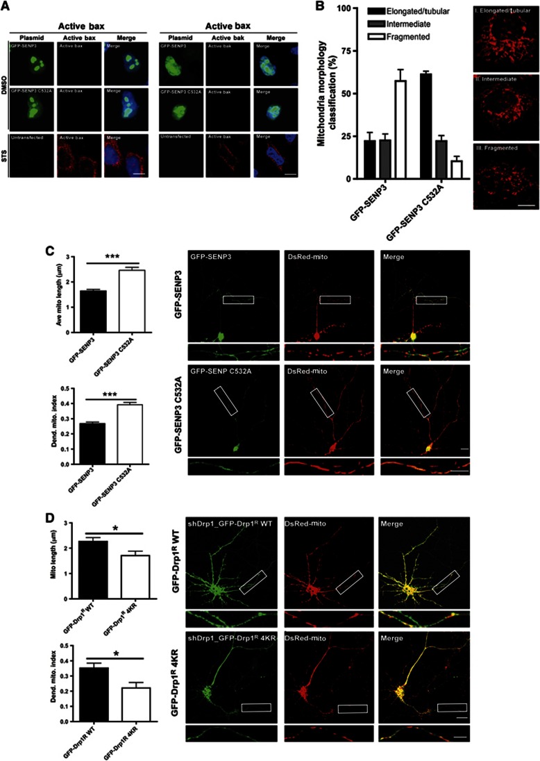
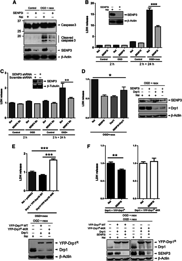
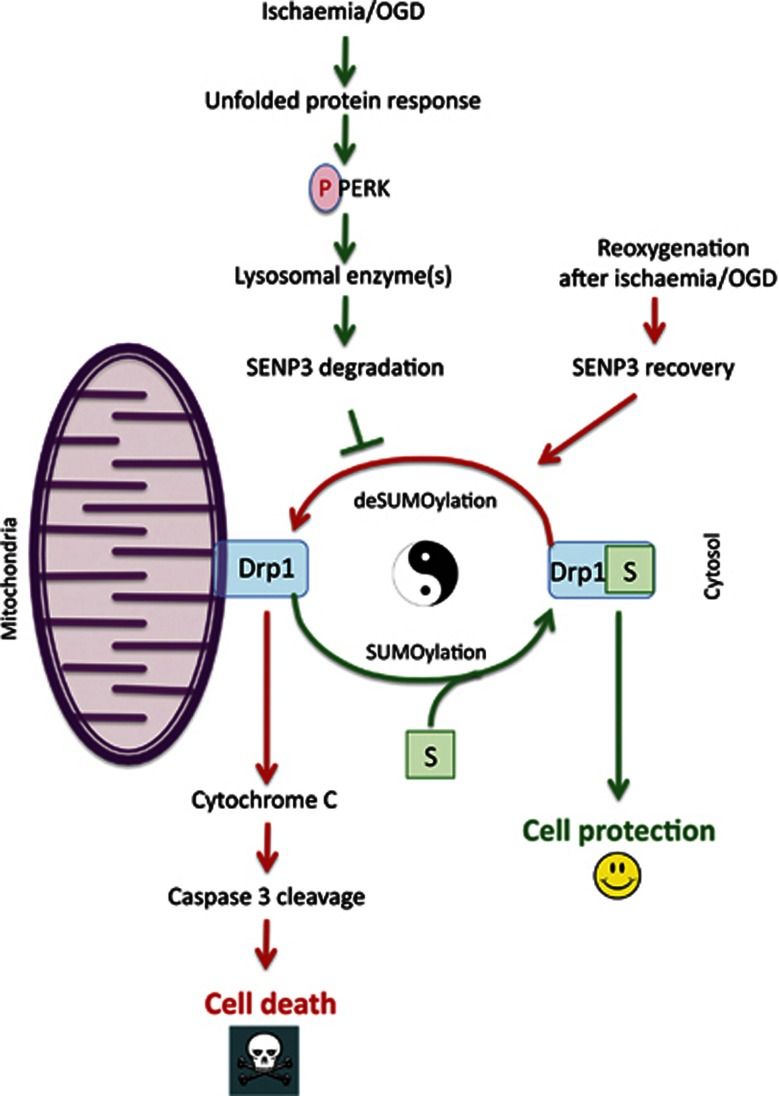
Global increases in small ubiquitin-like modifier (SUMO)-2/3 conjugation are a neuroprotective response to severe stress but the mechanisms and specific target proteins that determine cell survival have not been identified. Here, we demonstrate that the SUMO-2/3-specific protease SENP3 is degraded during oxygen/glucose deprivation (OGD), an in vitro model of ischaemia, via a pathway involving the unfolded protein response (UPR) kinase PERK and the lysosomal enzyme cathepsin B. A key target for SENP3-mediated deSUMOylation is the GTPase Drp1, which plays a major role in regulating mitochondrial fission. We show that depletion of SENP3 prolongs Drp1 SUMOylation, which suppresses Drp1-mediated cytochrome c release and caspase-mediated cell death. SENP3 levels recover following reoxygenation after OGD allowing deSUMOylation of Drp1, which facilitates Drp1 localization at mitochondria and promotes fragmentation and cytochrome c release. RNAi knockdown of SENP3 protects cells from reoxygenation-induced cell death via a mechanism that requires Drp1 SUMOylation. Thus, we identify a novel adaptive pathway to extreme cell stress in which dynamic changes in SENP3 stability and regulation of Drp1 SUMOylation are crucial determinants of cell fate.


Conflict of interest statement

The authors declare that they have no conflict of interest.

Figures

Figure 1
Figure 1
OGD increases global SUMO-2/3 conjugation and decreases SENP3 in neurons. (A) Lysates of primary cortical neurons were blotted for SUMO-2/3 and β-tubulin after OGD (1 h). (B) sumo-2 mRNA levels are decreased and sumo-3 mRNA levels remain unchanged after OGD (1 h) in primary cortical neurons. (C) Levels of SENP3 are reduced in neurons after OGD (30 min). (D) OGD (30 min) does not alter SENP1, SENP2 or SENP5 levels in neurons. (E) senp3 mRNA levels are not reduced by OGD (1 h). For (B) and (E) significance was determined using a two-tailed Paired t-test; for sumo-2 mRNA (n=9; **P<0.01), for sumo-3 mRNA (n=7) and for senp3 mRNA (n=8). Source data for this figure is available on the online supplementary information page.
Figure 2
Figure 2
PERK activation is required for decreased SENP3. (A) PERK is phosphorylated (p-PERK) in neurons by OGD (30 min). Blots were probed with phospho-PERK, PERK and β-tubulin antibodies. (B) Overexpression of HA-PERK causes loss of Flag-SENP3 in HEK293 cells, which is blocked by overexpression of the PERK inhibitor p58IPK. Lysates were blotted with Flag, phospho-PERK and HA antibodies. (C) The kinase activity of PERK is required for SENP3 removal in HEK293 cells. HA-PERK, but not kinase-dead HA-PERK K618A decreases SENP3. The top panel shows the effect of PERK on overexpressed Flag-SENP3, while the lower panel shows endogenous SENP3. (D) PERK is required for OGD-induced loss of SENP3. Confluent wild-type and PERK−/− MEFs were subjected to OGD (2 h), harvested and the lysates probed as indicated. (E) Overexpression of HA-PERK in HEK293 cells reduces levels of SENP3 but not of SENP1, SENP2 or SENP5. Source data for this figure is available on the online supplementary information page.
Figure 3
Figure 3
SENP3 regulates cytochrome c release via deSUMOylation of Drp1. (A) Overexpression of Flag-SENP3, but not inactive C532A mutant causes cytochrome c release in HEK293 cells. The cytosol fraction or whole cell lysate was blotted for cytochrome c, Flag, and the cytosolic marker GAPDH, or β-actin. (B) Overexpressing SENP3 decreases mitochondrial cytochrome c in HeLa cells. Fixed cells were immunostained to assess the localization of cytochrome c in mitochondria labelled with MitoTracker. Representative images showing GFP-SENP3 (top panel) and GFP-SENP3 C532A expressing cells (lower panel, Green: SENP3; Red: MitoTracker; Blue: cytochrome c; scale bar: 10 μm). White arrows indicate transfected cells with percentages denoting the relative proportions of transfected cells with significantly reduced cytochrome c in mitochondria for wild-type versus inactive mutant overexpression (n=75 cells for GFP-SENP and n=113 for GFP-SENP3 C532A). (C) SENP3-mediated cytochrome c release is Drp1 dependent. HEK293 cells expressing Flag-SENP3 were treated with Mdivi-1 (50 μM) for 4 h. The cytosol fraction or whole cell lysate was blotted for cytochrome c, Smac/Diablo, Flag, and GAPDH, or β-actin. (D) Drp1 is SUMO-2-ylated in HEK293 cells expressing Flag-Ubc9, His-SUMO-2 and HA-Drp1. Lysate was incubated with Ni2+ beads to precipitate His-SUMO-2-ylated proteins (His PD). (E) Overexpression of SENP3 decreases Drp1 SUMOylation. Constructs expressing HA-Drp1, Flag-Ubc9, His-SUMO-2 and either GFP or GFP-SENP3 were transfected into HEK293 cells. (F) SENP3 knockdown enhances Drp1 SUMOylation. Either non-specific siRNA (Nsi) or SENP3 siRNA (SENP3i) together with constructs expressing HA-Drp1, Flag-Ubc9 and His-SUMO-2 were co-expressed in HEK293 cells. (G) OGD enhances Drp1 SUMOylation. Two days post transfection, HEK293 cells were exposed to OGD (2 h). In (CF), His-pulldown and lysate samples were blotted as indicated with antibodies against HA, Flag, His, GFP, SENP3 and β-actin. (H) OGD (2 h) for primary cortical neurons increases SUMOylation of endogenous Drp1. (I) Preventing Drp1 SUMOylation results in cytochrome c release. YFP-Drp1R WT or Drp1R 4KR were expressed in HEK293 cells after knockdown of endogenous Drp1. Source data for this figure is available on the online supplementary information page.
Figure 4
Figure 4
SENP3 regulates mitochondrial association of Drp1. (A) Knockdown of Drp1 decreases cytosolic cytochrome c. (B) Knockdown of SENP3 decreases the mitochondrial association of endogenous Drp1 and reduces levels of cytosolic cytochrome c in HEK293 cells (n=3; **P<0.01). (C) SENP3 knockdown decreases mitochondrial localization of Drp1 in HeLa cells. Fixed cells were immunostained to assess the co-localization of endogenous Drp1 with mitochondria. Representative images showing control (top panel, Nsi) and SENP3 knockdown cells (lower panel, SENP3i; Green: Drp1; Red: MitoTracker; Yellow: co-localization of Drp1 and MitoTracker; Magenta: SENP3; Blue: Hoechst; scale bar: 10 μm). The region of interest defined by the white box is enlarged in the middle panels (scale bar: 5 μm). Right hand panels illustrate the individual channel data. The histogram shows the comparative levels of Drp1 localization at mitochondria (n=36 cells for Nsi and n=27 cells for SENP3i; **P<0.01). (D) SENP3 overexpression increases localization of Drp1 at mitochondria in HEK293 cells (n=3; *P<0.05). (E) SUMOylation affects Drp1 dynamics. YFP-Drp1R WT or non-SUMOylatable Drp1R 4KR was expressed in HeLa cells after knockdown of endogenous Drp1 and the cells subjected to FRAP analysis. The left hand images show the representative cells sampled and the subsequent image panels are enlargements of frames at the specified time points (Green: YFP-DRP1R; Red: MitoTracker; Yellow: co-localization. Scale bar: 10 μm for first frame, 5 μm for enlarged frames). The area defined by the white box is the photobleached region of interest. Recovery curves for Drp1R WT (black) and Drp1R 4KR (white) are shown and the values presented in the table. Values=mean±s.e.m. (n=12 cells per condition). (F) Non-SUMOylatable Drp1 shows enhanced mitochondrial association. YFP-Drp1R WT or Drp1R 4KR was expressed in HeLa cells after knockdown of endogenous Drp1, and cytosolic fraction, mitochondrial fraction, or whole cell lysates were blotted as indicated. Source data for this figure is available on the online supplementary information page.
Figure 5
Figure 5
SENP3 regulates Drp1-mediated mitochondrial fission. (A) Overexpression of SENP3 does not cause Bax/Bak activation in HeLa cells whereas staurosporine (STS; 1 μM; 2 h) potently activates Bax/Bak. Representative images show immunostaining for active Bax (left panels) or active Bak (right panels) in GFP-SENP3 or GFP-SENP C532A transfected cells or untransfected staurosporine-treated cells (Green: GFP-SENP3, Red: active Bax/Bak; Blue: Hoechst; Scale bar: 10 μm). (B) Overexpression of SENP3 promotes mitochondrial fission in HeLa cells. Cells overexpressing GFP-SENP3 or inactive C532A mutant were stained with MitoTracker and mitochondrial morphology classified into one of three categories: (I) Elongated/Tubular, (II) Intermediate or (III) Fragmented. There was a significantly increased proportion of cells with fragmented mitochondria in GFP-SENP3 compared to C532A overexpressing cells (P<0.0001, chi square test; n=99 cells for GFP-SENP3 and n=86 cells for GFP-SENP3 C532A). (C) Representative images showing GFP-SENP3 expressing (upper panels) and GFP-SENP3 C532A expressing neurons (lower panels) co-transfected with Mito-DsRed (scale: 10 μm, white box ROI magnified below). Data were quantified and expressed as a dendritic mitochondrial index (total mitochondrial length/dendrite length). Values bar charts are shown as mean±s.e.m. n=29 cells for GFP-SENP3 and n=27 for C532A, ***P<0.0001). (D) Representative images showing knockdown of endogenous Drp1 and rescue with GFP-Drp1R WT or non-SUMOylatable Drp1R 4KR in neurons. Non-SUMOylatable Drp1R 4KR increases mitochondrial fission visualized using Mito-DsRed (scale bar: 10 μm, white box ROI magnified below). Mitochondria morphology analysis was performed as in (C). Values in bar charts are shown as mean±s.e.m. (n=8 cells for GFP-Drp1R WT and n=7 cells for GFP-Drp1R 4KR), *P<0.05.
Figure 6
Figure 6
SENP3 regulation of Drp1 SUMOylation plays a critical role in cell death following reoxygenation. (A) SENP3 knockdown decreases OGD plus reoxygenation-induced caspase 3 cleavage in HEK293 cells. Two days post transfection with Nsi or SENP3i HEK293 cells were subjected to OGD (2 h) and then reoxygenation (24 h). Lysates were blotted as indicated. (B) SENP3 knockdown decreases LDH release from HEK293 cells after OGD plus reoxygenation. Cells were treated as in (A) and culture media was sampled after OGD (2 h) and after 24 h reoxygenation (2 h+24 h). (C) SENP3 knockdown decreases LDH release in neurons following OGD plus reoxygenation. Neurons were infected with retrovirus containing either scrambled shRNA (Scr) or SENP3 shRNA (SENP3 sh), subjected to OGD (2 h) and then reoxygenation (24 h), and media sampled for LDH as in (B). Inset panels in (B) and (C) show immunoblots of cell lysates confirming SENP3 knockdown. (D) Knockdown of both SENP3 and Drp1 is not additive on OGD plus reoxygenation-evoked LDH release from HEK293 cells. Immunoblots (right panel) confirm knockdown of SENP3 and/or Drp1. (E) Drp1 knockdown and rescue with non-SUMOylatable Drp1R 4KR increases LDH release in response to OGD plus reoxygenation in HEK293 cells (upper panel). Immunoblots (lower panel) confirm knockdown and rescue of Drp1. (F) SENP3 knockdown does not reduce LDH release induced by OGD plus reoxygenation in Drp1R 4KR-rescued cells. Immunoblots (lower panels) confirm knockdown of Drp1/SENP3 and rescue of Drp1. In (BF), values are presented as mean±s.e.m. (n?5 replicates for each group; *P<0.05; **P<0.01; ***P<0.001). Source data for this figure is available on the online supplementary information page.
Figure 7
Figure 7
Schematic of proposed cell death/survival pathway. During ischaemic stress, the UPR kinase PERK is activated, which leads to lysosome-mediated degradation of the SUMO-2/3-specific deSUMOylating enzyme SENP3. The absence of SENP3 prolongs Drp1 SUMOylation, favouring localization in the cytosol and reducing Drp1-mediated cytochrome c release. Following reoxygenation, however, SENP3 levels recover, promoting mitochondrial association of Drp1 and cell death.

Comment in

References

    1. Anderson DB, Wilkinson KA, Henley JM (2009) Protein SUMOylation in neuropathological conditions. Drug News Perspect 22: 255–265 - PMC - PubMed
    1. Bekes M, Prudden J, Srikumar T, Raught B, Boddy MN, Salvesen GS (2011) The dynamics and mechanism of SUMO chain deconjugation by SUMO-specific proteases. J Biol Chem 286: 10238–10247 - PMC - PubMed
    1. Boya P, Andreau K, Poncet D, Zamzami N, Perfettini JL, Metivier D, Ojcius DM, Jaattela M, Kroemer G (2003) Lysosomal membrane permeabilization induces cell death in a mitochondrion-dependent fashion. J Exp Med 197: 1323–1334 - PMC - PubMed
    1. Boya P, Kroemer G (2008) Lysosomal membrane permeabilization in cell death. Oncogene 27: 6434–6451 - PubMed
    1. Cassidy-Stone A, Chipuk JE, Ingerman E, Song C, Yoo C, Kuwana T, Kurth MJ, Shaw JT, Hinshaw JE, Green DR, Nunnari J (2008) Chemical inhibition of the mitochondrial division dynamin reveals its role in Bax/Bak-dependent mitochondrial outer membrane permeabilization. Dev Cell 14: 193–204 - PMC - PubMed

Publication types

MeSH terms