. 2013 Apr;123(4):1571-9.
doi: 10.1172/JCI63685. Epub 2013 Mar 25.

Lymphatic vasculature mediates macrophage reverse cholesterol transport in mice

Affiliations

Lymphatic vasculature mediates macrophage reverse cholesterol transport in mice

Catherine Martel et al. J Clin Invest. 2013 Apr.

Abstract

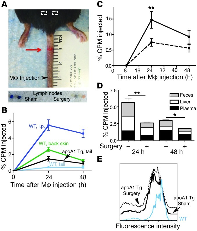
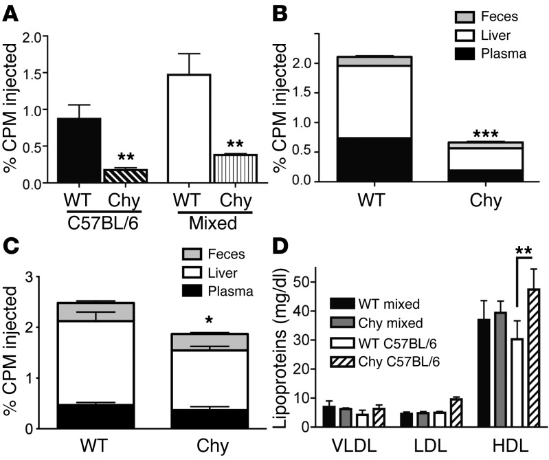
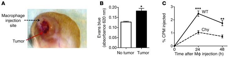
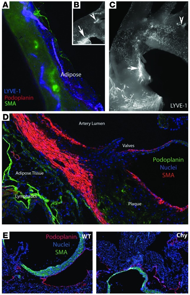
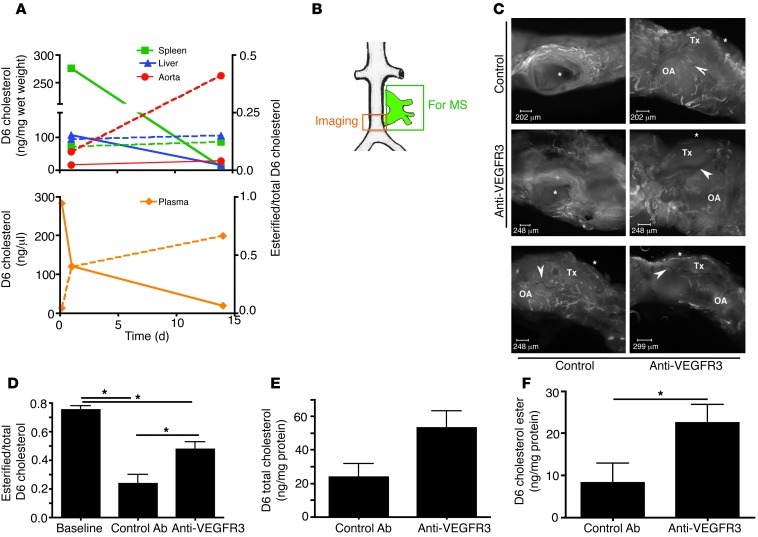
Reverse cholesterol transport (RCT) refers to the mobilization of cholesterol on HDL particles (HDL-C) from extravascular tissues to plasma, ultimately for fecal excretion. Little is known about how HDL-C leaves peripheral tissues to reach plasma. We first used 2 models of disrupted lymphatic drainage from skin--1 surgical and the other genetic--to quantitatively track RCT following injection of [3H]-cholesterol-loaded macrophages upstream of blocked or absent lymphatic vessels. Macrophage RCT was markedly impaired in both models, even at sites with a leaky vasculature. Inhibited RCT was downstream of cholesterol efflux from macrophages, since macrophage efflux of a fluorescent cholesterol analog (BODIPY-cholesterol) was not altered by impaired lymphatic drainage. We next addressed whether RCT was mediated by lymphatic vessels from the aortic wall by loading the aortae of donor atherosclerotic Apoe-deficient mice with [2H]6-labeled cholesterol and surgically transplanting these aortae into recipient Apoe-deficient mice that were treated with anti-VEGFR3 antibody to block lymphatic regrowth or with control antibody to allow such regrowth. [2H]-Cholesterol was retained in aortae of anti-VEGFR3-treated mice. Thus, the lymphatic vessel route is critical for RCT from multiple tissues, including the aortic wall. These results suggest that supporting lymphatic transport function may facilitate cholesterol clearance in therapies aimed at reversing atherosclerosis.


Figures

Figure 1
Figure 1. Surgical separation of lymphatic vessels and its impact on RCT.
(A) Image of the mouse tail following surgical separation of lymphatic vessels (red arrow). White boxes depict approximate location of tail-draining lymph nodes. These nodes (lower photomicrographs) were removed after Evans blue dye was injected into the tail; enlarged images of the 2 lymph nodes are shown from a control mouse receiving a sham operation (Sham) and 1 that underwent lymphatic separation (Surgery). Similar results were obtained in 4 other analyses (n = 5) using Evans blue dye to verify repeated success of the procedure. (B) [3H]-Cholesterol–loaded macrophages were injected into the peritoneum (i.p.), in skin of the tail, or the lower back of WT or Apoa1 transgenic mice. Tritium counts in the plasma are charted. (C) Tritium counts in plasma in control mice (solid line) or in those subjected to lymphatic separation (dotted line) after [3H]-cholesterol–loaded macrophages were injected into the tail upstream (with respect to lymph flow) of the lymphatic separation as shown in A. (D) Compiled tritium counts in plasma, liver, and feces from the experimental design used in C. (E) Cholesterol efflux was monitored as the loss of fluorescence from gated CD45.1 macrophages loaded ex vivo with BODIPY-cholesterol and retrieved 24 hours later from the tails of WT or Apoa1 transgenic mice with or without lymphatic separation. All data represent the mean ± SEM from at least 2 experiments performed with 5 replicates per experimental group. *P < 0.05; **P < 0.01. CPM, counts per minute.
Figure 2
Figure 2. RCT in Chy mutant mice.
(A) [3H]-Cholesterol–loaded macrophages were injected into the rear footpads of Chy mice or control littermates and tritium counts were assessed in plasma at 24 hours. Two background strains were compared; injected macrophages were made from the bone marrow of mice on the same background as the recipients into which they were injected. (B) Compiled tritium counts in plasma (black), liver (white), and feces (gray) from C57BL/6 Chy and WT mice euthanized 24 hours after [3H]-cholesterol–loaded macrophages were injected into the rear footpad. (C) Compiled tritium counts in plasma (black), liver (white), and feces (gray) from C57BL/6 Chy and WT mice euthanized 24 hours after [3H]-cholesterol–loaded macrophages were injected into the scapular area of the back skin. (D) Lipoprotein profiles in the plasma of Chy mice and littermate controls from the mixed or C57BL/6 backgrounds. Each panel represents 2 experiments performed with 5 replicates per experimental group (mean ± SEM). Statistics compared Chy mice with their control littermates in each case. *P < 0.05; **P < 0.01; ***P < 0.001.
Figure 3
Figure 3. Impact of vascular leakage on RCT through lymphatic vessels.
(A) LLC cells were injected into the ear pinnae of Chy and control littermates (mice on a mixed background were used). Tumors grew for 3 weeks before [3H]-cholesterol–loaded macrophages were injected as shown. (B) Increased vascular permeability was documented using Evans blue dye injected i.v. with extraction of ear tissue 30 minutes later to quantify dye recovery using a spectrophotometer. (C) Analysis of tritium counts in the plasma 24 and 48 hours later. Tumor-bearing mice were housed by requirement under biohazard containment that precluded the use of individual caging needed to collect feces. Each panel represents 2 experiments performed with 5 replicates per experimental group. Data represent the mean ± SEM comparing indicated conditions (B) or Chy and control mice at each time point (C). *P ≤ 0.05; **P ≤ 0.01; ***P ≤ 0.001.
Figure 4
Figure 4. Lymphatic vessel distribution in the mouse aorta and aortic sinus.
(A) The descending aorta is seen through its autofluorescence in the green channel. Vessels staining positively for podoplanin and LYVE-1 are seen on the adventitial side of the aorta, weaving in and out of adjacent adipose tissue. These vessels lack smooth muscle actin (SMA) coverage, though some nearby blood vessels are positive. LYVE-1+ SMA lymphatic vessels are absorptive lymphatic capillaries, rather than lymphatic collecting vessels. (B and C) LYVE-1 staining in the region of the aortic arch. Arrows point to a lymphatic capillary along the lesser curvature; arrowheads point out lymphatic capillaries near arteries that branch off the arch. (D) Cross-sectional analysis of the aortic sinus in an Apoe–/– mouse reveals lymphatic vessels under the areas where plaque typically develops. (E) Both WT littermates and Chy mice develop lymphatic vessels in the aorta as depicted in these cross sections of the aortic sinus.
Figure 5
Figure 5. Role of lymphatic vessels in RCT from aortae.
(A) Kinetics of D6-cholesterol were analyzed in duplicate 1 or 14 days after liposomes containing D6-cholesterol were administered i.v. Solid lines correspond to the left-hand y-axis that plots ng of D6-cholesterol detected in aortae. Data depicted by dotted lines correspond to the right-hand y-axis that plots the esterified/total D6-cholesterol ratio. (B) Schematic diagram of the aortic transplant positioned in the abdominal aorta. Green box depicts the portion of tissue collected for mass spectrometric (MS) analysis; orange box shows the portion of tissue collected for imaging lymphatic regrowth. (C) Images of 2 independent pairings of mice transplanted on the same day, 1 treated with control mAb and the other with anti-VEGFR3 mAb; stained for LYVE-1 (white vessels). Stars depict location of the lumen of the transplanted aortic arch where it connects with the original aorta. Arrowheads point to surgical sutures that demarcate the boundary between the transplant (Tx) and the original aorta (OA). Upper images show a pairing from 2 different orientations and lower images show a side view of a different surgical pair. Scale bars: 250 μm. (D) Ester/total D6-cholesterol achieved at baseline prior to transplantation and ratios recovered in 5 pairs of transplants 4 weeks after surgery, treated with apoE vector at 2 weeks and with control or anti-VEGFR3 throughout (E and F). Total (E) and esterified (F) D6-cholesterol recovered in pairs treated with control or anti-VEGFR3 mAb. D and E depict data as the mean ± SD (n = 5–9 for each bar). *P ≤ 0.05.

Comment in

  • Lymphatic vessels clean up your arteries.
    Fernández-Hernando C. Fernández-Hernando C. J Clin Invest. 2013 Apr;123(4):1417-9. doi: 10.1172/JCI68657. Epub 2013 Mar 25. J Clin Invest. 2013. PMID: 23524960 Free PMC article.

References

    1. Potteaux S, et al. Suppressed monocyte recruitment drives macrophage removal from atherosclerotic plaques of Apoe–/– mice during disease regression. . J Clin Invest. 2011;121(5):2025–2036. doi: 10.1172/JCI43802. - DOI - PMC - PubMed
    1. Spann NJ, et al. Regulated accumulation of desmosterol integrates macrophage lipid metabolism and inflammatory responses. Cell. 2012;151(1):138–152. doi: 10.1016/j.cell.2012.06.054. - DOI - PMC - PubMed
    1. Duewell P, et al. NLRP3 inflammasomes are required for atherogenesis and activated by cholesterol crystals. Nature. 2010;464(7293):1357–1361. doi: 10.1038/nature08938. - DOI - PMC - PubMed
    1. Hermansson A, et al. Inhibition of T cell response to native low-density lipoprotein reduces atherosclerosis. J Exp Med. 2010;207(5):1081–1093. doi: 10.1084/jem.20092243. - DOI - PMC - PubMed
    1. Small DM, Bond MG, Waugh D, Prack M, Sawyer JK. Physicochemical and histological changes in the arterial wall of nonhuman primates during progression and regression of atherosclerosis. J Clin Invest. 1984;73(6):1590–1605. doi: 10.1172/JCI111366. - DOI - PMC - PubMed

Publication types

MeSH terms

Substances