. 2013 Apr;123(4):1513-30.
doi: 10.1172/JCI64551. Epub 2013 Mar 25.

The TGR5 receptor mediates bile acid-induced itch and analgesia

Affiliations

The TGR5 receptor mediates bile acid-induced itch and analgesia

Farzad Alemi et al. J Clin Invest. 2013 Apr.

Abstract

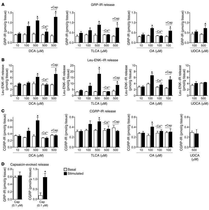
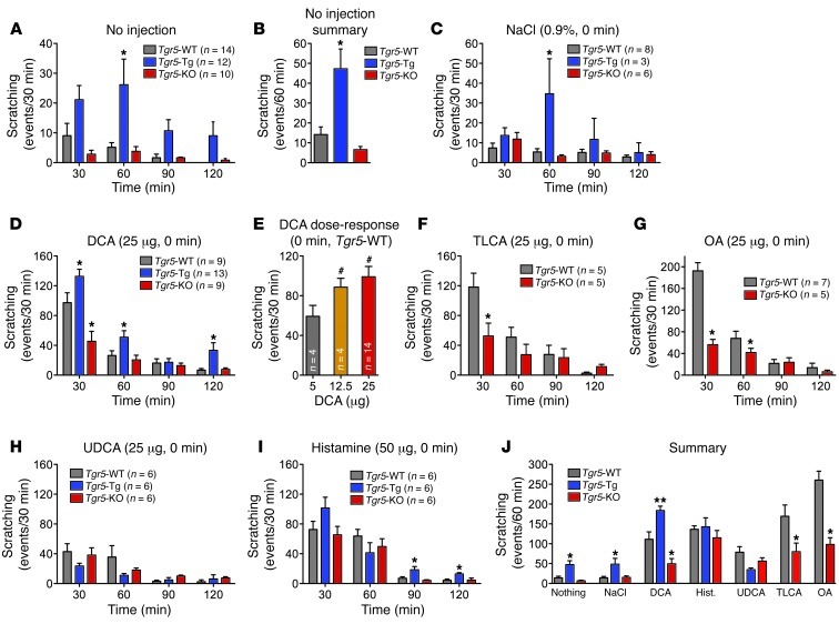
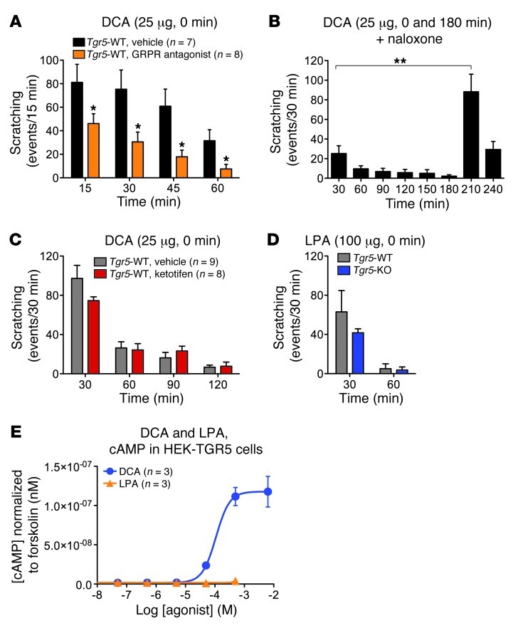
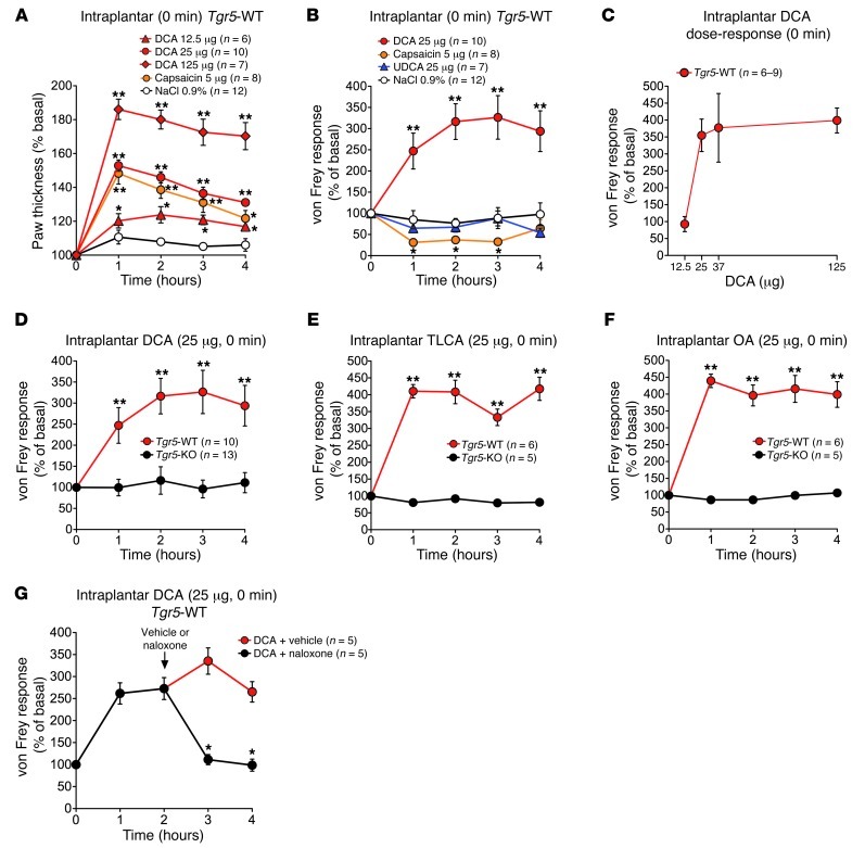
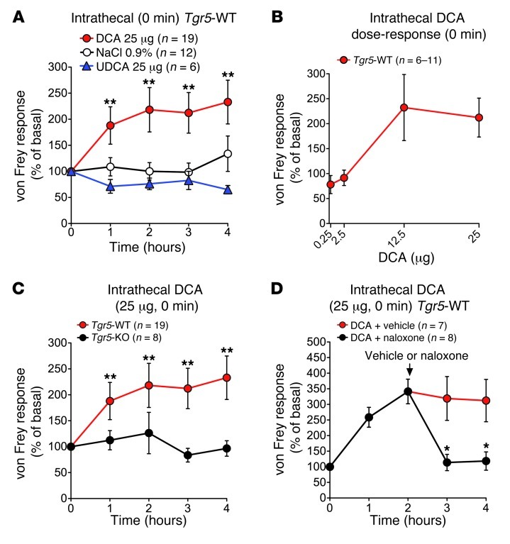
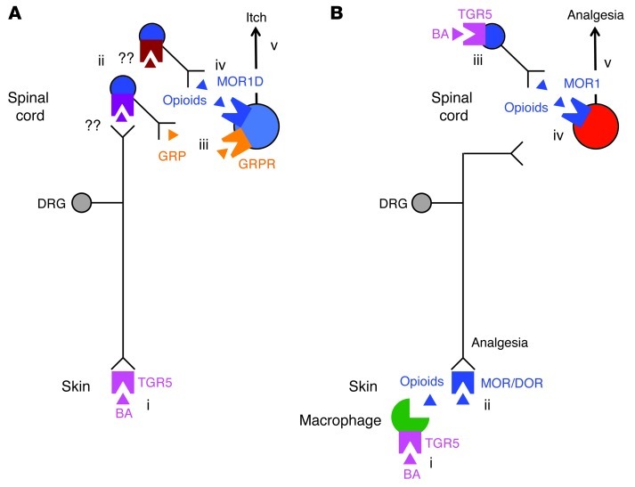
Patients with cholestatic disease exhibit pruritus and analgesia, but the mechanisms underlying these symptoms are unknown. We report that bile acids, which are elevated in the circulation and tissues during cholestasis, cause itch and analgesia by activating the GPCR TGR5. TGR5 was detected in peptidergic neurons of mouse dorsal root ganglia and spinal cord that transmit itch and pain, and in dermal macrophages that contain opioids. Bile acids and a TGR5-selective agonist induced hyperexcitability of dorsal root ganglia neurons and stimulated the release of the itch and analgesia transmitters gastrin-releasing peptide and leucine-enkephalin. Intradermal injection of bile acids and a TGR5-selective agonist stimulated scratching behavior by gastrin-releasing peptide- and opioid-dependent mechanisms in mice. Scratching was attenuated in Tgr5-KO mice but exacerbated in Tgr5-Tg mice (overexpressing mouse TGR5), which exhibited spontaneous pruritus. Intraplantar and intrathecal injection of bile acids caused analgesia to mechanical stimulation of the paw by an opioid-dependent mechanism. Both peripheral and central mechanisms of analgesia were absent from Tgr5-KO mice. Thus, bile acids activate TGR5 on sensory nerves, stimulating the release of neuropeptides in the spinal cord that transmit itch and analgesia. These mechanisms could contribute to pruritus and painless jaundice that occur during cholestatic liver diseases.


Figures

Figure 1
Figure 1. TGR5 expression and localization in mouse DRG.
(A) Single-cell RT-PCR analysis of DRG neurons from C57BL/6 mice. Small-diameter neurons were selected, and Tgr5, Grp, Trpa1, and Trpv1 mRNA was amplified. Results from 10 neurons are shown (78 neurons, 7 mice). Neurons 1, 2, 6, and 8 coexpressed Tgr5, Grp, Trpa1, and Trpv1. No transcripts were amplified from bath fluid. (B) Proportion of small-diameter neurons expressing Tgr5 (36%), Grp (50%), Trpa1 (59%), and Trpv1 (77%). Of the Tgr5-expressing neurons, 39% coexpressed Grp, 41% coexpressed Trpa1, and 32% coexpressed Trpv1. Tgr5, Grp, Trpa1, and Trpv1 were all coexpressed by 22% of small-diameter neurons. (C) Localization of TGR5-IR, Hu-IR, CGRP-IR, SP-IR, GRP-IR, and IB4-FITC binding in DRG (thoracic, lumbar, and sacral) of C57BL/6 mice. Arrowheads denote neurons coexpressing markers; arrowheads with asterisks denote lack of marker coexpression. TGR5-IR was prominently expressed in small-diameter Hu-positive neurons, most of which coexpressed CGRP-IR, SP-IR, or GRP-IR. TGR5-IR was rarely expressed in neurons that bound IB4-FITC. (D) Cross-sectional area of the TGR5-IR population (50-μm2 bins), which indicated that 50% of TGR5-IR neurons were 150–250 μm2. (E) Controls for specific detection of TGR5-IR. TGR5-IR was prominently detected in small-diameter DRG neurons of Tgr5-WT mice (arrowheads). TGR5-IR of small-diameter neurons of Tgr5-KO mice was markedly diminished (arrowheads with asterisks), although the background fluorescence of larger-diameter neurons was retained. Preadsorption of the TGR5 antibody with the receptor fragment used for immunization abolished TGR5-IR in DRG of C57BL/6 mice. There was no staining when the primary antibody was replaced with normal rabbit (Rb) IgG. Scale bars: 50 μm.
Figure 2
Figure 2. Expression and localization of TGR5 in mouse spinal cord and skin.
(A) Amplification of Tgr5 and Grp from mouse spinal cord and gall bladder. Shown are representative gels from 3 or 4 mice. (B) Localization of TGR5-IR, Hu-IR, CGRP-IR, SP-IR, GRP-IR, and IB4-FITC binding in the dorsal horn (dashed outline) of the spinal cord (sacral and lumbar) of C57BL/6 mice. Arrowheads denote neurons coexpressing markers; arrowheads with asterisks denote lack of marker coexpression. TGR5-IR was prominently localized in Hu-positive neurons in laminae I, II, and X. There was no clear colocalization of TGR5-IR with CGRP-IR, SP-IR, GRP-IR, or IB4-FITC in nerve fibers. Preadsorption of the TGR5 antibody abolished staining. (C) In mouse skin, TGR5-IR localized to dermal macrophages (arrows), which were identified based on appearance and location. Preadsorption of the TGR5 antibody with the receptor fragment used for immunization abolished TGR5-IR in spinal neurons and dermal macrophages. Scale bars: 50 μm.
Figure 3
Figure 3. Effects of DCA on intrinsic excitability of DRG neurons from Tgr5-WT and Tgr5-KO mice.
(A) Representative perforated current clamp recordings in response to 500 ms current injection at rheobase (left) and 2× rheobase (right). Recordings were made from the same neuron from a Tgr5-WT mouse before (control; top) and after (bottom) incubation with DCA (100 μM, 10 minutes). Square waves represent the electrical stimulus applied to the cell (500 ms). (B) Summary data showing rheobase and AP discharge frequency at 2× rheobase of neurons from Tgr5-WT and Tgr5-KO mice. Recordings were made before (control) and after incubation with DCA (100 μM, 10 minutes). Rheobase and AP discharge frequency were normalized to control values to account for variability in control responses between experiments. DCA decreased rheobase and increased AP discharge frequency at 2× rheobase in neurons from Tgr5-WT mice, but had no effect on neurons from Tgr5-KO mice. (C) Summary data showing that DCA did not affect resting membrane potential or input resistance of neurons from Tgr5-WT mice. *P < 0.05, ***P < 0.005 vs. control; paired t test. The number of neurons is indicated in parenthesis in each bar. Neurons were obtained from ≥3 mice.
Figure 4
Figure 4. Effects of graded concentrations of BAs and a TGR5 agonist on intrinsic excitability of DRG neurons from C57BL/6 mice.
(AC and E) Summary data showing rheobase and AP discharge frequency at 2× rheobase. Recordings were made before (control) and after incubation with DCA (A), TLCA (B), OA (C), or UDCA (E) (10, 30, or 100 μM, 10 minutes). Rheobase and AP discharge frequency were normalized to control values to account for variability in control responses between experiments. DCA (100 μM) decreased rheobase and increased AP discharge frequency, whereas TLCA (10 and 100 μM) only decreased rheobase. OA caused a robust and concentration-dependent decrease in rheobase and increase in AP discharge frequency. UDCA had no effect on rheobase or AP discharge frequency. (D) Representative recording of the membrane potential of a neuron immediately before and after incubation with OA (100 μM, 10 minutes). OA exposure resulted in spontaneous AP discharge (no input current). **P < 0.001, ***P < 0.0001 vs. control, 1-way ANOVA and Dunnett post-hoc test. The number of neurons is indicated in parenthesis in each bar. Neurons were obtained from ≥3 mice.
Figure 5
Figure 5. Effects of BAs, a TGR5-selective agonist, and capsaicin on GRP-IR, Leu-ENK–IR, and CGRP-IR release from rat spinal cord with attached dorsal roots.
Release of GRP-IR (A), Leu-ENK–IR (B), and CGRP-IR (C) was measured from superfused segments of rat spinal cord with attached dorsal roots (combined cervical, thoracic, and lumbar-sacral regions) under basal conditions and after superfusion with DCA, TLCA, OA, or UDCA (10, 100, and/or 500 μM, 60 minutes superfusion). DCA, TLCA, and OA stimulated GRP-IR, Leu-ENK–IR, and CGRP-IR release over basal. UDCA stimulated GRP-IR release only at the highest concentration (500 μM). Removal of extracellular calcium (–Ca2+) prevented DCA-, TLCA- and OA-stimulated release. Preincubation with capsaicin (+Cap) did not affect DCA-, TLCA-, or OA-evoked GRP-IR or Leu-ENK–IR release, but prevented CGRP-IR release. (D) Capsaicin did not stimulate GRP-IR release, but strongly stimulated CGRP-IR release. *P < 0.05 vs. basal in the same tissue; paired t test. n = 4–5.
Figure 6
Figure 6. Effects of BAs on scratching behavior in Tgr5-WT, Tgr5-KO, and Tgr5-Tg mice.
Results are expressed as the number of scratching events for the indicated time periods. (A and B) Spontaneous scratching in untreated mice. (A) Frequency of spontaneous scratching events during 30-minute intervals over 120 minutes of recording. (B) Frequency of spontaneous scratching events per 60 minutes, averaged over 120 minutes of recording. Tgr5-Tg mice exhibited increased frequency of spontaneous scratching compared with Tgr5-WT and Tgr5-KO mice. (C) Vehicle control (0.9% NaCl, intradermal to the nape of the neck) did not stimulate scratching, although scratching was generally more frequent in Tgr5-Tg mice. (D) DCA (25 μg, intradermal) robustly stimulated scratching in Tgr5-WT mice, and scratching was exacerbated in Tgr5-Tg mice and suppressed in Tgr5-KO mice. (E) DCA (5–25 μg) stimulated dose-dependent scratching in Tgr5-WT mice. (F and G) TLCA and OA (25 μg) stimulated scratching in Tgr5-WT mice, which was markedly attenuated in Tgr5-KO mice. (H) UDCA (25 μg) had a small stimulatory effect that was not different among Tgr5-Tg, Tgr5-KO, and Tgr5-WT mice. (I) Histamine (50 μg) stimulated scratching to a similar extent in Tgr5-Tg, Tgr5-KO, and Tgr5-WT mice. (J) Summarized results showing the frequency of scratching events during the first 60 minutes after injection. *P < 0.05, **P < 0.01 vs. Tgr5-WT; #P < 0.05 vs. 5 μg DCA; ANOVA and Student-Newman-Keuls post-hoc test. n is indicated.
Figure 7
Figure 7. Mechanisms of BA-stimulated scratching in mice.
(A) The GRPR antagonist [Tyr4, D-Phe12]-bombesin or vehicle (control) were injected intrathecally 10 minutes before intradermal injection of DCA (25 μg) to Tgr5-WT mice. GRPR antagonist attenuated DCA-stimulated scratching at all time points compared with vehicle. (B) Naloxone was administered intravenously 30 minutes before the first intradermal injection of DCA into Tgr5-WT mice (at 0 minutes). Naloxone attenuated the scratching response to the first DCA challenge. DCA administered at 180 minutes, when naloxone was cleared, strongly stimulated scratching. (C) Ketotifen or vehicle was administered intravenously 5 minutes before intradermal injection of DCA (25 μg) to Tgr5-WT mice. Ketotifen had no effect on the scratching response to DCA. (D) LPA (100 μg) was injected intradermally. Scratching frequency was similar in Tgr5-WT and Tgr5-KO mice. (E) cAMP generation in HEK293 cells expressing human TGR5. DCA stimulated concentration-dependent cAMP formation, whereas LPA did not stimulate cAMP generation (n = 3 experiments, in triplicate). *P < 0.05 vs. vehicle, **P < 0.01 as indicated; unpaired t test. n is indicated.
Figure 8
Figure 8. Peripheral mechanisms of BA- and TGR5-induced mechanical analgesia and edema in Tgr5-WT and Tgr5-KO mice.
Test agents were injected into the plantar surface of the hind paw. Responses to stimulation of the plantar surface of the paw with von Frey filaments of graded stiffness and paw thickness were recorded. Results are expressed as percent basal value. An increased von Frey response indicates that a stiffer filament was required to induce withdrawal (mechanical analgesia), whereas a decreased response indicates that a less stiff filament was required to induce withdrawal (mechanical hyperalgesia). (A) Intraplantar injection of DCA (12.5–125 μg) or capsaicin (5 μg) increased paw thickness in Tgr5-WT mice, indicative of inflammatory edema. (B) Intraplantar DCA (25 μg), but not UDCA (25 μg), caused analgesia, whereas capsaicin (5 μg) caused hyperalgesia, in Tgr5-WT mice. (C) Intraplantar DCA (12.5, 25, 37.5, and 125 μg) stimulated dose-dependent analgesia measured at 3 hours in Tgr5-WT mice. (D) Intraplantar DCA (25 μg) caused analgesia in Tgr5-WT, but not Tgr5-KO, mice. (E and F) TLCA and OA (25 μg) caused analgesia in Tgr5-WT, but not Tgr5-KO, mice. (G) Naloxone or vehicle control was administered by intraplantar injection 2 hours after intraplantar injection of DCA (25 μg) to Tgr5-WT mice. Naloxone rapidly reversed the mechanical analgesia. *P < 0.05, **P < 0.01 vs. respective vehicle control (A, B, and G) or vs. Tgr5-KO (D, E, and F); ANOVA and Student-Newman-Keuls (A and B) or unpaired t test (DF, and G). n is indicated.
Figure 9
Figure 9. Central mechanisms of BA- and TGR5-induced mechanical analgesia in Tgr5-WT and Tgr5-KO mice.
Test agents were injected intrathecally. Responses to stimulation of the plantar surface of the paw with von Frey filaments of graded stiffness were recorded. Results are expressed as percent basal value. (A) Intrathecal injection of DCA (25 μg), but not UDCA (25 μg), caused analgesia in Tgr5-WT mice. (B) Intrathecal DCA (0.25, 2.5, 12.5, and 25 μg) stimulated dose-dependent analgesia measured at 3 hours in Tgr5-WT mice. (C) Intrathecal DCA (25 μg) caused analgesia in Tgr5-WT, but not Tgr5-KO, mice. (D) Naloxone or vehicle control was administered systemically 2 hours after intrathecal injection of DCA (25 μg) to Tgr5-WT mice. Naloxone rapidly reversed the mechanical analgesia. *P < 0.05 vs. respective vehicle control (A and D) or Tgr5-KO (C); ANOVA and Student-Newman-Keuls test (A) or unpaired t test (C and D). n is indicated.
Figure 10
Figure 10. Hypothesized mechanisms of BA- and TGR5-induced itch and analgesia.
(A) Mechanisms of itch. BAs in the skin activate TGR5 on sensory nerve endings (i), which increases neuronal excitability and stimulates release of unknown transmitters from the central projections of sensory nerves in dorsal horn of the spinal cord (ii). The transmitters induce release of GRP (iii) and opioids (iv) from spinal neurons. GRP activates GRPR and opioids activate the MOR1D/GRPR heterodimer on itch-selective spinal neurons. Activated GRPR induces itch (v). (B) Mechanisms of analgesia. BAs in the skin activate TGR5 on dermal macrophages (i) to stimulate release of opioids that activate MORs and δ-opioid receptors (DOR) on sensory nerve endings to induce peripheral mechanisms of analgesia (ii). BAs in the spinal cord activate TGR5 on spinal neurons (iii) to stimulate release of opioids that activate MOR1 on pain-selective spinal neurons (iv). Activated MOR1 induces central mechanisms of analgesia (v).

Comment in

References

    1. European Association for the Study of the Liver. EASL Clinical Practice Guidelines: management of cholestatic liver diseases. J Hepatol. 2009;51(2):237–267. doi: 10.1016/j.jhep.2009.04.009. - DOI - PubMed
    1. Bergasa NV. The itch of liver disease. Semin Cutan Med Surg. 2011;30(2):93–98. doi: 10.1016/j.sder.2011.04.009. - DOI - PubMed
    1. Fitzgerald JE, White MJ, Lobo DN. Courvoisier’s gallbladder: law or sign? World J Surg. 2009;33(4):886–891. doi: 10.1007/s00268-008-9908-y. - DOI - PubMed
    1. Moretti EW, Robertson KM, Tuttle-Newhall JE, Clavien PA, Gan TJ. Orthotopic liver transplant patients require less postoperative morphine than do patients undergoing hepatic resection. J Clin Anesth. 2002;14(6):416–420. doi: 10.1016/S0952-8180(02)00390-2. - DOI - PubMed
    1. Nelson L, Vergnolle N, D’Mello C, Chapman K, Le T, Swain MG. Endogenous opioid-mediated antinociception in cholestatic mice is peripherally, not centrally, mediated. J Hepatol. 2006;44(6):1141–1149. doi: 10.1016/j.jhep.2005.11.043. - DOI - PubMed

Publication types

MeSH terms