. 2013 Apr;123(4):1798-808.
doi: 10.1172/JCI66220. Epub 2013 Mar 25.

Sudden unexpected death in a mouse model of Dravet syndrome

Affiliations

Sudden unexpected death in a mouse model of Dravet syndrome

Franck Kalume et al. J Clin Invest. 2013 Apr.

Abstract

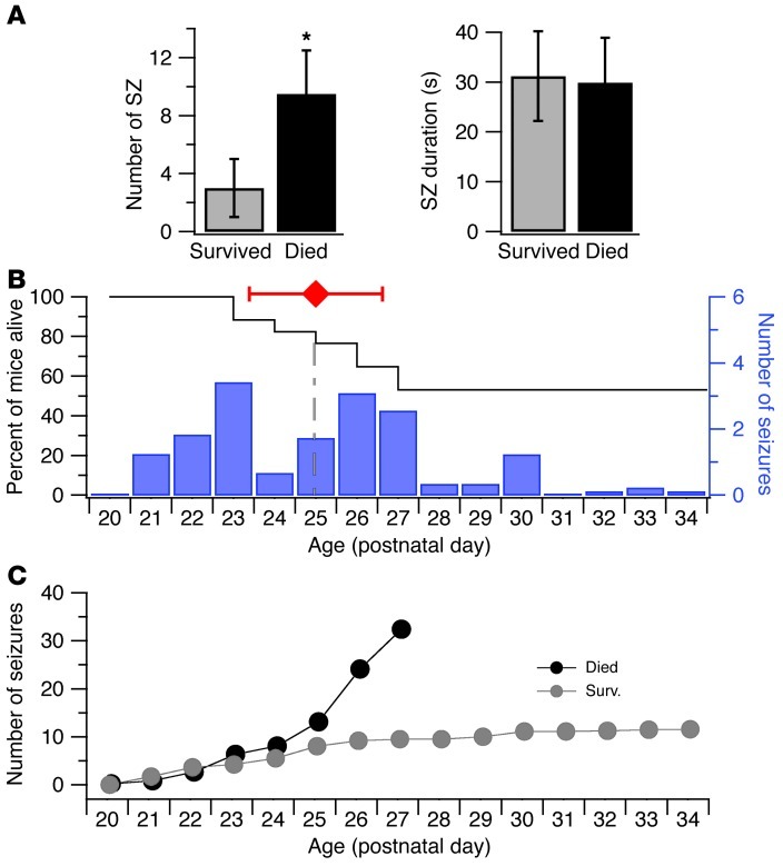
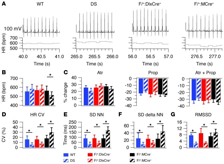
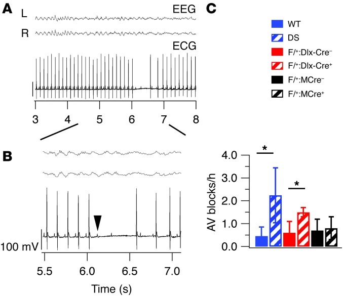
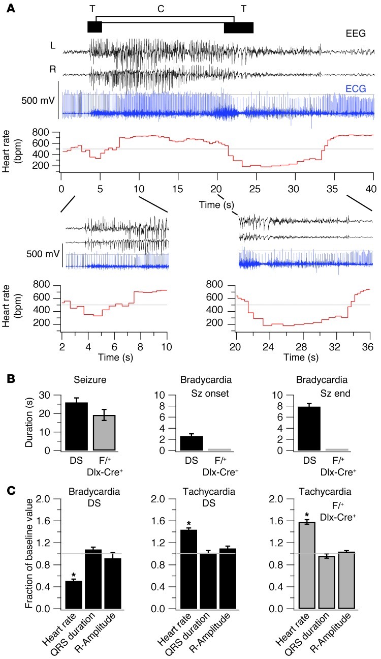
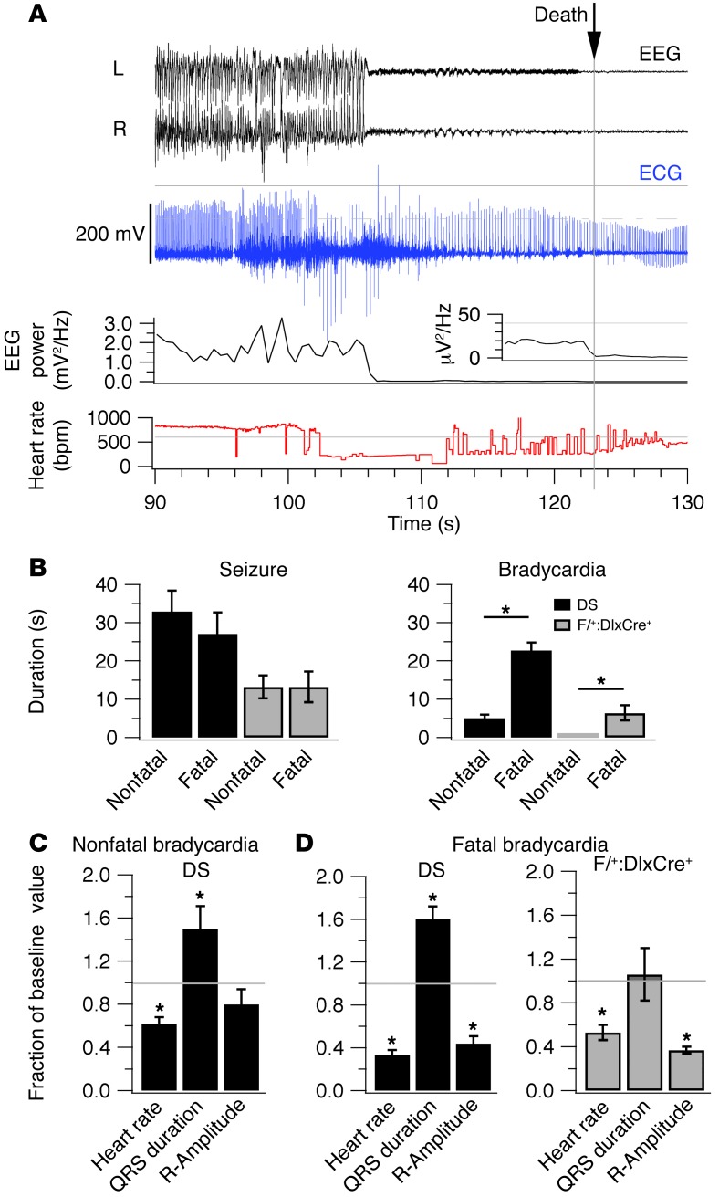
Sudden unexpected death in epilepsy (SUDEP) is the most common cause of death in intractable epilepsies, but physiological mechanisms that lead to SUDEP are unknown. Dravet syndrome (DS) is an infantile-onset intractable epilepsy caused by heterozygous loss-of-function mutations in the SCN1A gene, which encodes brain type-I voltage-gated sodium channel NaV1.1. We studied the mechanism of premature death in Scn1a heterozygous KO mice and conditional brain- and cardiac-specific KOs. Video monitoring demonstrated that SUDEP occurred immediately following generalized tonic-clonic seizures. A history of multiple seizures was a strong risk factor for SUDEP. Combined video-electroencephalography-electrocardiography revealed suppressed interictal resting heart-rate variability and episodes of ictal bradycardia associated with the tonic phases of generalized tonic-clonic seizures. Prolonged atropine-sensitive ictal bradycardia preceded SUDEP. Similar studies in conditional KO mice demonstrated that brain, but not cardiac, KO of Scn1a produced cardiac and SUDEP phenotypes similar to those found in DS mice. Atropine or N-methyl scopolamine treatment reduced the incidence of ictal bradycardia and SUDEP in DS mice. These findings suggest that SUDEP is caused by apparent parasympathetic hyperactivity immediately following tonic-clonic seizures in DS mice, which leads to lethal bradycardia and electrical dysfunction of the ventricle. These results have important implications for prevention of SUDEP in DS patients.


Figures

Figure 1
Figure 1. Spontaneous seizures and sudden unexpected deaths in DS mice.
(A) Incidence and duration of spontaneous generalized tonic-clonic convulsions 24 hours prior to death. Continuous short-term videos of DS mice in home cages were collected from P25 to P28. Times of death and convulsions in the 24 hours prior to each death were identified during off-line visual inspection of records. Incidence and duration of convulsions were calculated. Left bar graph illustrates higher incidence of seizures observed in mice that died (9.5 ± 3 seizures) than in those that survived (3 ± 2 seizures, P < 0.05). Right graph shows seizure durations (31.2 ± 9 s in mice that died vs. 29.9 ± 9 s in mice that survived, P > 0.05). *P < 0.05. Data are mean ± SD. (B) Survival plot (black line, left axis) and bar graph of daily convulsion incidence (right axis) in DS mice illustrating higher mortality and convulsion incidence in the fourth postnatal week of the mice. Continuous long-term videos of DS mice in home cages were collected from P20 to P34. Deaths and convulsions were identified during off-line inspections of records. Daily number of fatalities and seizure incidence were counted. (C) Graph of cumulative numbers of convulsions in mice studied in B showing a history of elevated number of convulsions experienced by mice that died sporadically during the monitoring period (P20–P34) than by those that survived.
Figure 2
Figure 2. Cardiovascular effects of global or conditional KO of Scn1a.
(A) Representative resting ECG traces with computed instantaneous heart rates. ECG traces were collected during 8-hour video-EEG-ECG recordings, and stable segments devoid of movement artifacts, electrical noise, ectopic beats, or arrhythmias were analyzed. (B) Resting heart rates (HR) calculated from the same ECG records used in A. (C) Change in resting heart rate (%) after acute i.p. administration of atropine (atr) (1 mg/kg), propranolol (prop) (4 mg/kg), and combined atropine (1 mg/kg) plus propranolol (4 mg/kg). Continuous video-EEG-ECG records were obtained before, during, and after drug treatment. Peak effects on ECG were observed around 30 minutes after injection. (DG) Bar graphs of indices of resting heart-rate variability in Scn1a KO mice and their respective controls. The values of the coefficients of variation of resting heart rate (HR CV) (D), SD of normal R-R intervals (SD NN) (E), SD of δ NN (F), and root mean square of differences between adjacent normal R-R intervals (RMSSD) (G) were depressed for DS and F/+:Dlx-Cre+ mice but elevated values for F/+:MCre+ mice. These indices were calculated from the same records used in A. *P < 0.05.
Figure 3
Figure 3. Frequency of AV block.
Increased interictal frequency of AV blocks in DS mice and forebrain interneuron-specific Scn1a-KO (F/+:Dlx-Cre+) mice, but not in cardiac-specific Scn1a-KO (F/+:MCre+) mice. The 8 hours of continuous video-EEG-ECG recordings were considered for assessments of AV blocks. ECG traces were visually inspected to identify these arrhythmias. (A) Representative simultaneous resting EEG-ECG records from a DS mouse showing regular ECG rhythm and a type-2, second-degree AV block, characterized by skipped QRS complexes without prolongation of preceding PR intervals. All preceding PR intervals in this figure had identical duration, 30.9 ms. (B) Magnified segments of the traces in A. Arrowhead indicates an example of a P wave. (C) Bar graph of interictal AV block frequencies in Scn1a mutant mice and respective control mice illustrating higher frequency of AV blocks in DS (2.24 ± 1.2 vs. 0.6 ± 0.4 AV blocks/h in controls) and F/+:Dlx-Cre+ mice (1.5 ± 0.2 AV blocks/h vs. 0.7 ± 0.5 AV blocks/h in controls), not in F/+:MCre+ mice (0.8 ± 0.5 AV blocks/h vs. 0.7 ± 0.6 AV blocks/h in controls). *P < 0.05.
Figure 4
Figure 4. Thermally induced seizures and bradycardia.
Continuous video-EEG-ECG records were obtained before, during, and after thermal seizure inductions in DS and F/+:Dlx-Cre+ mice as described in Methods. (A) Representative combined EEG-ECG records (black, blue) and computed instantaneous heart rate (red). The seizure was characterized by generalized spike-and-wave discharges and Racine 5 generalized tonic-clonic convulsions observed on video. Bradycardia was defined by a fall of instantaneous heart rate more than 1 SD below the mean (gray line). Tachycardias were defined by a rise of instantaneous heart rate more than 1 SD above the mean. Horizontal bars indicate the tonic (T) and clonic (C) phases of the seizure. (Lower traces) Magnified segments of records containing ictal bradycardias. (B) Seizure and bradycardia durations in 19 DS mice (black) and 5 F/+:Dlx-Cre+ (gray) mice that did not die. Left shows seizure (sz) durations: 26 ± 2.4 ms for deaths; 19.2 ± 3.0 ms for survivors. Middle and right show bradycardia durations: 2.6 ± 0.4 ms, seizure onset; 7.9 ± 0.6 ms, seizure termination in survivors. No bradycardia was observed in F/+:Dlx-Cre+ survivors. (C) Fractional changes in ECG intervals caused by thermal seizures in 19 DS (black) and 5 F/+:Dlx-Cre+ (gray) survivors, calculated as ratio of ictal bradycardia/baseline values. Left shows bradycardia in DS mice. QRS duration, 1.08 ± 0.04; R wave amplitude, 0.92 ± 0.1. Middle shows tachycardia in DS mice. QRS duration, 1.02 ± 0.04; R-wave amplitude, 1.1 ± 0.04. Right shows tachycardia in F/+:Dlx-Cre+ mice. QRS duration, 0.96 ± 0.04; R wave amplitude, 1.04 ± 0.02. No bradycardia was observed in F/+:Dlx-Cre+ survivors. *P < 0.05.
Figure 5
Figure 5. Ictal bradycardia and premature death.
(A) Representative traces of EEG-ECG records (black, blue), instantaneous heart rate, and instantaneous EEG power before death, illustrating the seizure and bradycardia preceding death. Time of death was defined as the moment when the power of the EEG fell to zero value, which coincided with cessation of ambulatory and respiration movements. Inset shows magnification of power trace below. (B) Left shows seizure durations in 4 DS (black) and 4 F/+:Dlx-Cre+ mice (gray) that died. In DS mice, the duration of seizures was 32.9 ± 5.5 ms for fatal seizures vs. 27.1 ± 5.6 ms for nonfatal seizures (P > 0.05). In F/+:Dlx-Cre+ mice, duration was 13.2 ± 3.0 for fatal seizures vs. 13.2 ± 4.0 ms for nonfatal (P > 0.05). Right shows bradycardia duration. DS mice, 22.8 ± 2.0 ms in fatal seizures vs. 5.1 ± 1.0 ms in nonfatal ones; F/+:Dlx-Cre+ mice, no nonfatal seizures; bradycardia duration for fatal seizures, 6.4 ± 2.0 ms. (C) Fractional changes in ECG parameters during nonfatal bradycardia in DS mice (n = 4), calculated as the ratio ictal bradycardia/baseline. (D) Fractional changes in ECG parameters in fatal bradycardia. Left shows DS mice (n = 4). Right shows F/+:Dlx-Cre+ mice (n = 4). *P < 0.05.
Figure 6
Figure 6. Effects of atropine and N-methylscopolamine on bradycardia and death.
Thermal seizures were induced in DS mice treated with autonomic nervous system blockers, and ECG traces were examined for ictal bradycardia. (A) DS mice without ictal bradycardia (%) after acute i.p. treatment with saline, atropine (1 mg/kg), or saline on 3 consecutive days. (B) DS mice without ictal bradycardia (%) after acute i.p. treatment with saline, propranolol (4 mg/kg), or saline on 3 consecutive days. (C) DS mice without ictal bradycardia (%) after acute i.p. treatment with saline, atropine plus propranolol, or saline on 3 consecutive days. (D) DS mice without ictal bradycardia (%) after acute i.p. treatment with atropine, N-methyl scopolamine (NMS), or saline on 3 consecutive days. (E) DS mice that died sporadically (%) during chronic treatment with atropine, NMS, or saline via osmotic pump from P21–P28. **P < 0.01 (χ2 test).

Comment in

  • Sudden death in epilepsy: of mice and men.
    Friedman D, Chyou J, Devinsky O. Friedman D, et al. J Clin Invest. 2013 Apr;123(4):1415-6. doi: 10.1172/JCI67759. Epub 2013 Mar 25. J Clin Invest. 2013. PMID: 23524959 Free PMC article.

References

    1. Annegers JF, Coan SP. SUDEP: overview of definitions and review of incidence data. Seizure. 1999;8(6):347–352. doi: 10.1053/seiz.1999.0306. - DOI - PubMed
    1. Aurlien D, Larsen JP, Gjerstad L, Tauboll E. Increased risk of sudden unexpected death in epilepsy in females using lamotrigine: a nested, case-control study. Epilepsia. 2012;53(2):258–266. doi: 10.1111/j.1528-1167.2011.03334.x. - DOI - PubMed
    1. Shorvon S, Tomson T. Sudden unexpected death in epilepsy. Lancet. 2011;378(9808):2028–2038. doi: 10.1016/S0140-6736(11)60176-1. - DOI - PubMed
    1. Hitiris N, Suratman S, Kelly K, Stephen LJ, Sills GJ, Brodie MJ. Sudden unexpected death in epilepsy: a search for risk factors. Epilepsy Behav. 2007;10(1):138–141. doi: 10.1016/j.yebeh.2006.11.010. - DOI - PubMed
    1. Marini C, et al. Idiopathic epilepsies with seizures precipitated by fever and SCN1A abnormalities. Epilepsia. 2007;48(9):1678–1685. doi: 10.1111/j.1528-1167.2007.01122.x. - DOI - PubMed

Publication types

MeSH terms