. 2013 May 15;126(Pt 10):2213-24.
doi: 10.1242/jcs.120519. Epub 2013 Mar 22.

Brain and muscle Arnt-like 1 is a key regulator of myogenesis

Affiliations

Brain and muscle Arnt-like 1 is a key regulator of myogenesis

Somik Chatterjee et al. J Cell Sci. .

Abstract

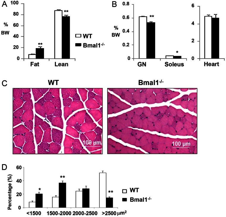
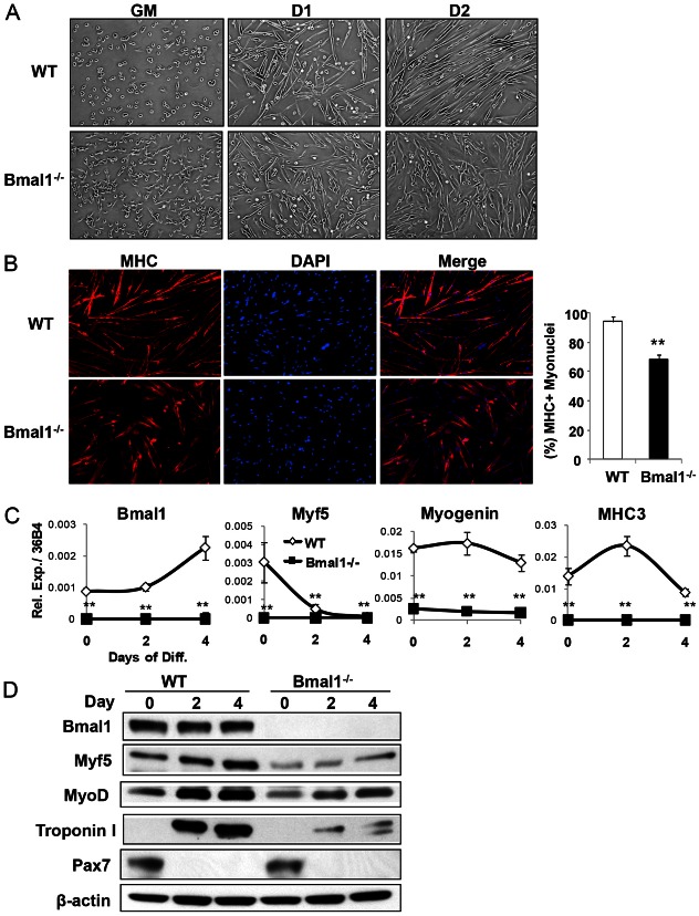
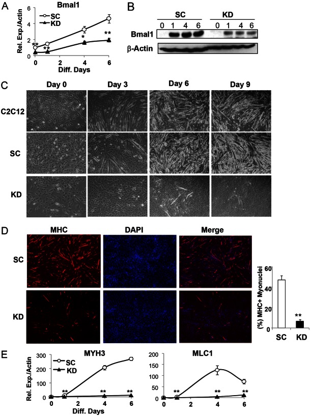
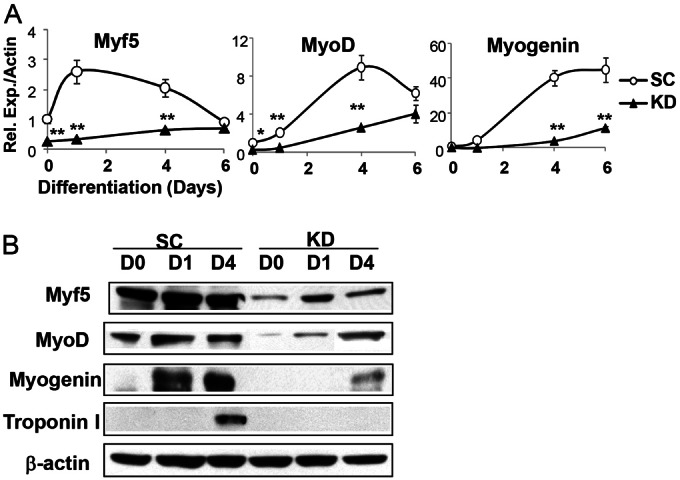
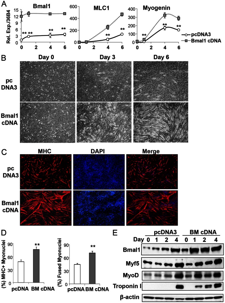
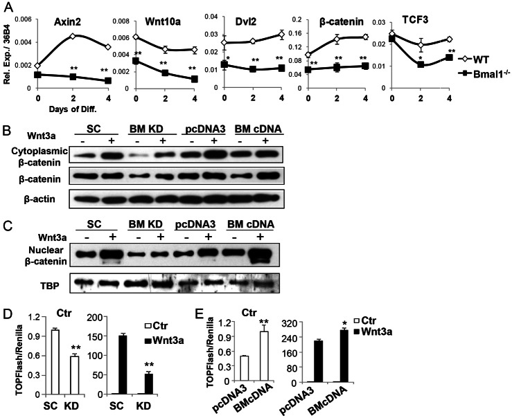
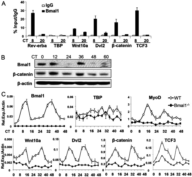
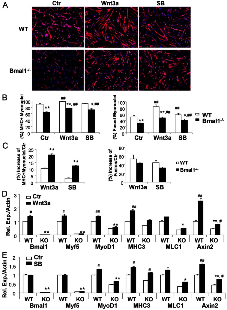
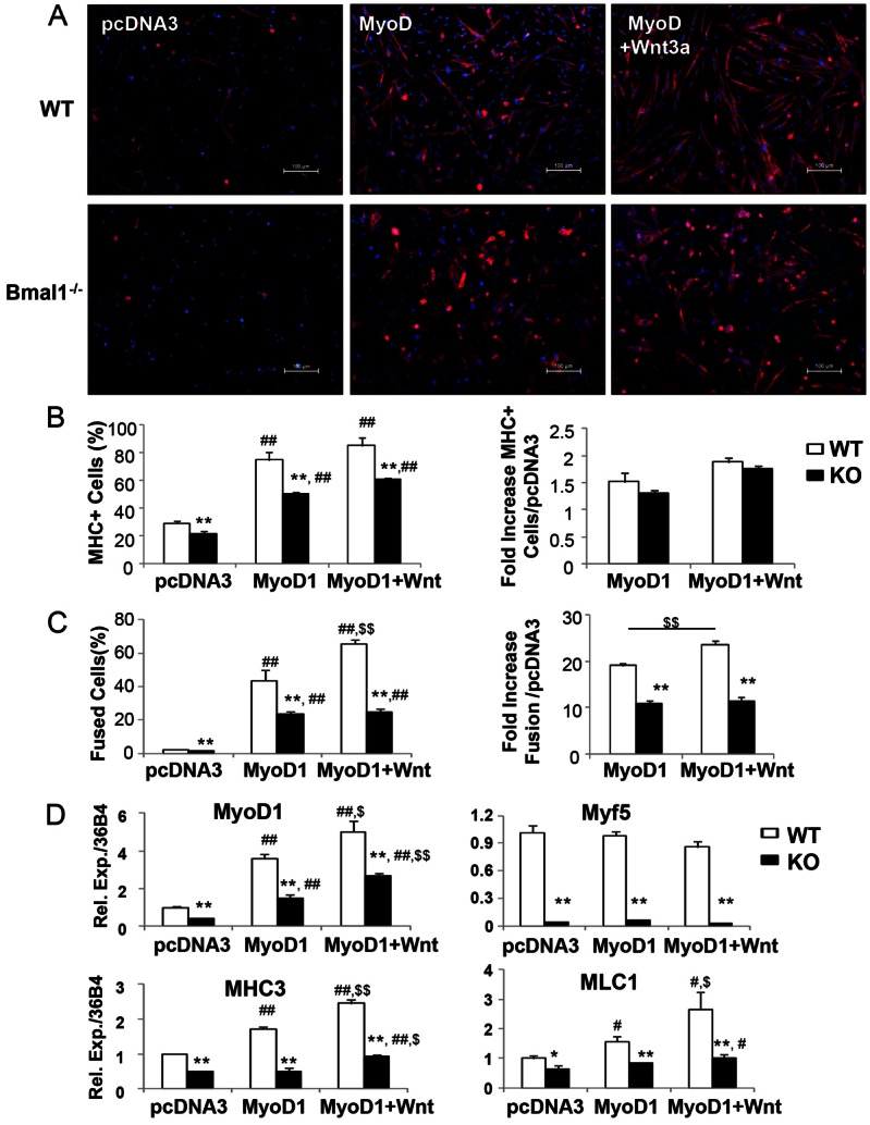
The circadian clock network is an evolutionarily conserved mechanism that imparts temporal regulation to diverse biological processes. Brain and muscle Arnt-like 1 (Bmal1), an essential transcriptional activator of the clock, is highly expressed in skeletal muscle. However, whether this key clock component impacts myogenesis, a temporally regulated event that requires the sequential activation of myogenic regulatory factors, is not known. Here we report a novel function of Bmal1 in controlling myogenic differentiation through direct transcriptional activation of components of the canonical Wnt signaling cascade, a major inductive signal for embryonic and postnatal muscle growth. Genetic loss of Bmal1 in mice leads to reduced total muscle mass and Bmal1-deficient primary myoblasts exhibit significantly impaired myogenic differentiation accompanied by markedly blunted expression of key myogenic regulatory factors. Conversely, forced expression of Bmal1 enhances differentiation of C2C12 myoblasts. This cell-autonomous effect of Bmal1 is mediated by Wnt signaling as both expression and activity of Wnt components are markedly attenuated by inhibition of Bmal1, and activation of the Wnt pathway partially rescues the myogenic defect in Bmal1-deficient myoblasts. We further reveal direct association of Bmal1 with promoters of canonical Wnt pathway genes, and as a result of this transcriptional regulation, Wnt signaling components exhibit intrinsic circadian oscillation. Collectively, our study demonstrates that the core clock gene, Bmal1, is a positive regulator of myogenesis, which may represent a temporal regulatory mechanism to fine-tune myocyte differentiation.

Keywords: Circadian clock; Myogenesis; Wnt signaling.


Figures

Fig. 1.
Fig. 1.
Reduced muscle mass in Bmal1-null (Bmal1−/−) mice. (A) Percentage of total lean and fat mass of 12-week-old mice (n = 8–10) determined by NMR analysis. BW, body weight. (B) Weights of isolated gastrocnemius (GN), soleus and heart muscles. (C) Representative micrographs of cross sections of quadriceps muscle from WT and Bmal1−/− mice, stained with Hematoxylin and Eosin. (D) Measurement of quadriceps muscle fiber cross-section area of 150 fibers in three representative fields for each animal (n = 4) in respective groups. *P<0.05, **P<0.01, Bmal1−/− versus WT by Student’s t-test.
Fig. 2.
Fig. 2.
Bmal1 deficiency impairs primary myoblast differentiation. (A) Phase-contrast images of isolated primary myoblasts from WT and Bmal1−/− mice during myogenesis ex vivo (10× magnification). GM, growth medium (day 0). (B) Immunofluorescence staining of myosin heavy chain (MHC) at day 2 of differentiation (10× magnification). Quantification of MHC-positive myonuclei from five representative fields (300–350 myonuclei/field) of three independent experiments is shown on the right. (C) Expression of myogenic factors and mature myocyte markers, determined by qPCR analysis (n = 3). Values are normalized to 36B4 as an internal control. (D) Immunoblot analysis of myogenic factors in WT and Bmal1−/− myoblasts. **P<0.01 Bmal1−/− versus WT by Student’s t-test.
Fig. 3.
Fig. 3.
Stable knockdown of Bmal1 suppresses myogenic differentiation of C2C12 myoblasts. (A) qPCR analysis of Bmal1 mRNA and (B) immunoblot analysis of protein level in stable knockdown (KD) and scrambled control (SC) cells. (C) Phase-contrast micrographs of the morphological changes during differentiation for 9 days in 2% horse serum. (D) MHC immunostaining at day 4 of differentiation (4× magnification). (Right panel) Quantification of MHC-positive myonuclei in five representative fields (800–1000 myonuclei/field) of four independent experiments. (E) qPCR analysis of mature myocyte marker genes, MLC1 and MHC3. Values are expressed as fold change compared with SC on day 0, after normalization to β-actin. *P<0.05, **P<0.01, Bmal1 KD versus SC control.
Fig. 4.
Fig. 4.
Silencing of Bmal1 attenuates myogenic transcription factor expression. (A) qPCR analysis (n = 3) and (B) immunoblot analysis of myogenic factors in Bmal1 SC and KD C2C12 myoblasts. Gene expression values are expressed as fold change compared with SC on day 0, after normalization to β-actin.
Fig. 5.
Fig. 5.
Forced expression of Bmal1 promotes C2C12 myoblast differentiation. (A) qPCR analysis of myogenic markers. (B) Phase-contrast micrographs of the morphological changes during differentiation of control (pcDNA3) and stable Bmal1-expressing cells (Bmal1 cDNA) (4× magnification). Gene expression values are expressed as fold change compared with empty vector control after normalization to 36B4. (C,D) MHC immunostaining (C) at day 4 of differentiation, and quantification of MHC-positive myonuclei and percentage of fusion (D) in five representative fields (1000–1200 myonuclei/field) from four independent experiments (10× magnification) . **P<0.01 Bmal1 cDNA versus pcDNA control. (E) Representative immunoblot analysis of myocyte-specific and myogenic factors from four independent experiments. BM cDNA, stable overexpression of Bmal1.
Fig. 6.
Fig. 6.
Bmal1 regulates genes of the Wnt signaling cascade. (A) Expression of Wnt pathway genes in Bmal1−/− primary myoblasts and WT controls, determined by qPCR analysis. Values are normalized to 36B4 as an internal control. (B,C) Representative immunoblot analysis of β-catenin cytoplasmic accumulation (B) and nuclear abundance (C) after treatments as indicated; from three independent experiments. BM cDNA, stable overexpression of Bmal1. (TBP was used as an internal loading control for nuclear proteins.) (D,E) TOPFlash luciferase assay under basal and Wnt3a-stimulated conditions in Bmal1 knockdown (D) and overexpression (E) in C2C12 myoblasts, with respective controls (n = 4). *P<0.05, **P<0.01.
Fig. 7.
Fig. 7.
Circadian regulation of Wnt pathway genes by Bmal1. (A) Chromatin immunoprecipitation (ChIP) analysis of Bmal1 occupancy of promoters of the Wnt pathway genes at CT8 and CT20 after serum shock synchronization in C2C12 myoblasts. CT, circadian time after serum shock. (B,C) Western blot (B) and qPCR analysis (C) of circadian expression patterns of Bmal1, TBP, MyoD and genes of the Wnt pathway in WT and Bmal1−/− primary myoblasts, starting at 1 hour after serum shock for 48 hours (n = 3/time point). Values are normalized to β-actin as an internal control. Data are representative of two independent experiments.
Fig. 8.
Fig. 8.
Activation of the Wnt pathway partially restores myogenesis in Bmal1-deficient primary myoblasts. (A) MHC immunostaining (10× magnification), (B) quantification of MHC-positive myonuclei and fusion of myonuclei (350–500 total nuclei/field), and (C) percentage increase of MHC-positive myonuclei or fusion after Wnt3a or SB-216763 treatment compared to the control condition in day-1 differentiated WT and Bmal1−/− myoblasts. Cells were treated with Wnt3a (40%) or SB-216763 (6 µM) for 8 hours prior to myogenic induction, and representative images of four independent experiments are shown. (D,E) qPCR analysis of myogenic marker gene and Wnt target gene expression of 2-day differentiated primary myoblasts treated with (D) Wnt3a or (E) SB-216763 (n = 4). Results are expressed as fold change compared with the no treatment control after normalization to β-actin. *P<0.05 and **P<0.01 Bmal1−/− versus WT with Wnt3a or SB-216763 treatment; #P<0.05, ##P<0.01 Wnt3a or SB-216763 treatment versus controls, respectively.
Fig. 9.
Fig. 9.
Effect of forced expression of MyoD on myogenesis in primary myoblasts. (A) MHC immunostaining (10× magnification), (B) quantification of MHC-positive myonuclei (left panel) and fold increase of MHC-positive myonuclei compared to vector (pcDNA3) control (right panel), and (C) fusion of myonuclei (left panel) and fold increase of fusion compared to vector (pcDNA3) control in WT and Bmal1−/− myoblasts transiently transfected with pcDNA3 or MyoD cDNA in the absence or presence of Wnt3a at day 1 of differentiation. Representative images of three independent experiments are shown, and 350–400 total nuclei/field were counted. (D) qPCR analysis of myogenic genes (n = 3). Values are expressed as fold change compared with vector alone controls after normalization to 36B4. *P<0.05 and **P<0.01 Bmal1−/− versus WT; #P<0.05, ##P<0.01 MyoD1 overexpression or MyoD1 overexpression with Wnt3a treatment versus pcDNA3 controls; $P<0.05, $$P<0.01 MyoD1 overexpression with Wnt3a treatment versus MyoD1 alone, by Student’s t-test.

Similar articles

Cited by

References

    1. Andrews J. L., Zhang X., McCarthy J. J., McDearmon E. L., Hornberger T. A., Russell B., Campbell K. S., Arbogast S., Reid M. B., Walker J. R. et al.(2010). CLOCK and BMAL1 regulate MyoD and are necessary for maintenance of skeletal muscle phenotype and function. Proc. Natl. Acad. Sci. USA 107, 19090–19095 10.1073/pnas.1014523107 - DOI - PMC - PubMed
    1. Balsalobre A., Damiola F., Schibler U. (1998). A serum shock induces circadian gene expression in mammalian tissue culture cells. Cell 93, 929–937 10.1016/S0092-8674(00)81199-X - DOI - PubMed
    1. Bass J., Takahashi J. S. (2010). Circadian integration of metabolism and energetics. Science 330, 1349–1354 10.1126/science.1195027 - DOI - PMC - PubMed
    1. Blau H. M., Pavlath G. K., Hardeman E. C., Chiu C. P., Silberstein L., Webster S. G., Miller S. C., Webster C. (1985). Plasticity of the differentiated state. Science 230, 758–766 10.1126/science.2414846 - DOI - PubMed
    1. Borello U., Berarducci B., Murphy P., Bajard L., Buffa V., Piccolo S., Buckingham M., Cossu G. (2006). The Wnt/beta-catenin pathway regulates Gli-mediated Myf5 expression during somitogenesis. Development 133, 3723–3732 10.1242/dev.02517 - DOI - PubMed

Publication types

Substances