. 2013 May 3;288(18):12828-39.
doi: 10.1074/jbc.M113.459396. Epub 2013 Mar 22.

Crystal structure and computational characterization of the lytic polysaccharide monooxygenase GH61D from the Basidiomycota fungus Phanerochaete chrysosporium

Affiliations

Crystal structure and computational characterization of the lytic polysaccharide monooxygenase GH61D from the Basidiomycota fungus Phanerochaete chrysosporium

Miao Wu et al. J Biol Chem. .

Abstract

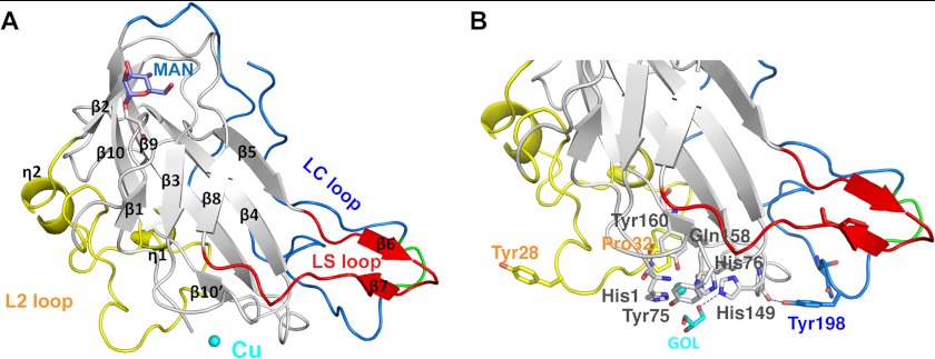
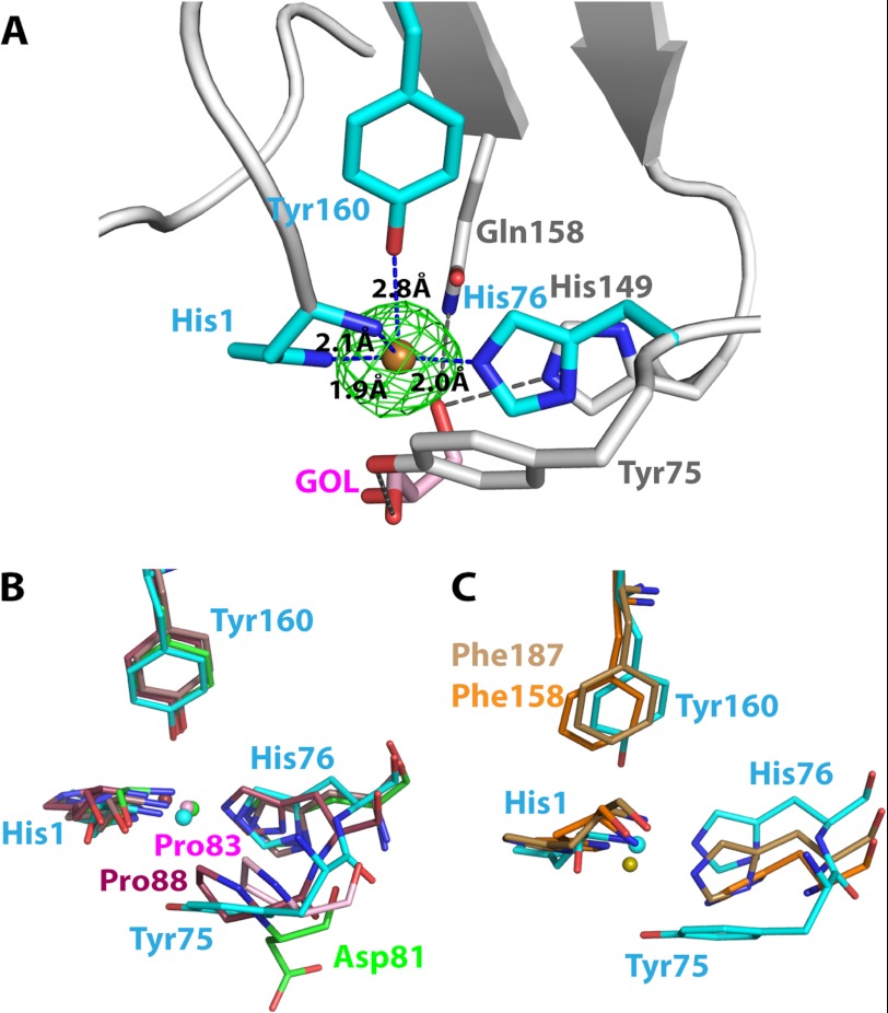
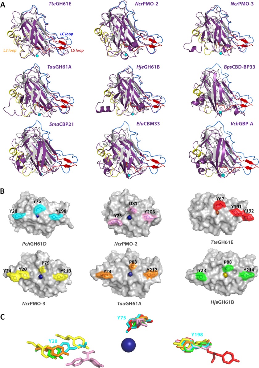
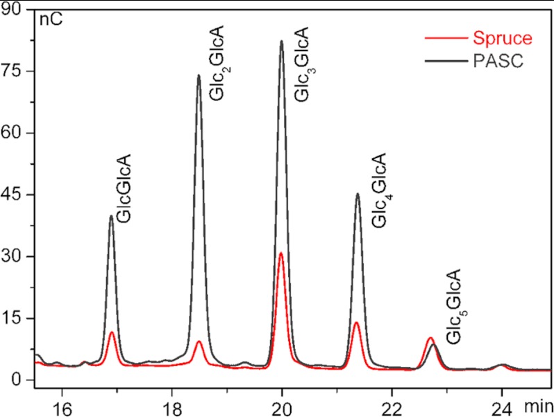
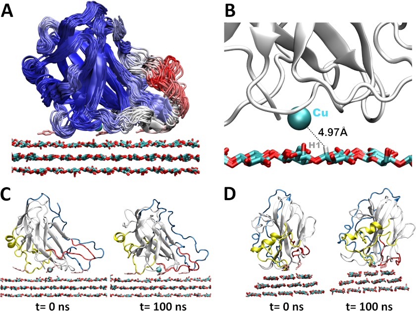
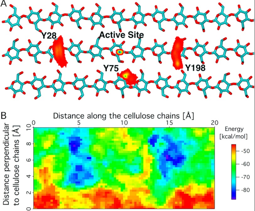
Carbohydrate structures are modified and degraded in the biosphere by a myriad of mostly hydrolytic enzymes. Recently, lytic polysaccharide mono-oxygenases (LPMOs) were discovered as a new class of enzymes for cleavage of recalcitrant polysaccharides that instead employ an oxidative mechanism. LPMOs employ copper as the catalytic metal and are dependent on oxygen and reducing agents for activity. LPMOs are found in many fungi and bacteria, but to date no basidiomycete LPMO has been structurally characterized. Here we present the three-dimensional crystal structure of the basidiomycete Phanerochaete chrysosporium GH61D LPMO, and, for the first time, measure the product distribution of LPMO action on a lignocellulosic substrate. The structure reveals a copper-bound active site common to LPMOs, a collection of aromatic and polar residues near the binding surface that may be responsible for regio-selectivity, and substantial differences in loop structures near the binding face compared with other LPMO structures. The activity assays indicate that this LPMO primarily produces aldonic acids. Last, molecular simulations reveal conformational changes, including the binding of several regions to the cellulose surface, leading to alignment of three tyrosine residues on the binding face of the enzyme with individual cellulose chains, similar to what has been observed for family 1 carbohydrate-binding modules. A calculated potential energy surface for surface translation indicates that P. chrysosporium GH61D exhibits energy wells whose spacing seems adapted to the spacing of cellobiose units along a cellulose chain.

Keywords: Biofuel; CBM33; Carbohydrate-binding Protein; Copper Monooxygenase; GH61; Glycoside Hydrolases; Lytic Polysaccharide Monooxygenase; Molecular Dynamics; Phanerochaete chrysosporium; Structural Biology.


Figures

FIGURE 1.
FIGURE 1.
Features of the PchGH61D crystal structure. A, schematic representation of the PchGH61D structure with the bound copper atom, depicted as a sphere in cyan. The L2 loop (residues 17–57) is colored in yellow, the short LS loop containing a β-hairpin in red (residues 109–124), and the C-terminal LC loop in blue (residues 170–217). All secondary structure elements of the enzyme are labeled according to their position in the protein sequence. The glycosylated residue Ser-11 and the attached O-linked mannose residue are shown in a stick representation in gray and slate blue, respectively. B, close up view of the structure showing potentially important residues at the proposed substrate-binding surface and the metal binding site in a stick representation with the same color coding as in A. The flexible portion of the LC loop (residues 201–204) is colored in green in both panels.
FIGURE 2.
FIGURE 2.
Copper K-edge fluorescence scan of the PchGH61D crystal. The scan demonstrates that copper is the bound metal.
FIGURE 3.
FIGURE 3.
The metal binding site of LPMOs. A, close up view of the PchGH61D in the vicinity of the copper binding site (PDB code 4B5Q). The green FoFc map of the copper atom is contoured at 0.41 e/Å3 (3σ). Cyan-colored residues are coordinated to the copper atom. A glycerol molecule was modeled below the copper atom at the active site, colored in pink (denoted GOL). The bound glycerol molecule is stabilized by His-149, Gln-158, and Tyr-75 (in gray) by hydrogen bonds. B, superposition of the metal binding sites of PchGH61D (PDB code 4B5Q; cyan) with the metal binding sites of NcrPMO-2 (4EIR; green), TauGH61A (3ZUD; pink), and HjeGH61B (2VTC; maroon). The metal ions were modeled as Cu2+ in the first three structures and as Ni2+ in the HjeGH61B structure. C, comparison of the metal binding site of PchGH61D (cyan) with the corresponding non-occupied metal binding sites of SmaCBP21 (2BEM; brown) and EfaCBM33 (4A02; orange).
FIGURE 4.
FIGURE 4.
Structural comparison of LPMOs. A, superimposed structures of PchGH61D (gray) with other LPMOs (purple): NcrPMO2 (PDB code 4EIR); TteGH61E (3EJA); NcrPMO-3 (4EIS); TauGH61A (3ZUD); HjeGH61B (2VTC); BpsCBD-BP33 (3UAM); SmaCBP21 (2BEM); EfaCBD-CBM33 (4A02); and VchGlc-binding protein A (2XWM). Yellow, blue, and red regions correspond to the L2 loop, LC loop, and LS loop, respectively, in the PchGH61D structure. B, aromatic residues (Tyr-28, Tyr-75, and Tyr-198) on the flat substrate binding surface of PchGH61D are shown on the molecular surface in cyan. The corresponding residues or additional aromatic residues on the surface of other GH61s are colored as follows. Pink, NcrPMO2 (PDB code 4EIR); red, TteGH61E (3EJA); yellow, NcrPMO-3 (4EIS); orange, TauGH61A (3ZUD); green, HjeGH61B (2VTC). The residue numbers are indicated beside the depicted residues. C, superposition of the residues shown in B with the corresponding color, in a stick representation. Tyr-25 in NcrPMO-2 occurs in two conformations in pink.
FIGURE 5.
FIGURE 5.
High performance anion exchange chromatography; chromatogram showing soluble aldonic acids (degree of polymerization 2–6) obtained upon incubation of 0.1% (w/v) PASC or 0.5% (w/v) steam-exploded spruce with 34 μg/ml PchGH61D in 25 μm sodium acetate, pH 5.3, 1.5 mm ascorbic acid for 20 h at 50 °C. Note the difference between the relative amounts of products released from the two substrates. Control reactions without enzyme yielded no detectable aldonic acids (data not shown).
FIGURE 6.
FIGURE 6.
Simulation results for PchGH61D on the hydrophobic surface of cellulose. A, cluster view of PchGH61D with snapshots taken every 5 ns, colored by RMSF from blue (low) to red (high). The tyrosine side chains (Tyr-28, Tyr-75, and Tyr-198) are shown in pink stick format in the conformation obtained after 100 ns. B, the copper (shown as a cyan sphere) fluctuates at ∼5 Å from the hydrogen atom on the C1 carbon during the MD simulation. C, side view of PchGH61D on the cellulose surface at t = 0 ns and t = 100 ns. The loops are colored as in Fig. 1. D, back view of PchGH61D on the cellulose surface at t = 0 ns and t = 100 ns.
FIGURE 7.
FIGURE 7.
A, histogram of the tyrosine residues (Tyr-28, Tyr-75, and Tyr-198) and the PchGH61D active site positions on the cellulose surface. The bottom two layers of cellulose are not shown, and the cellulose chains are truncated, both for visual clarity. The color code denotes the position on a 0.1 × 0.1-Å grid on the cellulose surface, with red being low density and blue being the highest density. B, PES of PchGH61D on cellulose. The x direction is along the chains of cellulose, and the y direction is perpendicular to the cellulose chains. Energy minima are found over the putative site of attack, with ∼10-Å separation (i.e. a distance corresponding to a cellobiose unit).

References

    1. Cantarel B. L., Coutinho P. M., Rancurel C., Bernard T., Lombard V., Henrissat B. (2009) The Carbohydrate-Active EnZymes database (CAZy). An expert resource for glycogenomics. Nucleic Acids Res. 37, D233–D238 - PMC - PubMed
    1. Vaaje-Kolstad G., Westereng B., Horn S. J., Liu Z., Zhai H., Sørlie M., Eijsink V. G. (2010) An oxidative enzyme boosting the enzymatic conversion of recalcitrant polysaccharides. Science 330, 219–222 - PubMed
    1. Forsberg Z., Vaaje-Kolstad G., Westereng B., Bunæs A. C., Stenstrøm Y., MacKenzie A., Sørlie M., Horn S. J., Eijsink V. G. (2011) Cleavage of cellulose by a CBM33 protein. Protein Sci. 20, 1479–1483 - PMC - PubMed
    1. Phillips C. M., Beeson W. T., Cate J. H., Marletta M. A. (2011) Cellobiose dehydrogenase and a copper-dependent polysaccharide monooxygenase potentiate cellulose degradation by Neurospora crassa. ACS Chem. Biol. 6, 1399–1406 - PubMed
    1. Quinlan R. J., Sweeney M. D., Lo Leggio L., Otten H., Poulsen J.-C., Johansen K. S., Krogh K. B., Jørgensen C. I., Tovborg M., Anthonsen A., Tryfona T., Walter C. P., Dupree P., Xu F., Davies G. J., Walton P. H. (2011) Insights into the oxidative degradation of cellulose by a copper metalloenzyme that exploits biomass components. Proc. Natl. Acad. Sci. 108, 15079–15084 - PMC - PubMed

Publication types

Associated data