. 2013 Jan 3;4(1):180-188.
doi: 10.1021/jz301893w. Epub 2012 Dec 18.

Hysteresis as a Marker for Complex, Overlapping Landscapes in Proteins

Affiliations

Hysteresis as a Marker for Complex, Overlapping Landscapes in Proteins

Benjamin T Andrews et al. J Phys Chem Lett. .

Abstract

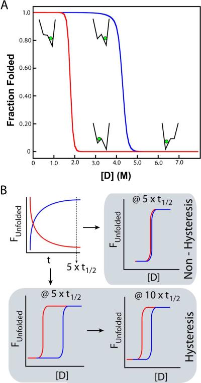
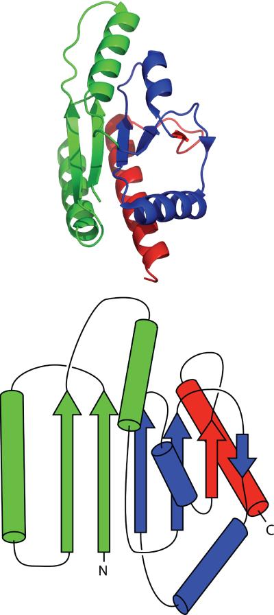
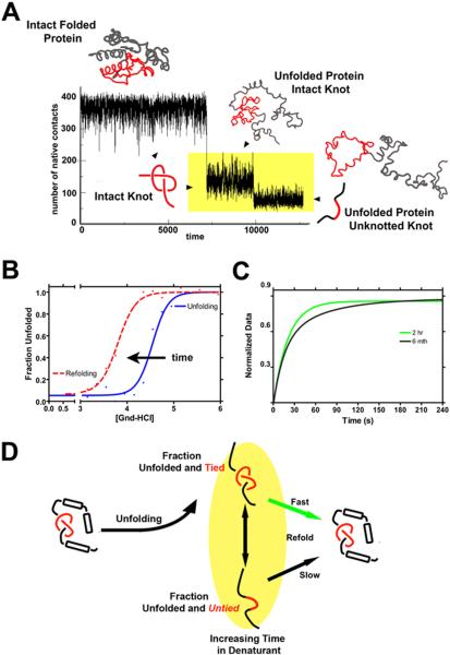
Topologically complex proteins fold by multiple routes as a result of hard-to-fold regions of the proteins. Oftentimes these regions are introduced into the protein scaffold for function and increase frustration in the otherwise smooth-funneled landscape. Interestingly, while functional regions add complexity to folding landscapes, they may also contribute to a unique behavior referred to as hysteresis. While hysteresis is predicted to be rare, it is observed in various proteins, including proteins containing a unique peptide cyclization to form a fluorescent chromophore as well as proteins containing a knotted topology in their native fold. Here, hysteresis is demonstrated to be a consequence of the decoupling of unfolding events from the isomerization or hula-twist of a chromophore in one protein and the untying of the knot in a second protein system. The question now is- can hysteresis be a marker for the interplay of landscapes where complex folding and functional regions overlap?

Keywords: Energy Landscape; Interplay; Knotted Proteins; Protein Folding.


Figures

Figure 1
Figure 1
(A) A rough energy landscape can lead to observed hysteresis. Non-coincidence of equilibrium unfolding (blue) and refolding (red) curves are evidence of a complex energy landscape. Hysteresis is a property in which a system does not immediately respond to the stresses applied to it, which may arise from a bifurcation in the energy landscape, which leads to a bi-stable system. Here, the observed equilibrium is dependent not only on the final conditions, but also the initial conditions (memory of the system). A simplified landscape is shown at selected parts of the hysteresis curves to show how a hysteresis cycle can arise. (B) Hysteresis is a kinetic effect manifesting itself within “equilibrium” data. At 5 half-lives, folding (red) and unfolding (blue) are considered complete. In a non-hysteretic (simple) folding scheme (upper right schematic), equilibrium curves overlay and are stable. In proteins that exhibit hysteresis (lower left panel), “equilibrium” folding and unfolding curves are non-equivalent after 5 half-lives, and may continue to drift (lower right) as a second, non-folding kinetic step limits denaturation.
Figure 2
Figure 2
GFP exists as a highly regular β-barrel surrounding the fluorescent chromophore (green). A splay diagram presents and numbers the β-strands as discussed in the text.
Figure 3
Figure 3
The thermophilic methyltransferase 1O6D is a knotted protein. A splay diagram simplifies the structure to focus on the knotted topology.
Figure 4
Figure 4
Theoretical and Experimental Data both Suggest Time-Dependent Unfolding of the Knotted Polypeptide Chain (A) The mechanism of unfolding shows two distinct steps, where unfolding of the secondary structure occurs first, followed by untying of the knot. The unfolding and untying events appear on distinctly different timescales and are highlighted in yellow. (B) Denaturant-induced unfolding (blue) and refolding (red dashes) measured by circular dichroism (CD) spectroscopy. The unfolding (blue) and refolding (red dashes) transitions for 1O6D show apparent hysteresis (the nonsuperimposability of the curves), consistent with the uncoupling of unfolding and untying of the knotted protein and the shift in the folded ensemble. The fit of the data was to a two-state model. Given enough time, these curves would coalesce. (C) Observed experimental refolding kinetics as a function of time in the denatured state, monitored by CD spectroscopy. Protein was unfolded at 6.0M denaturant (Gnd-HCl) for the given amount of time, and refolding was initiated by dilution to a final Gnd-HCl concentration of 3.2M. As predicted, changes are observed in the folding kinetics, consistent with untying the knot in the unfolded ensemble, and occur over a period of 6 months. The fit of the data was to a single-state (green trace) and two-state (black trace) model, respectively. (D) A schematic drawing of the “double-jump” experiment used in (C) to test the effect of the persistence of the knot in the denatured state on the refolding kinetics. In this scenario, extended times in the denatured state are necessary for untying of the unfolded protein.

References

    1. Levinthal C. Are There Pathways for Protein Folding? Extrait du Journal De Chimie Physique. 1968;65(1):44–7.
    1. Weissman JS. All roads lead to Rome? The multiple pathways of protein folding. Chem Biol. 1995;2(5):255–60. - PubMed
    1. Dill KA, Chan HS. From Levinthal to pathways to funnels. Nature structural biology. 1997;4(1):10–9. - PubMed
    1. Onuchic JN, Luthey-Schulten Z, Wolynes PG. Theory of protein folding: the energy landscape perspective. Annu Rev Phys Chem. 1997;48:545–600. - PubMed
    1. Onuchic JN, Wolynes PG. Theory of protein folding. Current opinion in structural biology. 2004;14(1):70–5. - PubMed