. 2013 Feb 14;2(1):e000034.
doi: 10.1161/JAHA.112.000034.

Impaired collagen biosynthesis and cross-linking in aorta of patients with bicuspid aortic valve

Affiliations

Impaired collagen biosynthesis and cross-linking in aorta of patients with bicuspid aortic valve

Dick Wågsäter et al. J Am Heart Assoc. .

Abstract

Background: Patients with bicuspid aortic valve (BAV) have an increased risk of developing ascending aortic aneurysm. In the present study, collagen homeostasis in nondilated and dilated aorta segments from patients with BAV was studied, with normal and dilated aortas from tricuspid aortic valve (TAV) patients as reference.

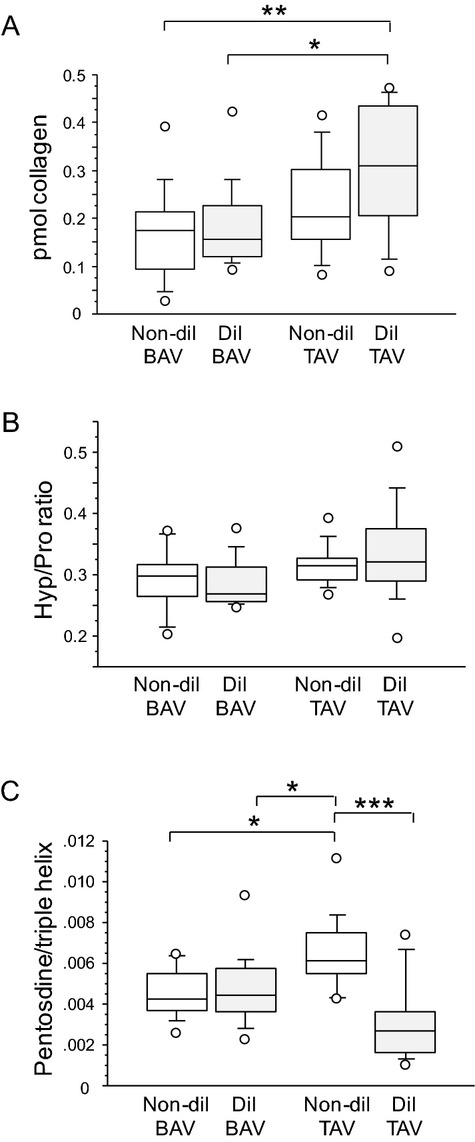
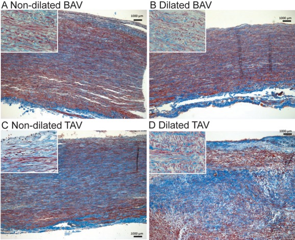
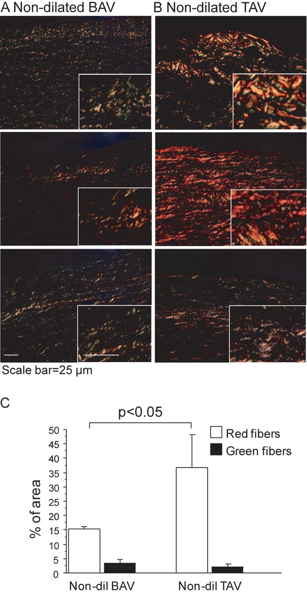
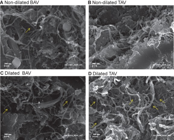
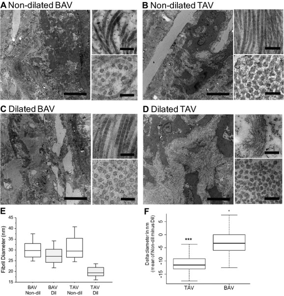
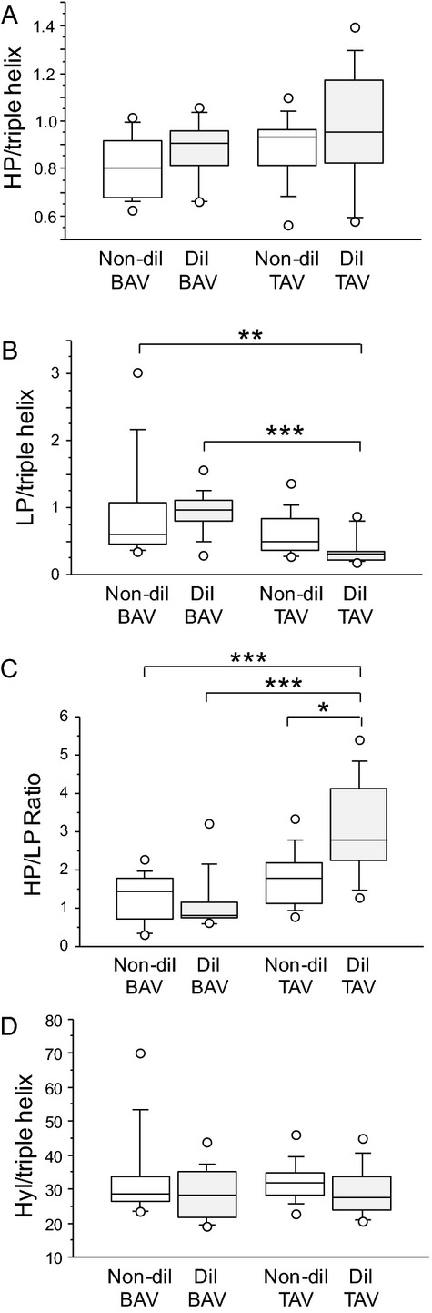
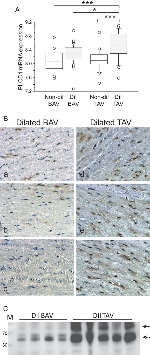
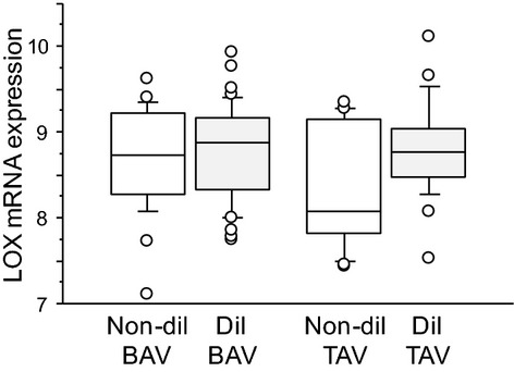
Methods and results: Ascending aortas from 56 patients were used for biochemical and morphological analyses of collagen. mRNA expression was analyzed in 109 patients. Collagen turnover rates were similar in nondilated and dilated aortas of BAV patients, showing that aneurysmal formation in BAV is, in contrast to TAV, not associated with an increased collagen turnover. However, BAV in general was associated with an increased aortic collagen turnover compared with nondilated aortas of TAV patients. Importantly, the ratio of hydroxylysyl pyridinoline (HP) to lysyl pyridinoline (LP), 2 distinct forms of collagen cross-linking, was lower in dilated aortas from patients with BAV, which suggests that BAV is associated with a defect in the posttranslational collagen modification. This suggests a deficiency at the level of lysyl hydroxylase (PLOD1), which was confirmed by mRNA and protein analyses that showed reduced PLOD1 expression but normal lysyl oxidase expression in dilated aortas from patients with BAV. This suggests that impaired collagen cross-linking in BAV patients may be attributed to changes in the expression and/or activity of PLOD1.

Conclusions: Our results demonstrate an impaired biosynthesis and posttranslational modification of collagen in aortas of patients with BAV, which may explain the increased aortic aneurysm formation in BAV patients.


Figures

Figure 1.
Figure 1.
Collagen content (A, B) and turnover (C) in nondilated and dilated aortas from patients with BAV and TAV. *P<0.05, **P<0.01, ***P<0.001 after adjustment for age. BAV nondilated (Non‐dil), n=12; BAV dilated (Dil), n=16; TAV nondilated, n=15; TAV dilated, n=13. BAV indicates bicuspid aortic valve; TAV, tricuspid aortic valve; Hyp/Pro, hydroxyproline/proline.
Figure 2.
Figure 2.
Masson trichrome staining of dilated and nondilated aortas from patients with BAV and TAV. Scale bar=1000 μm. Representative photomicrographs from 16 patients in total are shown. BAV indicates bicuspid aortic valve; TAV, tricuspid aortic valve.
Figure 3.
Figure 3.
Picrosirius red staining of nondilated aortas from 3 randomly selected patients with BAV (A) and TAV (B). Green fibers indicate immature thin fibers and orange‐red fibers indicate mature, thicker, and better‐aligned collagen fibers. Magnifications of the stained areas are shown in right corner in each figure. All figures have been taken using the same conditions and settings. Scale bar=25 μm. C, Quantification of picrosirius red staining. Error bars are indicated by SEM. BAV nondilated (Non‐dil), n=3; TAV nondilated (Dil), n=3. BAV indicates bicuspid aortic valve; TAV, tricuspid aortic valve.
Figure 4.
Figure 4.
Scanning electron microscopy of collagen structure in ascending aortas. Nondilated aortas from patients with BAV (A) and TAV (B); dilated aortas of patients with BAV (C) and TAV (D). Representative photomicrographs from a total of 10 patients are shown. Thin collagen fibers are indicated in yellow. Bar=300 nm. BAV indicates bicuspid aortic valve; TAV, tricuspid aortic valve.
Figure 5.
Figure 5.
Transmission electron microscopy of collagen structure in ascending aortas. Nondilated (Non‐dil) aortas from patients with BAV (A) and TAV (B); dilated (Dil) aortas of patients with BAV (C) and TAV (D). Images represent overview, collagen fibers, and cross‐sectioned fibers. Bars are 5 μm, 200 nm, and 100 nm, respectively. Representative photomicrographs from a total of 4 patients are shown. E and F, Quantification of fibril diameters; 100 randomly selected fibrils from 4 patients were measured. ***P<0.001. BAV indicates bicuspid aortic valve; TAV, tricuspid aortic valve.
Figure 6.
Figure 6.
Cross‐linking of collagen in nondilated (Non‐dil) and dilated (Dil) aortas from patients with BAV and TAV. A, Hydroxylysylpyridinoline (HP); B, lysylpyridinoline (LP); C, HP:LP ratio; D, hydroxylysine (Hyl) residues. *P=0.05, **P<0.01, ***P<0.001 after adjustment for age. BAV nondilated, n=12; BAV dilated, n=16; TAV nondilated, n=15; TAV dilated, n=13. BAV indicates bicuspid aortic valve; TAV, tricuspid aortic valve.
Figure 7.
Figure 7.
Decreased PLOD1 expression in dilated aortas of BAV patients. A, mRNA expression of PLOD1 in intima‐media of ascending aortas from patients with BAV and TAV. BAV nondilated (Non‐dil), n=24; BAV dilated (Dil), n=45; TAV nondilated, n=17; TAV dilated, n=23. *P<0.05, ***P<0.001 after adjustment for age. B, PLOD1 expression in media region of dilated ascending aortas from BAV (n=3) and TAV (n=3). C, Western blot of PLOD1 using protein extracts from dilated ascending aortas from BAV (N=4) and TAV (N=5). Arrow denotes PLOD1 protein with molecular weight of 83 kDa; broken arrow shows upregulated band of lower molecular weight. BAV indicates bicuspid aortic valve; TAV, tricuspid aortic valve.
Figure 8.
Figure 8.
No difference in LOX mRNA expression between BAV and TAV. mRNA expression of LOX in intima/media of ascending aortas from patients with BAV and TAV. P>0.10 after adjustment for age. BAV nondilated (Non‐dil), n=24; BAV dilated (Dil), n=45; TAV nondilated, n=17; TAV dilated, n=23. BAV indicates bicuspid aortic valve; TAV, tricuspid aortic valve.

References

    1. Fedak PW, David TE, Borger M, Verma S, Butany J, Weisel RD. Bicuspid aortic valve disease: recent insights in pathophysiology and treatment. Expert Rev Cardiovasc Ther. 2005; 3:295-308 - PubMed
    1. Yasuda H, Nakatani S, Stugaard M, Tsujita‐Kuroda Y, Bando K, Kobayashi J, Yamagishi M, Kitakaze M, Kitamura S, Miyatake K. Failure to prevent progressive dilation of ascending aorta by aortic valve replacement in patients with bicuspid aortic valve: comparison with tricuspid aortic valve. Circulation. 2003; 108suppl 1:II291-II294 - PubMed
    1. Nistri S, Sorbo MD, Marin M, Palisi M, Scognamiglio R, Thiene G. Aortic root dilatation in young men with normally functioning bicuspid aortic valves. Heart. 1999; 82:19-22 - PMC - PubMed
    1. Nkomo VT, Enriquez‐Sarano M, Ammash NM, Melton LJ, III, Bailey KR, Desjardins V, Horn RA, Tajik AJ. Bicuspid aortic valve associated with aortic dilatation: a community‐based study. Arterioscler Thromb Vasc Biol. 2003; 23:351-356 - PubMed
    1. Nistri S, Sorbo MD, Basso C, Thiene G. Bicuspid aortic valve: abnormal aortic elastic properties. J Heart Valve Dis. 2002; 11:369-373‐ - PubMed

Publication types

MeSH terms