

Insights into the structure and assembly of the Bacillus subtilis clamp-loader complex and its interaction with the replicative helicase
- PMID: 23525462
- PMCID: PMC3643586
- DOI: 10.1093/nar/gkt173
Insights into the structure and assembly of the Bacillus subtilis clamp-loader complex and its interaction with the replicative helicase
Abstract
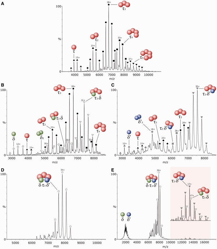
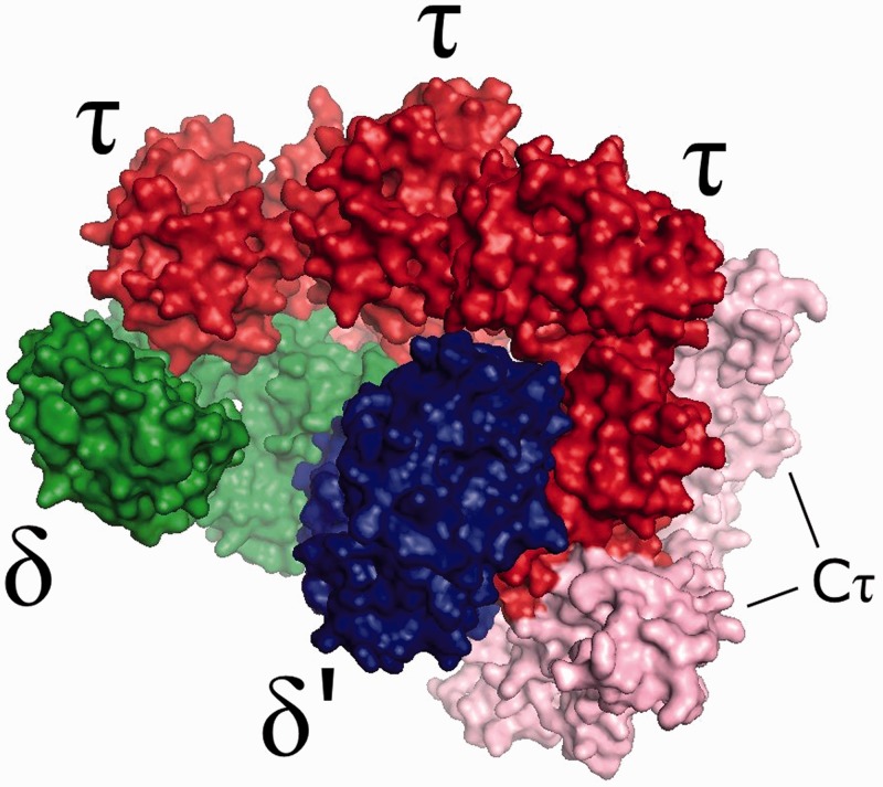
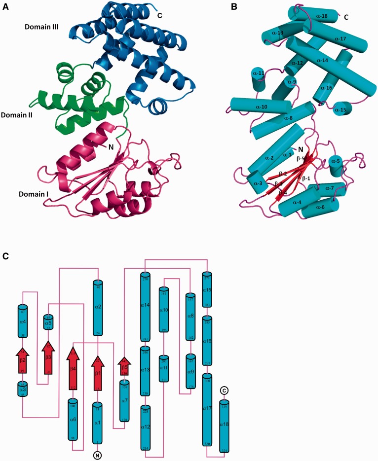
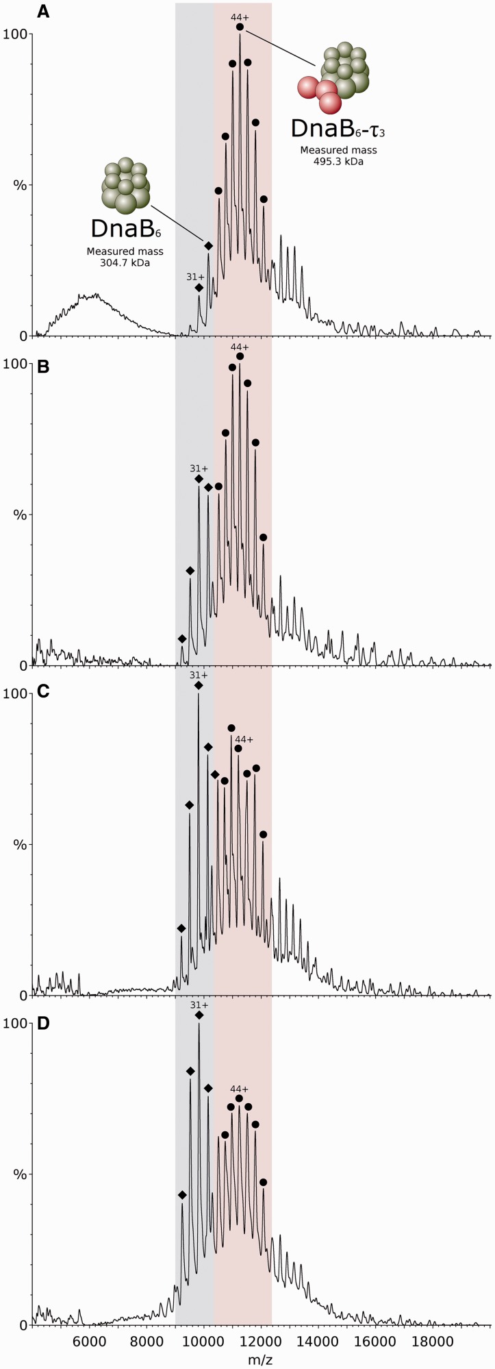
The clamp-loader complex plays a crucial role in DNA replication by loading the β-clamp onto primed DNA to be used by the replicative polymerase. Relatively little is known about the stoichiometry, structure and assembly pathway of this complex, and how it interacts with the replicative helicase, in Gram-positive organisms. Analysis of full and partial complexes by mass spectrometry revealed that a hetero-pentameric τ3-δ-δ' Bacillus subtilis clamp-loader assembles via multiple pathways, which differ from those exhibited by the Gram-negative model Escherichia coli. Based on this information, a homology model of the B. subtilis τ3-δ-δ' complex was constructed, which revealed the spatial positioning of the full C-terminal τ domain. The structure of the δ subunit was determined by X-ray crystallography and shown to differ from that of E. coli in the nature of the amino acids comprising the τ and δ' binding regions. Most notably, the τ-δ interaction appears to be hydrophilic in nature compared with the hydrophobic interaction in E. coli. Finally, the interaction between τ3 and the replicative helicase DnaB was driven by ATP/Mg(2+) conformational changes in DnaB, and evidence is provided that hydrolysis of one ATP molecule by the DnaB hexamer is sufficient to stabilize its interaction with τ3.
Figures
Similar articles
-
The clamp-loader-helicase interaction in Bacillus. Atomic force microscopy reveals the structural organisation of the DnaB-tau complex in Bacillus.J Mol Biol. 2004 Feb 13;336(2):381-93. doi: 10.1016/j.jmb.2003.12.043. J Mol Biol. 2004. PMID: 14757052 Free PMC article.
-
Allosteric regulation of the primase (DnaG) activity by the clamp-loader (tau) in vitro.Mol Microbiol. 2009 Apr;72(2):537-49. doi: 10.1111/j.1365-2958.2009.06668.x. Mol Microbiol. 2009. PMID: 19415803 Free PMC article.
-
Helicase binding to DnaI exposes a cryptic DNA-binding site during helicase loading in Bacillus subtilis.Nucleic Acids Res. 2006;34(18):5247-58. doi: 10.1093/nar/gkl690. Epub 2006 Sep 26. Nucleic Acids Res. 2006. PMID: 17003052 Free PMC article.
-
Structural Insight Into the Function of DnaB Helicase in Bacterial DNA Replication.Proteins. 2025 Feb;93(2):420-429. doi: 10.1002/prot.26746. Epub 2024 Sep 4. Proteins. 2025. PMID: 39230358 Review.
-
ATP Analogues for Structural Investigations: Case Studies of a DnaB Helicase and an ABC Transporter.Molecules. 2020 Nov 12;25(22):5268. doi: 10.3390/molecules25225268. Molecules. 2020. PMID: 33198135 Free PMC article. Review.
Cited by
-
Primase is required for helicase activity and helicase alters the specificity of primase in the enteropathogen Clostridium difficile.Open Biol. 2016 Dec;6(12):160272. doi: 10.1098/rsob.160272. Open Biol. 2016. PMID: 28003473 Free PMC article.
-
Mechanistic insight into the assembly of the HerA-NurA helicase-nuclease DNA end resection complex.Nucleic Acids Res. 2017 Nov 16;45(20):12025-12038. doi: 10.1093/nar/gkx890. Nucleic Acids Res. 2017. PMID: 29149348 Free PMC article.
-
Analyzing Protein Architectures and Protein-Ligand Complexes by Integrative Structural Mass Spectrometry.J Vis Exp. 2018 Oct 15;(140):57966. doi: 10.3791/57966. J Vis Exp. 2018. PMID: 30371663 Free PMC article.
-
Nucleotide and partner-protein control of bacterial replicative helicase structure and function.Mol Cell. 2013 Dec 26;52(6):844-54. doi: 10.1016/j.molcel.2013.11.016. Mol Cell. 2013. PMID: 24373746 Free PMC article.
-
Replisomal coupling between the α-pol III core and the τ-subunit of the clamp loader complex (CLC) are essential for genomic integrity in Escherichia coli.J Biol Chem. 2025 Feb;301(2):108177. doi: 10.1016/j.jbc.2025.108177. Epub 2025 Jan 10. J Biol Chem. 2025. PMID: 39798872 Free PMC article.
References
-
- Johnson A, O’Donnell M. Cellular DNA replicases: components and dynamics at the replication fork. Annu. Rev. Biochem. 2005;74:283–315. - PubMed
-
- Kong XP, Onrust R, O’Donnell M, Kuriyan J. Three-dimensional structure of the beta subunit of E. coli DNA polymerase III holoenzyme: a sliding DNA clamp. Cell. 1992;69:425–437. - PubMed
-
- Indiani C, O’Donnell M. The replication clamp-loading machine at work in the three domains of life. Nat. Rev. Mol. Cell Biol. 2006;7:751–761. - PubMed
Publication types
MeSH terms
Substances
Grants and funding
LinkOut - more resources
Full Text Sources
Other Literature Sources
Molecular Biology Databases
