Review
. 2013 Jun;11(2):136-46.
doi: 10.1007/s11914-013-0140-9.

High-resolution peripheral quantitative computed tomography for the assessment of bone strength and structure: a review by the Canadian Bone Strength Working Group

Affiliations
Review

High-resolution peripheral quantitative computed tomography for the assessment of bone strength and structure: a review by the Canadian Bone Strength Working Group

Angela M Cheung et al. Curr Osteoporos Rep. 2013 Jun.

Abstract

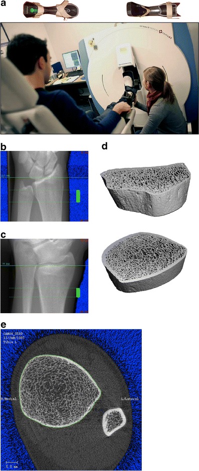
Bone structure is an integral determinant of bone strength. The availability of high resolution peripheral quantitative computed tomography (HR-pQCT) has made it possible to measure three-dimensional bone microarchitecture and volumetric bone mineral density in vivo, with accuracy previously unachievable and with relatively low-dose radiation. Recent studies using this novel imaging tool have increased our understanding of age-related changes and sex differences in bone microarchitecture, as well as the effect of different pharmacological therapies. One advantage of this novel tool is the use of finite element analysis modelling to non-invasively estimate bone strength and predict fractures using reconstructed three-dimensional images. In this paper, we describe the strengths and limitations of HR-pQCT and review the clinical studies using this tool.


Figures

Fig. 1
Fig. 1
The number of publications each year that utilized high-resolution peripheral quantitative computed tomography
Fig. 2
Fig. 2
a, A typical setup of HR-pQCT for patient measurements. At top, the casts used for securing the forearm and lower leg is shown. b, c, and d, The procedure for HR-pQCT analysis requires a ‘scout view’ x-ray of the radius or tibia (left) so that the operator can select the region of interest for scanning (solid green bar). Subsequently, three-dimensional data can be obtained in the scan region (right). e, A typical section from HR-pQCT showing the ultradistal tibia and fibula. The green line on the periosteal surface of the tibia is used to extract the bone of interest for subsequent three-dimensional analysis

References

    1. Tjong W, Kazakia GJ, Burghardt AJ, Majumdar S. The effect of voxel size on high-resolution peripheral computed tomography measurements of trabecular and cortical bone microstructure. Med Phys. 2012;39:1893–1903. doi: 10.1118/1.3689813. - DOI - PMC - PubMed
    1. Burghardt AJ, Pialat JB, Kazakia GJ, et al. Multicenter precision of cortical and trabecular bone quality measures assessed by high-resolution peripheral quantitative computed tomography. J Bone Miner Res. 2013;28:524–536. doi: 10.1002/jbmr.1795. - DOI - PMC - PubMed
    1. Sekhon K, Kazakia GJ, Burghardt AJ, et al. Accuracy of volumetric bone mineral density measurement in high-resolution peripheral quantitative computed tomography. Bone. 2009;45:473–479. doi: 10.1016/j.bone.2009.05.023. - DOI - PMC - PubMed
    1. MacNeil JA, Boyd SK. Improved reproducibility of high-resolution peripheral quantitative computed tomography for measurement of bone quality. Med Eng Phys. 2008;30:792–799. doi: 10.1016/j.medengphy.2007.11.003. - DOI - PubMed
    1. Engelke K, Stampa B, Timm W, et al. Short-term in vivo precision of BMD and parameters of trabecular architecture at the distal forearm and tibia. Osteoporos Int. 2012;23:2151–2158. doi: 10.1007/s00198-011-1829-1. - DOI - PubMed

Publication types