. 2012 Apr 10;45(7):3188-3196.
doi: 10.1021/ma300211t. Epub 2012 Mar 30.

Physiological levels of salt and polyamines favor writhe and limit twist in DNA

Affiliations

Physiological levels of salt and polyamines favor writhe and limit twist in DNA

Qing Shao et al. Macromolecules. .

Abstract

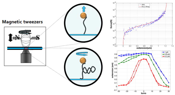
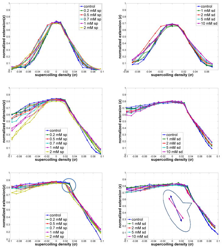
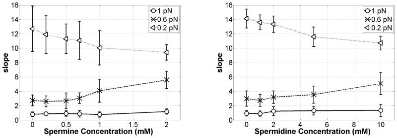
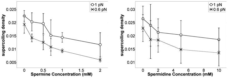
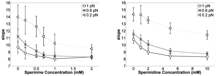
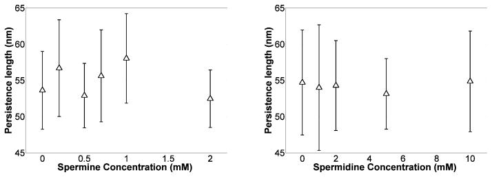
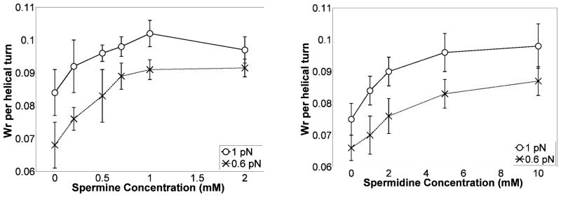
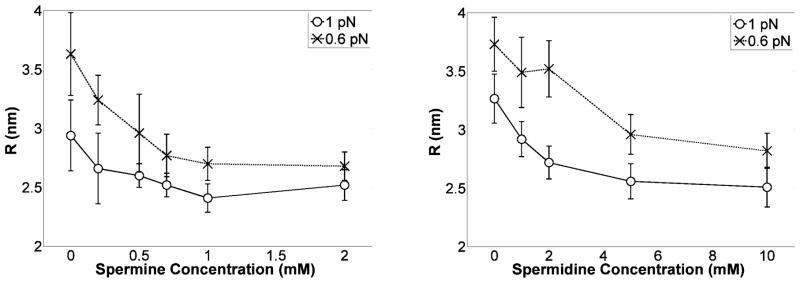
Quantitative analysis of single molecule experiments show that adding either of two natural polyamines, spermine or spermidine, produced more compact plectonemes in DNA in physiological concentrations of monovalent salt. They also promoted plectoneme formation at lower values of torsion in measurements of extension versus twist. Quantifying changes in the plectonemic DNA using some results from simple rod models suggested that exposure to polyamines reduced the radii and increased the densities of plectonemes. Thus, polyamines may limit the twist density by favoring writhe which maintains the B-form. Although polymerases may significantly stretch the double helix, denature DNA, and produce twist instead of writhe, natural polyamines stabilize base-pairing, limit twist to maintain the B-form, and promote supercoiling, which is conducive to replication and transcription and essential for DNA packaging.

Keywords: magnetic tweezers; single-molecule techniques; spermidine; spermine.


Figures

Figure 1
Figure 1
Structure of spermidine (top) and spermine (bottom). The basic amino (NH2) groups attract protons at physiological pH to become positively charged.
Figure 2
Figure 2
Stretching and twisting DNA with magnetic tweezers. Magnetic tweezers (left) can be used to stretch (upper middle) and twist (lower middle) single DNA molecules tethering para-magnetic beads to a glass surface. Systematically varying the separation between the magnets and the tethered bead generates a range of tensions as shown in a representative force vs. extension curve for a single DNA molecule (upper right). The red curve is a worm-like-chain fit to the data. Rotating the magnets to twist the DNA tether produces extension vs. twist curves for a DNA molecule at different tensions (lower right), from top to bottom: 1, 0.6, and 0.2 pN.
Figure 3
Figure 3
DNA extension vs. supercoiling density as a function of spermine (left) or spermidine (right) concentrations at 0.2 (upper), 0.6 (middle), and 1 pN (lower) of tension. “Knee-points” are encircled. The inset emphasizes the change in slope due to added spermidine. Typical measurement errors are shown in supplementary information on a copy of the curves for 0.6 pN with added spermine.
Figure 4
Figure 4
Average slope (dz/dσ, dimensionless) of the DNA extension vs. twist in Fig. 3 for under-wound DNA as a function of spermine (left) or spermidine (right) concentrations at 0.2, 0.6, and 1 pN of tension. Error bars represent standard deviations.
Figure 5
Figure 5
Twist values σ at which writhe first appeared in over-twisted DNA as a function of spermine (left) or spermidine (right) concentrations at 0.6 and 1 pN of tension. The σ values were defined using the linear extrapolation of extension vs. σ curves. Error bars represent standard deviations.
Figure 6
Figure 6
Average magnitude of the slope (dz/dσ, dimensionless) of DNA extension vs. twist for positively supercoiled DNA as a function of spermine (left) or spermidine (right) concentrations at 0.2, 0.6, and 1 pN of tension. Error bars represent standard deviations.
Figure 7
Figure 7
Average persistence length for eight DNA tethers at different spermine (left) or spermidine (left) concentrations. Polyamines did not significantly affect the bending stiffness of DNA. Error bars represent standard deviations.
Figure 8
Figure 8
Effect of spermine (left) or spermidine (right) concentrations on the writhe density in the plectonemic phase at 0.6 and 1 pN of tension. The increase in writhe density is a quantitive measure of plectoneme shrinking. Error bars represent standard deviations. Note that the model is valid for small fluctuations at tensions above 0.5 pN.
Figure 9
Figure 9
Radius of the helical plectoneme as a function of spermine (left) or spermidine (right) concentrations at 0.6 and 1 pN of tension. The radii of plectonemes decreased substantially as the polyamine concentration was increased, especially at low tension.

References

    1. Kusano T, Berberich T, Tateda C, Takahashi Y. Planta. 2008;228(3):367–81. - PubMed
    1. Wallace HM, Fraser AV, Hughes A. Biochemical Journal. 2003;376:1–14. - PMC - PubMed
    1. Grandgirard N, Ly-Sunnaram B, Ferrant D, Gandemer V, Edan C, Le Gall E, Moulinoux JP, Leray E, Goasguen JE. Leukemia Research. 2004;28(5):479–486. - PubMed
    1. Mcclellan JA, Boublikova P, Palecek E, Lilley DMJ. Proceedings of the National Academy of Sciences of the United States of America. 1990;87(21):8373–8377. - PMC - PubMed
    1. Wang JC. Nature Reviews Molecular Cell Biology. 2002;3(6):430–440. - PubMed

LinkOut - more resources